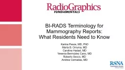
What Residents Need to Know Karina Pesce MD PhD María B Orruma MD Carolina Hadad MD Yesenia Bermúdez Cano MD Roberto Secco MD Andrea Cernadas MD Authors Affiliation Department of Breast Radiology ID: 913086
Download Presentation The PPT/PDF document "BI-RADS Terminology for Mammography Repo..." is the property of its rightful owner. Permission is granted to download and print the materials on this web site for personal, non-commercial use only, and to display it on your personal computer provided you do not modify the materials and that you retain all copyright notices contained in the materials. By downloading content from our website, you accept the terms of this agreement.
Slide1
BI-RADS Terminology for Mammography Reports: What Residents Need to Know
Karina Pesce, MD, PhD
María B. Orruma, MD
Carolina Hadad, MD
Yesenia Bermúdez Cano, MD
Roberto Secco, MD
Andrea Cernadas, MD
Slide2Authors’ Affiliation:
Department of Breast RadiologyHospital Italiano de Buenos AiresPres. Tte. Gral. Juan Domingo Perón 4190C1199 ABB, Buenos Aires, ArgentinaCorresponding Author:Karina Pesce, MD, PhDe-mail: drakarina.pesce@gmail.comPresented as an education exhibit (BR 176-ED-X) at the 2017 RSNA Annual Meeting. All authors have disclosed no relevant relationships.
Slide3The structure of the Breast Imaging Reporting and Data System (BI-RADS) lexicon lends itself to the consistent and rational evaluation of mammographic findings and facilitates resident and breast imaging fellowship training.
Both the American College of Radiologists (ACR) and the Society of Breast Imaging recommend that breast imaging education within residency and fellowship programs should require the use of BI-RADS terminology, assessment categories, and management recommendations.1In this presentation, we present the updated lexicon for mammography from the BI-RADS fifth edition,2 using examples of original cases to illustrate the BI-RADS terminology used to facilitate the education of resident radiologists. Introduction
Slide4Describe breast lesions by using the correct BI-RADS descriptors. Determine the appropriate BI-RADS category at mammography.Define the indications for using a BI-RADS category 3 at mammography.
Develop a skillful and practical approach to performing and interpreting mammograms using the BI-RADS lexicon.
Slide51
234The illustrative BI-RADS fifth edition2 is a product of years of collaboration among subsection heads, committees, and the ACR, and most importantly, input from users of the system.
Slide6Mammography Report Structure
Indication for examinationSuccinct description of the overall breast compositionClear description of any important findings with comparison to previous examinationsAssessmentManagementPerform mammographyRecall a screening-detected finding and evaluate a clinical findingSpecify the finding and its locationFollow-up as either a probably benign lesion or cancer, treated with breast conservation
1
Slide72
Descriptors of Breast Density Almost entirely fatty tissue
Scattered areas of fibroglandular densities
Heterogeneously dense tissue
Extremely dense tissue
The BI-RADS written descriptors
of breast density
should be used
in the radiologic report. Letters
or numbers should not be used.
a
b
c
d
(
a-d
) Mediolateral oblique (MLO) mammograms show the various types of breast tissue density according to the ACR.
Slide83
Findings Described in the BI-RADS Lexicon
Provide a clear description of any important findings.
It is assumed that most important findings are
(a)
of concern at screening,
(b)
inherently suspicious,
(c)
new, or
(d)
interpreted to be larger and/or more extensive when compared to the findings depicted at previous examinations.
*
Mass
Microcalcifications
Architectural distortion
Asymmetries
Slide9Higher
probability
of
a benign lesion
Higher
probability
of
malignancy
A mass is defined as a three-dimensional occupying lesion that is seen on two different mammographic projections.
What do residents need to know when describing a mass depicted at mammography?
Remember these
mnemonics:
Shape:
ROI
Margin:
COMIS
Masses Depicted at Mammography: Morphology
Slide10Higher
probability
of
benign
Higher
probability
of
a benign lesion
Higher
probability
of
malignancy
Associated features
to describe include:
Architectural distortion and edema
Duct changes
Skin thickening
Skin retraction
When describing the
location
of the mass, include its:
Laterality
Quadrant and clock-face description
Depth
Distance from the nipple
Masses Depicted at Mammography: Density, Calcifications, Associated Features, and Location
Mass
Slide11Typically Benign Microcalcifications
VascularRoundPopcorn-LikeRim
Management
Continued on next slide
Skin
Round
Dystrophic
Round
Vascular
Popcorn-like
Skin:
Typically lucent-centered and pathognomonic in appearance
Dystrophic
:
Irregular shape, usually > 1 mm in diameter, often with lucent centers
Round:
May be considered benign when diffuse and small (< 1 mm) and are frequently formed in the acini of lobules. When smaller than 0.5 mm, the term
punctate
should be used to describe these findings.
Vascular
:
Parallel tracks or linear tubular calcifications that are clearly associated with blood vessels
Popcorn-like
:
Classic large calcifications (> 2–3 mm at the greatest diameter); produced by an involuting fibroadenoma
Describing distribution may not be appropriate for typically benign calcifications.
Routine Mammography Screening
a
b
c
d
e
Mammograms obtained from different patients show typically benign microcalifications (arrow), including skin (
a
), dystrophic (
b
), round (
c
), vascular (
d
), and popcorn-like (
e
).
Slide12Round
Rim
Management
Large rodlike
Round
Milk-of-calcium
Suture
Rim
Large rodlike
: Associated with ductal ectasia; may form solid or discontinuous smooth linear rods, most of which are
0.5 mm or larger in diameter
Milk-of-calcium:
Manifestation of sedimented calcifications in macro- or microcysts, usually but not always grouped. On craniocaudal (CC) views, they are often less evident and appear as round smudgy deposits, while occasionally on MLO views and especially on 90° lateral (lateromedial/mediolateral [ML]) views they are more clearly defined and are often semilunar, crescent shaped, curvilinear (concave up), or linear, defining the dependent portion of the cysts.
Suture:
Calcium deposited on suture material
Rim:
T
hin benign calcifications that appear as calcium deposited on the surface of a sphere
Routine Mammography Screening
f
g
h
i
Typically
Benign Microcalcifications: Continued
Mammograms obtained from different patients show more typically benign microcalifications (arrow), including large rodlike (
f
), milk-of-calcium (
g
), suture (
h
), and rim (
i
).
Slide13RegionalPPV = 26%Scattered in a larger volume (> 2 cc) of breast tissue and not in the expected ductal distributionMicrocalcifications: DistributionGroupedPPV = 31%5 or more microcalcifications within 1 cm3, maximum 2 cm
Segmental
PPV = 62%
Calcium deposits in the ducts and branches of a segment or lobe
Linear
PPV 60%
Arrayed in a line is a finding suggestive of deposition along the ducts
The distribution of microcalcifications can indicate the positive predictive value (PPV) for malignancy in BI-RADS. The illustration depicts an MLO view of the breast that details the PPV with the corresponding type of distribution, along with corresponding insets of cropped mammograms.
Diffuse
PPV 0%
Slide14Suspicious Microcalcifications
Amorphous
Coarse
heterogeneous
Management
Tissue
diagnosis
Magnified areas of interest from mammograms show amorphous (arrows in
a
) and coarse heterogenous (arrow in
b
) calcifications. The patient depicted in
a
underwent a
stereotactic core biopsy, the results of which confirmed atypical apocrine adenosis.
The patient depicted in
b
underwent a
stereotactic core biopsy, the results of which confirmed a
high-grade intraductal carcinoma.
Morphology, distribution, associated features, and location of suspicious microcalcifications should be included in the radiologic report.
Amorphous:
PPV: 21% BI-RADS category 4B; so small and/or hazy in appearance that a more specific particle shape cannot be determined
Coarse heterogeneous:
PPV: 13% BI-RADS category 4B; irregular conspicuous calcifications that are generally 0.5 mm–1 mm in diameter and tend to coalesce but are smaller than dystrophic calcifications
a
b
Slide15Suspicious Microcalcifications: Continued
Fine
pleomorphic
Fine linear
or
fine-linear
branching
Management
Tissue
diagnosis
c
d
Mammograms show fine plemorphic (arrow in
c
) and fine linear or fine-linear branching (arrow in
d
) calcifications. Both patients underwent
stereotactic core biopsies, and the results of each confirmed
high-grade intraductal carcinoma
.
Morphology, distribution, associated features, and location of suspicious microcalcifications should be included in the radiologic report.
Fine pleomorphic:
PPV = 29%, BI-RADS category 4C; usually more conspicuous than amorphous forms and have discrete shapes. These irregular calcifications are distinguished from fine linear and fine linear branching forms by the absence of fine-linear particles. Fine pleomorphic calcifications vary in size and shape and are usually less than 0.5 mm in diameter.
Fine linear or fine-linear branching:
PPV = 70%, BI-RADS category 4C. Thin linear irregular calcifications that may be discontinuous and less than 0.5 mm in caliber. Occasionally, branching forms may be seen. Their appearance suggests filling of the lumen of a duct or ducts involved irregularly by breast cancer.
What is the next step?
Diagnostic evaluation and obtaining additional mammographic views at three-dimensional (3D) mammography and breast US.MLO mammogram shows architectural distortion (arrow).In the absence of history of trauma or surgery, architectural disortion is suspicious for malignancy or radial scar.
Architectural Distortion
Slide17(a, b)
Mammograms show a focal asymmetry (arrow).
Focal asymmetry
is evaluated relative to the corresponding location in the contralateral breast and represents a relatively small amount of dense fibroglandular tissue over a confined portion of the breast (less than one quadrant).
A two-view finding that lacks convex borders (may or may not contain interspersed fat)
occupies less than one quadrant of the breast, similar in appearance to that depicted on CC and MLO views.
Clinical Pearl
Diagnosing asymmetry
involves a careful
analysis
and the
comparison
with at least one (often
more) previous examination.
It should include a diagnostic evaluation with additional mammographic views and breast US images, and 3D mammography may also be performed for the evaluation.
(
a
) MLO mammogram shows an area of asymmetry (arrow).
(
b
) CC mammogram does not show the asymmetry.
Asymmetry
: An area of dense fibroglandular tissue that is visible on only one mammographic projection. Most of these findings represent summation artifacts and superimposition of normal breast structures.
A one-view
finding
that
lacks
convex
borders that may
or
may
not
contain interspersed fat occupies less than
one quadrant of the breast.
ab
ab
Asymmetry
Slide18MLO mammograms obtained in 2017 (a) and 2018 (b) show the development of an asymmetry (arrow). Developing asymmetry:
This is a focal asymmetry that is new, larger, and more conspicuous than that depicted on a previous examination.Approximately 15% of developing asymmetries are found to be malignant, so these cases warrant further imaging evaluation and biopsy unless found to be characteristically benign at further workup.
(
a, b) Bilateral MLO mammograms show a global asymmetry (arrow). Global asymmetry
: Global asymmetry is evaluated relative to the corresponding area in the contralateral breast and represents a large amount of dense fibroglandular tissue over a substantial portion of the breast (
at least one quadrant
).
There is no mass, architectural distortion, or associated suspicious calcifications.
Global asymmetry usually represents a normal variant.
a
a
b
b
Asymmetry: Continued
Slide19Mammogram shows a skin lesion (arrow). This finding may be described in the mammography report or annotated on the mammographic image when it projects over the breast (especially on two different projections) and may be mistaken for an intramammary lesion. A raised skin lesion that is large enough to be seen at mammography should be marked by the technologist with a radiopaque device designated for use as a marker for a skin lesion. Dots = marked raised skin lesion.
Mammograms show a solitary dilated duct (arrow). Solitary dilated duct is a rare mammographic finding, and it can be associated with noncalcified ductal carcinoma in situ (DCIS).3Solitary dilated duct appears to have a greater than 2% likelihood of malignancy, sufficiently high enough to suggest that a suspicious (BI-RADS category 4A) assessment may be appropriate.
Solitary dilated
duct3 Chang CB, Lvoff
NM, Leung JW, Brenner RJ, Joe BN, Tso HH, Sickles
EA.Solitary
dilated duct identified at mammography: outcomes analysis.
AJR Am J
Roentgenol
. 2010;194(2):378-382.
Skin
lesion
a
b
Continued on next slide
Slide20Skin
lesionMammogram shows a normal intrammary lymph node (arrow).A normal intramammary lymph node is a mass less than 1 cm in diameter that is well circumscribed and slightly lobulated. In addition, in most instances, a radiolucent cleft (which represents fat in the hilum of the node) is depicted. The typical location of these nodes is the upper outer quadrant of the breast.
Intramammary Lymph Node
Slide21Determining Lesion Location in the Breast
RIGHT
8’clock
anterior
Illustration shows the
right breast.
A complete set
of
lesion location
descriptors
should include:
Designation
of
the finding in the right
or
left
breast
Quadrant
and clock-face
notation
(
preferably
both
)
Depth (anterior, middle,
or
posterior third)
Distance
from
nipple
Superior
MLO view
MLO view
Lateral
CC view
CC view
Inferior
Medial
middle
posterior
Distance
from
the nipple
Illustration shows the
left breast.
Slide22Compare recent mammography examinations with previous examinations, if deemed appropriate by the interpreting physician
Assessing Location
Slide23A significant change in the fifth edition of BI-RADS that is relevant to all breast care providers is the separation of the BI-RADS assessment from patient management. 45
The separation was implemented to provide more flexibility for several specific clinical cases for which a seemingly discordant management recommendation might be appropriate for a given assessment
.
Assessment and Patient Management
Slide244
BI-RADS Assessment CategoriesBI-RADS 0: Needs additional image evaluation and/or prior mammograms for comparison
BI-RADS 1
: Negative (0%
likelihood
of
malignancy
)
BI-RADS 2
: Benign (0%
likelihood
of
malignancy
)
BI-RADS 3
: Probably
benign
(0 % to ≤ 2%
likelihood
of
malignancy
)
BI-RADS 4
:
Suspicious
abnormality
(
likelihood
of
malignancy
:
4A
=
2%–10%,
4B
=
10
%–50
%, and
4C =
50%–95%)
BI-RADS 5
:
Highly
suggestive
of malignancy (≥ 95% likelihood of malignancy)
BI-RADS 6
:
Known
biopsy-proven
malignancy
Management: BI-RADS Category 1
Bilateral MLO (a) and CC (b) mammograms show a normal examination. There are no findings to comment on.
Management
Routine mammography screening
a
b
Slide26Case 1: CC (a) and MLO (b) mammograms show dense breast tissue.
Assign a BI-RADS 1 category if there are no abnormal imaging findings in a patient with a palpable abnormality, (possibly a palpable cancer), but add a note in the report recommending surgical consultation or tissue diagnosis if clinically indicated. Negative Mammogram
With or without palpable breast nodule
In this case, assign the screening mammography a BI-RADS category 1.
BI-RADS Category 1
a
b
Slide27BI-RADS Category 2: Benign Assessment
Management
Routine mammography screening
Spectrum of Benign Findings
Slide28A focal asymmetry should be nonpalpable and should not have have a sonographic correlate.
These findings are generally depicted on baseline examinations.
IMPORTANT
A BI-RADS 3 should
not
be assigned at screening mammography. It
should
be assigned after a diagnostic workup has been
completed.
BI-RADS Category 3: Probably Benign
4
Continued on next slide
Focal asymmetry (arrows)
Slide29IMPORTANT
BI-RADS 3 should not be used when a radiologist is “not sure” whether a finding is benign or suspicious.“I don’t know.”“It is strange.”“I had a similar cancer case.”“I am not sure about it.”
The fifth edition of BI-RADS is more flexible than previous editions as it allows for a BI-RADS category 3 to be assigned (even if the lesion is not part of the three findings accepted as BI-RADS category 3) if the radiologist has had personal experience to support this assignment. Examples include:
Calcifications that are suggestive of early evolving fat necrosis
Developing calcifications (arrow in
a
) that seem to be most likely vascular, but not definitely
Lesions (arrows in
b
and
c
) in which stability evaluation is difficult owing to technical differences between an analog image (
b
) and a digital image (
c
). The can also occur when comparing images obtained using equipment from different vendors.
2012
2013
BI-RADS Category 3: Probably Benign
4
a
b
c
Slide30BI-RADS Category 3: Management Follow-up Algorithm
If stable, follow-up for 2 yearsIf the lesion increases in size (> 20% diameter) during follow-up, update the category to a BI-RADS category 4 and recommend biopsy. The increase in size (> 20%) applies to masses (likely fibroadenomas).
If the BI-RADS category 3 lesion disappears or becomes apparently benign before the 2-year follow-up, assign a
BI-RADS 2 category.
Interobserver
Variability
Disagreement on the use of BI-RADS descriptors from one examination to the next may lead to an upgrade in the BI-RADS categorization without any demonstrable change between examinations.
IMPORTANT
Slide31BI-RADS Category 4: Suspicious for Malignancy
Category 4A: Low suspicion for malignancy
Category 4B: Moderate suspicion for malignancy
Category 4C: High suspicion for malignancy
Management
Tissue Diagnosis
Division into BI-RADS 4A, 4B, 4C is an option.
BI-RADS 4A
Solitary
dilated
duct
BI-RADS 4B
Microcalcifications:
coarse
heterogeneous
BI-RADS 4C
Architectural
distortion
Mammographic Examples
Slide32Chance of malignancy ≥ 95%
Assigned for classic cases of malignancyIf the results of a biopsy indicate a benign lesion, recommend a repeat (usually surgical) biopsy.Recommendation should be noted as follows: “Biopsy should be performed in the absence of clinical contraindication.”New segmental fine-linear branching calcifications (arrow)
Irregular-shaped high-density mass (arrow) with skin retraction and calcificationsIrregular spiculated high-density mass (arrow) with associated
microcalcificationsExamples: Magnified Areas of Interest on Mammograms
BI-RADS
Category
5: High Risk for Malignancy
Slide33BI-RADS Category 6: Biopsy-Proven Malignancy
This category is reserved for examinations performed after the results of a biopsy indicate malignancy (eg, imaging performed after percutaneous biopsy but prior to complete surgical excision) in which there are no mammographic abnormalities other than the known cancer that might need additional evaluation.
When to use it:
Assign this category after monitoring response to neoadjuvant chemotherapy. BI-RADS category 6 is commonly assigned at preoperative MRI (when only the malignancy is depicted and there are no other suspicious findings).
a
b
ML (
a
) and CC (
b
) mammograms show a right inferior internal quadrant mass, skin edema, and axillary adenopathies
Slide34(a) MLO mammogram shows unilateral axillary adenopathy (arrow) with no suspicious findings in the breasts. Screening mammography = BI-RADS 0. Consequently, a careful search of the ipsilateral breast images is warranted.
Think:The presence of unilateral axillary adenopathy suggests occult breast carcinoma or, much less commonly, lymphoma, metastatic melanoma, ovarian cancer, or other metastatic cancers.BI-RADS Category 4 Why? In the absence of a known infectious or inflammatory cause, isolated unilateral axillary adenopathy is suspicious for malignancy.
Case 2: Unilateral Axillary Adenopathy
a
b
(
b
) Bilateral axillary US image shows asymmetry.
Bilateral US should be performed to confirm that the finding is asymmetric and/or unilateral.
Slide35(a) Bilateral MLO mammograms show bilateral axillary adenopathy (arrows) with no suspicious findings in the breasts in a patient with known lymphoma.
Clinical PearlIn this case, bilateral axillary adenopathy is presumed owing to the known diagnosis of lymphoma.BI-RADS Category 2Why? Because in this case, the BI-RADS assessment should be determined on the basis of the imaging findings depicted in the breasts, but the report also should indicate the presence of adenopathy and the known underlying disease.
Case 3: Bilateral Axillary Adenopathy
a
b
(
b
) US image shows axillary adenopathy with loss of the hilar cortical relationship.
Slide36Mammography is performed after an attempted complete surgical excision, and the results of a pathologic examination indicate positive resection margin.
(a, b) Mammograms show postsurgical scarring.If images show only postsurgical scarring (although the results of a pathologic examination are margin positive), then BI-RADS category 2 should be assigned. An extra note stating that the pathology report suggests a residual tumor should be included in the radiology report.Case 4: Postsurgical Scarring
a
b
Slide37ML (a) and CC (b) mammograms, magnification of the retroareolar section (c), and US image (
d) show no imaging findings of malignancy in a woman with clinically suspected Paget disease. (e) Photograph shows an erythematous scaly patch with oozing and crusting in the areola, which effaces the nipple.It is important to know the clinical manifestations of Paget disease because they can be the only signs of the breast cancer.
BI-RADS Category 2
Why?
Because in this situation, the BI-RADS assessment should be determined on the basis of the imaging findings depicted in the breasts.
Continued
on
next
slide
Case 5: Assessment-Management Discordance
Paget Disease
Symptoms
Itching, eczema, erythema of the nipple and areola, nipple erosion or ulceration, scaly or flaky skin, nipple retraction, bloody discharge from the nipple, or a combination of these
a
b
ML
CC
CC
c
d
e
Slide38Another case that involves assessment-management discordance occurs in a patient with clinical suspicion for Paget disease but no mammographic or US findings that suggest malignancy.
Performing breast MRI is the appropriate follow-up option.IMPORTANT!BI-RADS category 0 should not be used for diagnostic breast imaging findings that warrant further evaluation with MRI.BI-RADS Category 1Why? Because in this situation, the BI-RADS assessment should be determined on the basis of the imaging findings depicted in the breasts.
Assessment-Management Discordance: Continued
Slide39Skin Marking
To properly educate interpreting physicians, an institution should adopt a policy requiring the consistent use of two different shapes of radiopaque markers for marking palpable and skin lesions.No, because there has been no consensus on establishing the use of specific-shapedmarkers to represent palpable versus skin lesions.Nodule Palpable
Skin
lesion
Scar
Is
there
BI-RADS
guidance
concerning
the
standardization
of
breast
skin
markers
in
the
fifth edition?
Magnified areas of interest on mammograms demonstrate various methods of denoting skin markings.
Slide40Follow-up Postlumpectomy
20162017
2016
2017
Mammograms from 2016 (
a
) and 2017 (
b
) show an increase in architectural distortion (lines).
a
b
Slide41Mammography in Men
Do mammography examinations performed on men require a BI-RADS final assessment and/or numeric code?
All mammography examinations regardless of the patient’s sex
must have a final BI-RADS assessment category (but not a numeric code) in the mammography report. However, management recommendations may differ from those made for women because annual screening mammography is not usually appropriate for men.
(
a–c
) Mammograms obtained in a male patient show an irregular subareolar mass with spiculated margins.
a
b
c
Slide42Testing YourselfThe finding denoted by the arrow in the CC mammogram of the right breast is most likely which of the following?
A. An oil cyst.B. A calcified fibroadenoma.C. Fat necrosis.D. Milk-of-calciumcalcification.B. Popcorn-like calcifications (arrow) are findings compatible with calcified fibroadenoma.On the basis of the findings denoted by the arrow on the straight lateral (90°) magnification view from a mammogram, which BI-RADS category should be assigned?
D
. On straight lateral (90°) magnification view, layering the calcifications (arrow) would be necessary in order to make the diagnosis of benign milk-of-calcium calcifications.
As depicted on the accompanying mammogram, in which of the following cases would a BI-RADS category 6 assignment be
most
appropriate?
A. Suspicious abnormality; a biopsy should be performed.
B. Postprocedure mammography for marker placement.
C. Recent biopsy-proven breast cancer.
D. Previously treated breast cancer.
C
. BI-RADS category 6 is assigned to patients with a cancer diagnosis who have not yet been definitively treated.
A.BI-RADS 3.
B.BI-RADS 4.
C.BI-RADS 1.
D.BI-RADS 2.
Slide43Conclusion
Slide44Suggested ReadingsD’Orsi CJ, Kopans DB. Mammography interpretation: the BI-RADS method. Am Fam Physician 1997;55(5):1548–1550, 1552.D’Orsi CJ, Sickles EA, Mendelson EB, et al. ACR BI-RADS atlas, Breast Imaging Reporting and Data System. 5th ed. Reston, Va: American College of Radiology, 2013.Mercado CL. BI-RADS update. Radiol Clin North Am 2014;52(3):481–487.
Michaels AY, Birdwell RL, Chung CS, Frost EP, Giess CS. Assessment and management of challenging BI-RADS category 3 mammographic lesions. RadioGraphics 2016;36(5): 1261–1272.Rao AA, Feneis J, Lalonde C, Ojeda-Fournier H. Pictorial review of changes in the BI-RADS fifth edition. RadioGraphics 2016;36(3):623–639.Sickles EA, Philpotts LE, Parkinson BT, et al. American College of Radiology/Society of Breast Imaging curriculum for resident and fellow education in breast imaging. J Am Coll Radiol 2006;3(11):879–884.Tabár L, Vitak B, Chen TH, et al. Swedish two-county trial: impact of mammographic screening on breast cancer mortality during 3 decades. Radiology 2011;260(3):658–663.
Slide45References
Sickles EA, Philpotts LE, Parkinson BT, et al. American College of Radiology/Society of Breast Imaging curriculum for resident and fellow education in breast imaging. J Am Coll Radiol 2006;3(11):879–884.D’Orsi C, Sickles EA, Mendelson EB, Morris EA. Breast Imaging Reporting and Data System: ACR BI-RADS breast imaging atlas. 5th ed. Reston, Va: American College of Radiology, 2013Chang CB, Lvoff NM, Leung JW, et al. Solitary dilated duct identified at mammography: outcomes analysis. AJR Am J Roentgenol. 2010;194(2):378-382. Michaels AY, Birdwell RL, Chung CS, Frost EP, Giess CS. Assessment and management of challenging BI-RADS category 3 mammographic lesions. RadioGraphics 2016;36(5):1261–1272.