/
Injuries to the Thigh, Leg, and Knee… Injuries to the Thigh, Leg, and Knee…

Injuries to the Thigh, Leg, and Knee… - PowerPoint Presentation

popsmolecules
popsmolecules . @popsmolecules
Follow
343 views
Uploaded On 2020-06-16

Injuries to the Thigh, Leg, and Knee… - PPT Presentation

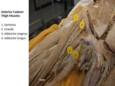
We will go over anatomy that covers bones ligaments tendons muscles nerves and blood vessels of the region We will discuss the kinesiology of movements created by the muscles though the major joints ID: 779214

athlete knee muscles joint knee athlete joint muscles patella ligament injuries

Share:

Link:

Embed:

Download Presentation from below link

Download The PPT/PDF document "Injuries to the Thigh, Leg, and Knee…" is the property of its rightful owner. Permission is granted to download and print the materials on this web site for personal, non-commercial use only, and to display it on your personal computer provided you do not modify the materials and that you retain all copyright notices contained in the materials. By downloading content from our website, you accept the terms of this agreement.


Presentation Transcript

Slide1

Injuries to the Thigh, Leg, and Knee…

Slide2

We will go over anatomy that covers bones, ligaments, tendons, muscles, nerves, and blood vessels of the region

We will discuss the kinesiology of movements created by the muscles though the major joints

The chapter continues with a description of soft-tissue injuries to the thigh that can become debilitating if not cared for properly

Including, contusions, strains, and various joint-related injuries

Slide3

This chapter will cover issues such as

osteochondritis

dissecans

, inflamed

bursae

, and patellar dislocation, along with injuries caused by chronic exercise

The chapter will also describe the four major ligaments of the knee and injuries to the knee joint that can be injured during sports participation

The chapter concludes with a discussion of prophylactic and functional knee bracing

Slide4

Anatomy Review…

The lower extremity is an area where many athletes experience some type of injury during their sports career

The bones of the extremity include the femur, tibia, fibula, patella, and those of the foot (Gray, 1974)

http://www.youtube.com/watch?v=qoiaUV7fGEI

Slide5

Anatomy Review…

The femur or thigh bone is the longest, strongest, and heaviest bone in the body

It has a rounded, ball-like head that attaches to the hip bone wit the help of ligaments

The head of the femur is attached to the shaft of the femur by a region known as the neck

The femur becomes flatter and wider as it proceeds toward the knee, where it articulates with the tibia

Slide6

Anatomy Review…

The thigh has a great deal of blood and nerve tissue, both

anteriorly

and

posteriorly

The anterior portion contains the long

saphenous

vein and several branches of the femoral nerve

The posterior section of the thigh are the deep femoral artery and the major nerve to the leg, the sciatic nerve

Most are quite well protected by the musculature of the thigh

Slide7

Anatomy Review…

The muscles of the thigh can be broken down into three basic regions

First, the anterior muscles of the thigh, commonly called the quadriceps have two functions

The

vastus

lateralis

,

vastus

intermedius

,

vastus

medialis

and rectus femoris work together to extend the leg at the knee jointThree of these muscles (VMO, intermedius, and lateralis) attach on the femur and run down the thigh to the quadriceps tendon

Slide8

Anatomy Review…

The rectus

femoris

attaches on the hip bone at the anterior inferior iliac spine and runs down the leg to the quadriceps tendon

The other muscle in the anterior portion of the thigh is the

satorius

It also attaches on the hip bone and runs somewhat diagonally down the thigh to the anterior medial portion of the

tibial

condyle

This muscle is responsible for flexing, abduction, and lateral rotating the thigh at the hip

Slide9

Anatomy Review…

The main muscles of the medial aspect of the thigh include the adductor

longus

, adductor

brevis

, adductor

magnus

and the

gracilis

These muscles attach on the pelvis and run to the femur

The main function of these muscles is to adduct the hip with flexion of the thigh

Slide10

Anatomy Review…

The third group of muscles in the thigh are in the posterior aspect of the thigh and are commonly known as the hamstrings

These include the

semitendinosus

,

semimembranosus

, and biceps

femoris

All these muscles attach on the pelvis and run down the leg to the tibia

The main function of this group of muscles is to flex the leg at the knee

Slide11

Anatomy Review…

http://www.youtube.com/watch?v=VdXAOWmbRuw

Slide12

Anatomy Review…

The knee is a very complex joint

It can be damaged through any number of accidents occurring during sports participation

The femur and the tibia articulate with each other to form the

tibiofemoral

joint

The patella and the femur articulate with each other to form the

patellofemoral

joint

Slide13

Anatomy Review…

The patella is a

sesamoid

bone, which means that it is totally enclosed within a tendon

In this case, the quadriceps tendon

The patella does not articulate with the tibia

Many ligaments support the knee joint

Slide14

Anatomy Review…

There are 4 ligaments that serve as primary stabilizers of this joint:

Tibial

or medial collateral ligament (MCL)

The fibular or lateral collateral ligament (LCL)

The anterior

cruciate

ligament (ACL)

Posterior

cruciate

ligament (PCL)

Slide15

Anatomy Review…

The

tibial

(medial) collateral ligament extends from the medial

epicondyle

of the femur down to the medial

condyle

of the tibia

The

tibular

(lateral) collateral ligament begins at the lateral

epicondyle

of the femur and extends to the head of the fibula

Slide16

Anatomy Review…

The fibular collateral ligament is the stronger of the two

Both ligaments help limit motion and/or disruption of the knee joint when movement at the joint is in a side-to-side direction

Valgus

(knock-knees)

Varus

(bow legs)

Slide17

Anatomy Review…

The

cruciate

ligaments, unlike the collateral ligaments, are situated on the inside of the joint

The ACL attaches on the anterior portion of the

intercondylar

area of the tibia and runs superiorly and

posteriorly

to the internal aspect of the lateral femoral head

Slide18

Anatomy Review…

The PCL attaches on the posterior aspect of the

intercondylar

area of the tibia and runs superiorly and

anteriorly

, passing the ACL on the medial side and attaching to the internal aspect of the medial femoral

condyle

The function of these two ligaments is primarily to reduce or prevent anterior and posterior displacement of the femur or the tibia

Slide19

Anatomy Review…

Two semicircular

fibrocartilaginous

disks, commonly called cartilage and more scientifically termed the menisci, are located within the space between the tibia and the femur

The menisci assist with the protection and nourishment of the knee joint, aid in the distribution of weight and stress applied to the joint surfaces, and help with the biomechanics of the joint

Slide20

Anatomy Review…

There are two the medial and lateral menisci

Tendons of the muscles mentioned earlier in the description of the thigh run across the knee

Between the tendons and bone are several

bursae

, which reduce the friction of muscle tendons rubbing over a prominent area of bone, thereby adding some padding for the exposed bony areas of the knee

Slide21

http://www.youtube.com/watch?v=_q-Jxj5sT0g

Slide22

Common Sports Injuries

Injuries can occur in any sport

This area can sustain injuries that are a result of overuse, trauma caused by an opponent, or trauma produced by the power and explosive movements required in some sports

Because the knee is part of a complex mechanical system that includes the foot, ankle, lower leg, hip, and pelvis, there are times when another part of this system causes problem that can eventually be exhibited in the knee

Slide23

Skeletal Injuries…Femoral Fractures

The femur is the longest bone ins the body and is therefore subject to being fractured

However, this requires a great deal of force and is not common occurrence in sports

If a fracture does occur to the shaft of the femur as a result of sports participation, the injury is quite obvious

The athlete should not attempt to walk on a femoral fracture

Slide24

Skeletal Injuries…Femoral Fractures

The athlete must be immediately transported to the nearest medical facility with the leg splinted and without bearing any weight on the affected limb

A femoral fracture requires urgent medical attention because the initial trauma can lead to multiple problems

Including a lack of circulation, nervous innervations, or shock and other medical issues

Slide25

Skeletal Injuries…Femoral Fractures

The neck of the femur can also be fractured

This occurs more often in sports than does a fracture of the shaft

Older children and teenagers are at greater risk for this injury because the fracture can potentially occur at the site of a growth plate

Among younger athletes these fractures can be the result of direct trauma or overuse

Slide26

Skeletal Injuries…Femoral Fractures

If direct trauma is the cause, the athlete typically had a foot planted and then got hit in the hip or upper thigh with a great deal of force

This injury needs to be evaluated ASAP by a physician

Once complication of a fracture in the neck of the femur is

avascular

necrosis (tissue death) of the femoral head

Caused by a blood supply to the bony portion of the femoral head

Slide27

Skeletal Injuries…Femoral Fractures

S&S

Pain at the site of injury

Difficulty ambulating on the affected leg

Swelling and/or deformity may occur

Athlete may report a traumatic event as the cause

The athlete may report having heard or felt a severe pop or snap at the time of injury

TX:

Be prepared to treat the athlete for shock if necessary

Splint the injured leg, preferably with a traction splint

Apply sterile dressings to any related open wounds

Monitor vital signs and circulation to the lower leg

Arrange for transport to the nearest medical facility

Slide28

Skeletal Injuries…Patella Fracture

Other skeletal problems include a fracture of the patella and dislocation of the knee or

tibiofemoral

joint

Although the patella can be fractured, this is not a common occurrence in sports

A patellar fracture is caused by violent trauma, and the athlete is incapacitated for a short period of time

There is a great deal of pain associated with this injury

Slide29

Skeletal Injuries…dislocation of

tibiofemoral

joint

A dislocation can sometimes compromise the blood flow to the lower leg

If there is a dislocation of the

tibiofemoral

joint, this is outwardly apparent, and the athlete will experience marked pain

Must be splinted, and the athlete must be referred to the nearest medical facility without delay

http://www.youtube.com/watch?v=-kRMSYelGTU

Slide30

Soft-tissue Injuries to the thigh

Most of the soft-tissue injuries to the thigh are either the result of contact with an opponent or explosive movement by the athlete causing a self-inflicted muscle strain

Many sports, such as football and hockey use some type of protective padding to prevent contact

However, complete prevention is not always possible, and injuries do occur

Slide31

Myositis

Ossificans

When an athlete receives a blow to the quadriceps muscle group, there is a contusion to the musculature from some other violent force (internally or externally), bleeding and damage often occur within the muscle fibers

Depending on the force of impact and the muscles involved, the contusion may be of varying degrees of severity

In any case, the athlete must be counseled about the care of this injury and the long term complications of improper care of a muscular contusion

Slide32

Myositis Ossificans

The initial muscular contusion causes bleeding

If not cared for properly, or if further damage occurs, there is an increase in the amount of blood lost in the same area

Over a long period of time, continued bleeding and insult to the area can result in calcification within the muscle, abnormal bone growth, and further

dissability

(Larson et al., 2002)

Slide33

Myositis Ossificans

S&S:

TX:

The athlete will report a forceful impact to the area

Muscular tightness and swelling may be present

Athlete has decreased ability to forcefully contract the muscle

Athlete has difficulties in ambulating with the affected leg

Apply ice and compression immediately

If the injury is severe, place the athlete on crutches

Have the athlete rest and avoid any contact with the area

The athlete must be allowed plenty of rest and time

Early controlled movement of the controlled contused muscle assist in regenerating the muscle

Slide34

Myositis Ossificans

The early mobilization in this case must be well controlled

The athlete should not be allowed to participate in full contact practice or competition until complete healing has occurred

The area should be padded if the athlete continues to participate

Moreover, the athlete should be well aware of the long-term consequences of continued trauma to the area

Slide35

Muscular Strains to the Thigh…

Most of the strains to athletes, however, are to the hamstrings and adductor muscles

Strains to the adductor muscles are commonly known as groin pulls

Most strains occur to the muscle itself and not the tendon

Such strains are usually the result of muscles being stretchered too far, which is the case with the adductor muscles

Slide36

Muscular Strains to the Thigh…

However, strains can be the result of miscommunication between agonistic muscles and antagonistic muscles

Agonistic muscle, muscles in a state of contraction as related to opposing muscles

Antagonistic muscle, muscles that counteract the action of agonistic muscles

http://www.youtube.com/watch?v=i2VG3HGBrBw

Slide37

Muscular Strains to the Thigh…

If the muscle is stretched too far, the fibers of the muscle are damaged and bleeding occurs

Which leads to loss of contractibility, stiffness, and impaired movement

In conjunction with agonistic and antagonistic muscles, the quadriceps musculature is contracting while the hamstrings are also contracting, causing the weaker muscle to be torn and damaged

Typically the hamstrings are the weaker of the two groups

Therefore, this is the musculature that is usually strained

Slide38

Muscular Strains to the Thigh…

Many athletes experience chronic tightness and repetitive strains to the muscles of the thigh adductor (groin) region

Specifically the adductor

brevis

,

longus

, and

magnus

muscles can exhibit problems

Especially in athletes participating in activities requiring multiple changes in speed and/or direction

Is it not uncommon for a track, soccer, football, or volleyball athlete to c/o tight, sore, or strained muscles

Slide39

Muscular Strains to the Thigh…

The groin muscles are critical movers in speed and change of direction movements and are not easy to warm up and stretch

Special attention must be given

Slide40

Muscular Strains to the Thigh…

These groin muscles can be debilitating if not cared for properly and quickly

Typically, when a strain to one or more of the groin muscles occurs, the athlete feels a sharp pain in the medial side of the thigh, possibility associated with a “tearing” feeling

Not long after the incident the athlete will c/o soreness, stiffness, and a lack of movement in the area

Slide41

Muscular Strains to the Thigh…

During the recovery, athletes need to implement a stretching program that specifically targets the adductor muscles

Stretching must be an integral part of the recovery from this and any other muscle strain injury because of the need to reduce scarring of the affected muscles

http://www.youtube.com/watch?v=ZY9LWbqEz5Q

Slide42

Muscular Strains to the Thigh…

S&S

TX:

A sharp pain in the affected muscle

Swelling and inflammation in the immediate area

Weakness and inability of the muscle to contract

After a few days there may be discoloration of the area

In severe cases, a visible defect is noted in the muscle

Apply ice and compression immediately

Have the athlete rest and use crutches if necessary

Have the athlete evaluated by a member of the medical team

Slide43

Patellofemoral Joint Injuries…

Several injuries to the

patellofemoral

joint, both chronic and acute, can become debilitating

Intervention is required if the athlete is to return to participation at peak level

Some of the problems causing injury are the result of faulty mechanics or growth in adolescents and are not caused by anything that could be prevented initially

Slide44

Osteochondritis

Dissecans

Also called “joint mice” because small pieces of bone that have been dislodged or chipped from the joint are floating within the joint capsule

In adolescents, OCD is the most common cause of a loose body in the joint space (

Hixon

& Gibbs, 2000)

This can lead to serious problems

When the joint surfaces are damaged and no longer make smooth contact with each other, further pain and joint damage are almost always inevitable

Slide45

Osteochondritis

Dissecans

The piece of bone does not always have to be freely floating within the joint space

It may be dislodged yet still attached to the original bone and causing painful movement

If in fact the piece of bone is freely floating within the joint space, it can cause a blocking or locking action that limits the movement at the knee joint

May juvenile athletes respond to conservative treatment, whereas others may require surgical intervention

http://www.youtube.com/watch?v=of0gg-zXERA

Slide46

Osteochondritis Dissecans

S&S

TX:

Chronic knee pain with exertion that is generalized, not specific

There may be chronic swelling present

The knee may lock if there is a loose body within the joint

The athlete may be unable to fully extend the extremity

The quadriceps group may atrophy (lose muscle tone)

One or both femoral

condyles

may be tender to palpation when the knee is flexed

Apply ice and compression

If the athlete has difficulty walking or the knee is locking, have the player use crutches

Have the athlete use a physician for proper treatment

Slide47

Inflamed Bursae

A bursa is a small fluid-filled sac located at a strategic point in the body that assist in the prevention of friction between bony surfaces, tendons, muscles, or skin

There are numerous

bursae

in the knee joint

However, only a few are commonly irritated

A bursa can become inflamed as a result of trauma or infection

Can also be due to chronic overuse and irritation of the bursa

Slide48

Inflamed Bursae

The

prepatellar

bursa is located just under the skin and above the patella and can be susceptible to direct trauma

The constant use of the legs and knees in some exercises creates too much friction in the area, and the

bursae

respond by becoming inflamed from direct trauma

Slide49

Inflamed Bursae

S&S:

TX:

Swelling and tenderness at the site

Increased pressure externally typically causes pain

The athlete may report direct trauma or a chronic buildup of swelling

Apply ice and compression

Reduce activity for a short period of time

In chronic cases, anti-inflammatory agents may be helpful

Slide50

Patellar Dislocation/Subluxation

When an athlete makes a quick, cutting motion to one side or another, a great deal of abnormal force is generated within the knee

As a result of this sudden abnormal force, the patella can move laterally instead of superiorly and inferiorly as it normally does

If the patella moves too far laterally, it can become dislocated (

subluxation

)

http://www.youtube.com/watch?v=qbFgl5iL_zw

Slide51

Patellar Dislocation/Subluxation

Whether the patella remains dislocated or returns to its normal position spontaneously tends to be related to the number of

tiems

this type of incident has occurred in the past

In many cases, if the athlete is a chronic

subluxor

, the patella will reduce without intervention

If it is the first time the patella has dislocated, it may or may not reduce itself

Slide52

Patellar Dislocation/Subluxation

S&S:

TX:

Athlete will report a great deal of pain and an abnormal movement of the patella when the injury occurred

There will be swelling

The knee and patella will be extremely tender, especially the medial aspect

There will be obvious deformity

Apply ice immediately

Compression and elevation will also be helpful

Splint the entire leg

Arrange for transport to the nearest medical facility

Slide53

Patellar Dislocation/Subluxation

When a patellar dislocation occurs, the patella most often moves laterally

In addition, when an athlete experiences a patellar dislocation most likely soft-tissue damage to the medial aspect of the knee accompanies it

Slide54

Osgood-Schlatter

Disease & Jumpers Knee

The attachment of the patellar tendon at the

tibial

tubercle can be the site of two similar problems associated with athletes who do a great deal of jumping, although jumping is not a prerequisite to experiencing the two

These two injuries can be confused with one another if the certified athletic trainer does not look carefully at the age of the athlete and the S&S the athlete is experiencing

The main difference in these two conditions is the exact location of the injury

Osgood-

schlatter

disease is typically a problem at the junction of the patellar tendon and the

tibial

tuberosity

in the adolescent athlete

Jumpers knee can exhibit itself at multiple sites within the patellar tendon along the entire tendon down to the

tibial

tuberosity

attachment

Slide55

Osgood-Schlatter

disease

Technically defined as an

osteochondritis

of the epiphysis of the

tibial

tuberosity

For this to occur there must be a growth plate at the site of the

tibial

tubercle; consequently, this condition is unique to children and adolescents

Constant jumping creates a pull on the patellar tendon and its attachment to the

tibial

tuberosityDuring the growth phase, there is an epiphyseal plate that is being pulled on simultaneously by the attachment of the patellar tendon at the tibiaIrritation causes inflammation and swelling to occur just below the patella

Slide56

Osgood-Schlatter disease

S&S:

TX:

Pain and tenderness about the patellar tendon complex

Swelling in the associated area

This swelling may be more localized to the

tibial

tuberosity

Decreased ability to use the quadriceps for running or jumping

If the inflammation continues, the area over the

tibial

tuberosity

may become more solid when palpated

Symptoms seem to be exacerbated by activity

Apply ice and compression to the area

Have the athlete see a physician ASAP

Rest is important until the inflammation subsides

Slide57

Jumpers Knee

Is also an irritation of the patellar tendon complex between its attachments on the tibia and the patella

This problem is common to the athlete who must jump a great deal as part of sports participation

Typically, the athlete experiences pain at one of three sites within this complex

The pain may be localized over the superior or inferior pole of the patella or at the

tibial

tuberosity

Slide58

Jumpers Knee

S&S:

TX:

Pain and tenderness about the patellar tendon complex

Swelling in the associated area

This swelling may spread from the patella to the

tibial

tuberosity

Decreased ability to use the quadriceps for running or jumping

Symptoms seem to be exacerbated by activity

Apply ice and compression to the area

Have the athlete see a physician for possible anti-inflammatory medications

Rest

Slide59

Patellofemoral Conditions

At times, athletes c/o nonspecific pain behind the patella

Sometimes this pain is caused by an increased Q angle, or it can be caused by any one of a number of other problems

Slide60

Patellofemoral Conditions

The Q angle is the difference between a straight line drawn from the anterior superior iliac spine and the center of the patella and one drawn from the center of the patella through the center of the tibia

The larger this angle, the greater the chance of the patella being pulled too far laterally during extension of the knee

Consequently, the patella rubs on the

condyle

of the femur, causing pain and irritation

It is generally accepted that this angle is larger in females because of the width of the pelvis (Magee, 2002)

It is an individual issue because this is often associated with patellar tracking, such as weak muscles or abnormal

patellafemoral

skeletal configuration

Slide61

Patellofemoral Conditions…

If there is abnormal

patellofemoral

configuration as a result of some skeletal, muscular, or mechanical dysfunction, this too can create

retropatellar

(behind the patella) pain of an idiopathic nature

Idiopathic is defined as the cause of a condition is unknown

This typically occurs in athletes such as runners or gymnasts who perform a great deal of repetitive movements in their sports activities

Slide62

Patellofemoral Conditions…

If this problem is allowed to continue, the possibility of

chondromalacia

exist

Chondromalacia

is a softening and wearing out of the posterior cartilage surface of the patella

This is detrimental to the athlete’s ability to perform in the future because there is associated pain and tenderness with this disorder that inhibits movement

http://www.youtube.com/watch?v=aouJ1p6vc6M

Slide63

Patellofemoral Conditions…

In the case of

retropatellar

pain and discomfort, the athlete c/o chronic pain and disability

There is immediate first aid care to be administered; however, the

athete

may gain some comfort from RICES and the use of an NSAID

Slide64

Menisci Injuries

The menisci have partial attachments to other structures about the knee joint such as the4

cruciate

ligaments, the tibia tubercles, and others

If a violent force injures the medial collateral ligament (MCL), there is also the possibility of damage to the medial meniscus because of a partial attachment between the two structures

Slide65

Menisci Injuries…

More commonly, a meniscus is damaged by being torn as a result of quick, sharp, cutting movements that occur when the foot is stabilized and does not turn with the body

The movement and others that cause excessive stress in abnormal planes can tear the meniscus at different points

Some athletes can function normally; others cannot completely extend the leg at the knee joint because of a tear in the meniscus that causes a blocking or locking effect

Slide66

Menisci Injuries

Slide67

Menisci Injuries…

S&S:

TX:

The athlete reports that a pop or snap was heard when the knee twisted

The athlete may not have any swelling, depending on the structures involved in the injury

The athlete may not c/o pain

Depending on the

severirity

of the injury, there may be a loss of ROM and/or movement with a blocking or locking effect

The

athelete

may be able to continue participation with the injury

The athlete may report a feeling of the knee “giving out” at times

Apply ice and compression

If the athlete has a blocked or locked knee, crutches should be used to aid in walking

Encourage the athlete to see a physician ASAP

Slide68

Menisci Injuries…

Meniscus injuries do not necessarily have to end an athlete’s playing season or career

New methods of surgery enable many athletes to return to participation relatively quickly

http://www.youtube.com/watch?v=eCMgRkhIQyk

Slide69

Knee Ligament Injuries…

Several ligaments can be damaged through trauma; however, only 4 of the main ligaments are discussed here

The four that are most commonly injured are the MCL, LCL, ACL, PCL

These ligaments are important stabilizers of the knee joint and are subject to many stresses, both internal and external

These ligaments can be traumatized and suffer first, second, or third degree sprains

Slide70

Knee Ligament Injuries…

The MOI by which ligaments can be injured include a broad range of maneuvers, from the athlete making a quick, sharp, cutting step and twisting the knee excessively to having an opposing lineman hit the knee from one side

Slide71

Collateral Ligament Injures

One of the more common injuries to knee ligaments in athletes is a sprain to the MCL

Can occur when an opponent is blocked or hits the athlete’s leg and knee from the outside

The opponent lands forcefully on the lateral side of the knee, resulting in the joint being pushed medially (

valgus

stress); this creates stress on the MCL beyond what it can withstand

Slide72

Collateral Ligament Injuries…

If just the

oppostive

mechanism occurs and an opponent lands on the inside of a player’s knee and pushes the joint laterally (

varus

stress), then the LCL is stressed beyond the normal level and sprained

Both of these ligament injuries render the knee unstable in side-to-side movements

Slide73

Collateral Ligament Injuries…

Because the knee is a hinge joint and little sideways movement occurs there, this would seem to create very few problems for the athlete

The collateral ligaments are impo9rtant in assisting the knee with overall stability, and injury to either of these structures does result in significant instability in the knee (

Levangie

&

Norkin

, 2005)

The more severe the ligament injury, the more unstable the knee is during movement and activity

Slide74

Cruciate Ligament Injuries…

The ACL can be injured by having the tibia moved forcefully in an anterior direction

This can occur when an athlete is making a very quick cutting motion on a hard surface, when an

athelte

gets hit from behind in the lower leg, or when the femur gets pushed backward while the tibia is held in place, as happens in contact sports

Slide75

Cruciate Ligament Injuries…

If the opposite occurs and the tibia is forced

posteriorly

, the PCL can be disrupted and injured.

The main function of these two ligaments is to stabilize the knee in anterior and/or posterior directions.

In addition, quick rotational forces can injure the ACL

A rotational injury can result from a noncontact mechanism.

Slide76

Cruciate Ligament Injuries…

For example, a football player may make a very quick change in direction with a firmly planted foot, and if the upper body goes off balance, it causes the knee to absorb potentially abnormal forces built up by the upper body twisting

http://www.youtube.com/watch?v=4RSH1Bv62Uo

If the circumstances are such that the soft-tissue structures in the knee cannot withstand the extra forces, these structures can be damaged

Slide77

Cruciate Ligament Injuries…

Work in conjunction with the collateral ligaments to create a stable knee; any time one of more of these ligaments is injured, the knee becomes unstable

A large majority of ACL injures are from non-contact mechanisms

There has been a lot of research done and most research has focused on the sport of soccer and specifically the female athlete

Slide78

Cruciate Ligament Injuries…

It appears that female soccer players are at a much higher risk of noncontact ACL injury when compared to males and players in other sports

Slide79

Cruciate Ligament Injuries…

One of the main causes of noncontact ACL injury is a change in direction or cutting movement combined with deceleration by the athlete

Other important mechanisms to be aware of are the anterior translation with forced rotation, landing in (or very near) knee full extension, pivoting with the knee in full extension and hyper flexion/extension of the knee

http://www.youtube.com/watch?v=LoFimQmMrbM

Slide80

Cruciate Ligament Injuries…

The authors suggest that that females are six times more prone to noncontact ACL injury

Alentorn-Geli

and associates (2009a) provide detailed and research-based arguments for five different bases for noncontact ACL injury

Slide81

Cruciate Ligament Injuries…

5 Different bases of noncontact ACL

Environmental bases

Surface, weather, footwear, and shoe-surface interface

Anatomic rationale

Mass, joint laxity, pelvis and trunk actions, Q angle, posterior

tibial

slope, notch width, and foot

pronation

studies

Hormones

The effect of sex hormones during the monthly menstrual cycle

Neuromuscular

Activity that continually occurs during movement including strength and recruitment of muscle fibers, joint stiffness, and muscular fatigue

Biomechanical

An analysis of the planes of movement

Slide82

Cruciate Ligament Injuries…

Most of the prevention programs target the female soccer player, but some programs encourage all players, male and female, to become involved in these prevention programs

By implementing a preseason conditioning and an in-season maintenance program, some noncontact ACL injuries can be avoided

Slide83

Cruciate Ligament Injuries

S&S:

TX:

Athlete reports that the knee was forced beyond its normal range

c/o pain at the site of injury

Swelling may occur in and around the knee

Athlete may have report having felt a pop or tear or having heard a snapping sound

Apply ice and compression immediately

If the knee is unstable, have the athlete walk with crutches

Have the athlete seek proper medical advice

May need surgery:

ACL

http://www.youtube.com/watch?v=TVnlW86TZ4g

Slide84

Cruciate Ligament Injuries…

At times, an athlete will receive a blow from the lateral side that injuries the MCL, ACL and the medial meniscus

This is called the female triad

Obviously, injuring all of these structures creates a very unstable knee

Slide85

Prevention