/
Department of Anatomy Government Medical College Nagpur Maharashtra Department of Anatomy Government Medical College Nagpur Maharashtra

Department of Anatomy Government Medical College Nagpur Maharashtra - PDF document

queenie
queenie . @queenie
Follow
342 views
Uploaded On 2022-08-16

Department of Anatomy Government Medical College Nagpur Maharashtra - PPT Presentation

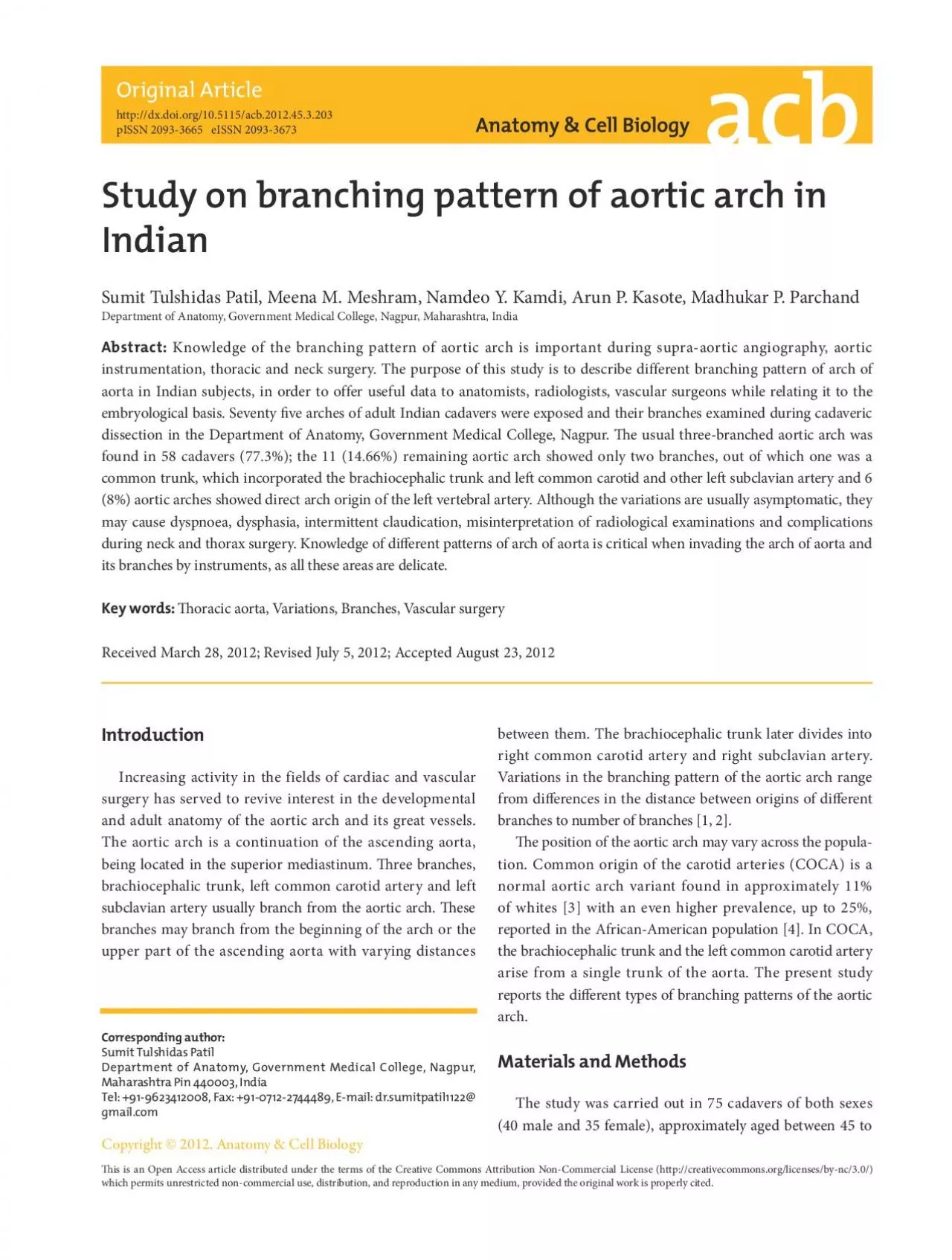
Abstract Knowledge of the branching pattern of aortic arch is important during supraaortic angiography aortic instrumentation thoracic and neck surgery The purpose of this study is to describe ID: 936140

arch aortic artery aorta aortic arch aorta artery pattern type branching common branches subclavian carotid trunk left variations arches

Share:

Link:

Embed:

Download Presentation from below link

Download Pdf The PPT/PDF document "Department of Anatomy Government Medical..." is the property of its rightful owner. Permission is granted to download and print the materials on this web site for personal, non-commercial use only, and to display it on your personal computer provided you do not modify the materials and that you retain all copyright notices contained in the materials. By downloading content from our website, you accept the terms of this agreement.


Presentation Transcript

Department of Anatomy, Government Medical College, Nagpur, Maharashtra, India Abstract: Knowledge of the branching pattern of aortic arch is important during supra-aortic angiography, aortic instrumentation, thoracic and neck surgery. The purpose of this study is to describe different branching pattern of arch of aorta in Indian subjects, in order to offer useful data to anatomists, radiologists, vascular surgeons while relating it to the embryological basis. Seventy ve arches of adult Indian cadavers were exposed and their branches examined during cadaveric Received March 28, 2012; Revised July 5, 2012; Accepted August 23, 2012 between them. The brachiocephalic trunk later divides into right common carotid artery and right subclavian artery. Variations in the branching pattern of the aortic arch range from dierences in the distance between origins of dierent branches to number of branches [1, 2]. e position of the aortic arch may vary across the popula tion. Common origin of the carotid arteries (COCA) is a normal aortic arch variant found in approximately 11% of whites [3] with an even higher prevalence, up to 25%, Increasing activity in the fields of cardiac and vascular surgery has served to revive interest in the developmental and adult anatomy of the aortic arch and its great vessels. The aortic arch is a continuation of the ascen

ding aorta, being located in the superior mediastinum. ree branches, brachiocephalic trunk, left common carotid artery and left subclavian artery usually branch from the aortic arch. ese branches may branch from the beginning of the arch or the upper part of the ascending aorta with varying distances Branching pattern of aortic arch http://dx.doi.org/10.5115/acb.2012.45.3.203 Anat Cell Biol 2012;45: 203-206 205 www.acbjournal.org comparison of these types of aorta, angiography images (Fig. 4) of aorta were obtained from intervention radiology department of our college. No other noticeable variations on the branching pattern of the great blood vessels were found. Discussion Knowledge of variations in the branching pattern of the aortic arch is of great importance in patients who have to undergo four vessel angiography, aortic instrumentation, or supra aortic thoracic, head and neck surgery [5]. In addition, knowledge of abnormal branches originating from the aortic arch is also important in the diagnosis of intracranial aneu rysms following subarachnoid haemorrhage [6]. A variant of origin and course of a great vessel arising from the aortic arch is of great clinical value, because lack of knowledge of these variations may lead to serious surgical complications during procedures occurring in the superior mediastinum and the root of neck. Var

iations in the branching pattern of the arch of aorta are likely to occur as a result of the altered development of certain aortic arch arteries during the embryonic period of gestation. The six pairs of aortic arches are a series of vessels that connect on each side the aortic sac with the corresponding dorsal aorta. At a later developmental stage, the aortic arches are both reduced in number and extensively transformed, and finally an asymmetric blood supply system is achieved. The rst and second aortic arches largely disappear by the time the third to sixth arches develop. The left limb of the aortic sac normally forms the part of the arch of aorta that intervenes between the origins of the brachiocephalic trunk and the le common carotid artery. If the aortic sac fails to bifurcate into right and le limbs, then the variations on the branching pattern of arch of aorta may occur, as observed in present study. The proximal part of the third aortic arch normally gets extended and absorbed into the le horn of aortic sac. If it gets absorbed into the right horn of the aortic sac, that also results in a variable branching pattern [7, 8]. Direct origin of the le vertebral artery from the upper convex surface of the arch of aorta between the origins of the le common carotid and left subclavian arteries may be explained as incr

eased absorption of embryonic tissue of the left subclavian artery between the origin of the arch of aorta and the vertebral artery. Variations, when there are more than three branches originating from the arch of aorta may include the vertebral arteries [9, 10]. According to Adachi [11], in about 80.0% of indivi duals, three branches arise from the arch of aorta: the bra chiocephalic trunk, left common carotid artery, and left subclavian artery. Adachi classied this branching pattern as type A. Another 11% have a common trunk incorporating the le common carotid artery and the brachiocephalic trunk leaving only two branches originating from the arch of aorta, Adachi’s type B. The pattern, type C, has the left vertebral artery, a fourth branch of the arch of aorta, originating proxi mal to the le subclavian artery. Williams and Edmonds [4] Table 1. Showing dierent types of aortic arches, total and split by sex Type Male Female Total % Type I 30 28 58 77.33 Type II (A+B) 6 5 11 14.66 Type II A 2 2 4 5.33 Type II B 4 3 7 9.33 Type III 4 2 6 8 Total 40 35 75 100 Fig. 4. Radiological images of aortic arch showing (A) type I, (B) type II, and (C) type III branching pattern of aortic arch. BCT, brachiocephalic trunk; GT, great trunk; LC, le common carotid; LS, le subclavian; LV, le vertebral artery; RS, right subclavia