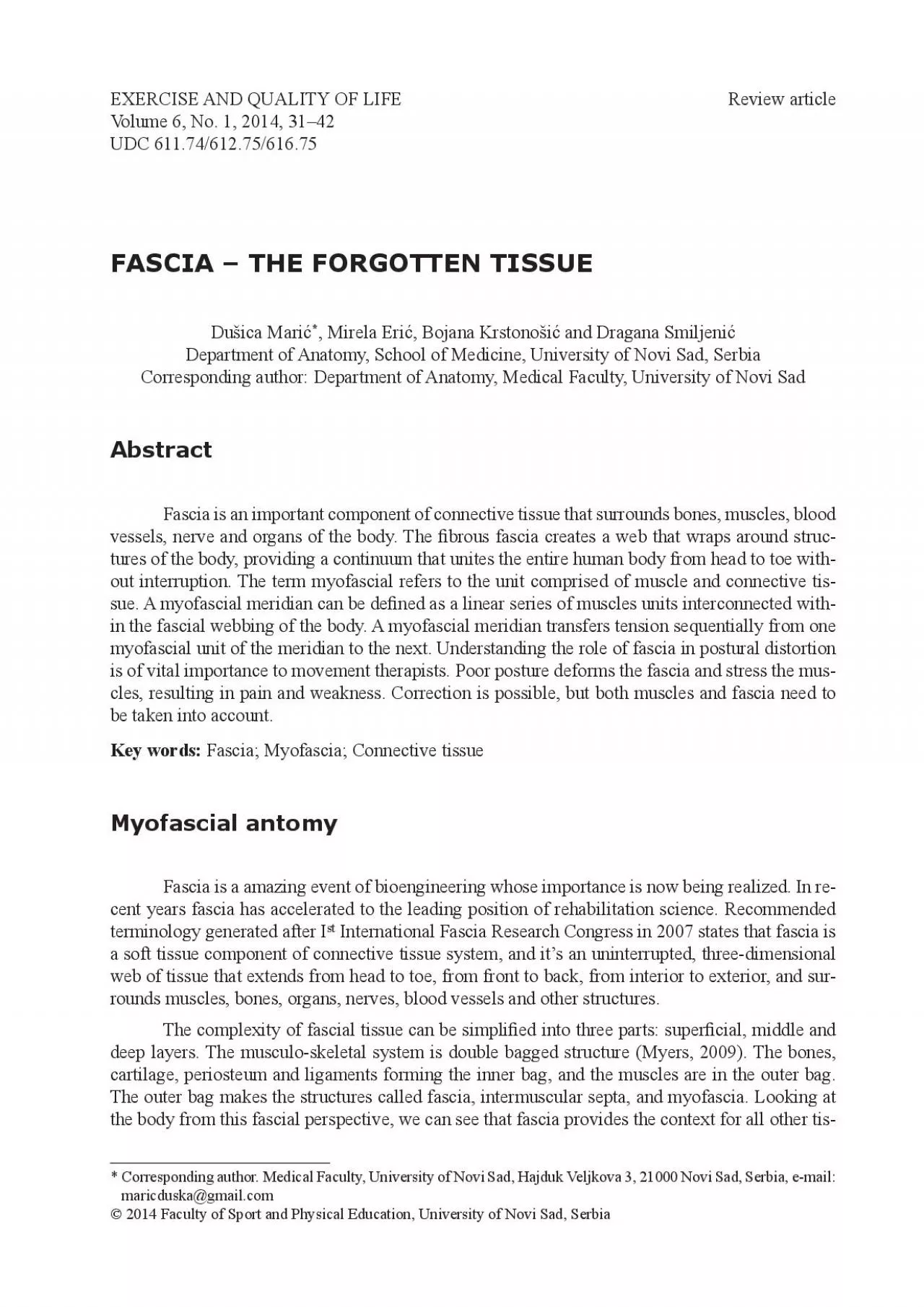
/
EXERCISE AND QUALITY OF LIFEVolume 6 No 1 2014 3142UDC 611746 EXERCISE AND QUALITY OF LIFEVolume 6 No 1 2014 3142UDC 611746

EXERCISE AND QUALITY OF LIFEVolume 6 No 1 2014 3142UDC 611746 - PDF document

queenie
queenie . @queenie
Follow
342 views
Uploaded On 2022-09-21

EXERCISE AND QUALITY OF LIFEVolume 6 No 1 2014 3142UDC 611746 - PPT Presentation

Corresponding author Medical Faculty University of Novi Sad Hajduk Veljkova 3 21000 Novi Sad Serbia email Fascia the forgotten tissue Dušica Marić Mirela Erić Bojana Krstonošić and ID: 954491

myofascial fascia fascial x00660069 fascia myofascial x00660069 fascial line pain front tissue muscle runs body muscles 2005 2008 arm

Share:

Link:

Embed:

Download Presentation from below link

Download Pdf The PPT/PDF document "EXERCISE AND QUALITY OF LIFEVolume 6 No ..." is the property of its rightful owner. Permission is granted to download and print the materials on this web site for personal, non-commercial use only, and to display it on your personal computer provided you do not modify the materials and that you retain all copyright notices contained in the materials. By downloading content from our website, you accept the terms of this agreement.


Presentation Transcript

EXERCISE AND QUALITY OF LIFEVolume 6, No. 1, 2014, 31–42UDC 611.74/612.75/616.75, Mirela Erić, Bojana Krstonošić and Dragana SmiljenićDepartment of Anatomy, School of Medicine, University of Novi Sad, SerbiaCorresponding author: Department of Anatomy, Medical Faculty, University of Novi SadFascia is an important component of connective tissue that surrounds bones, muscles, blood vessels, nerve and organs of the body. The �brous fascia creates a web that wraps around structures of the body, providing a continuum that unites the entire human body from head to toe without interruption. The term myofascial refers to the unit comprised of muscle and connective tissue. A myofascial meridian can be de�ned as a linear series of muscles units interconnected within the fascial webbing of the body. A myofascial meridian transfers tension sequentially from one myofascial unit of the meridian to the next. Understanding the role of fascia in postural distortion is of vital importance to movement therapists. Poor posture deforms the fascia and stress the muscles, resulting in pain and weakness. Correction is possible, but both muscles and fascia need to Fascia is a amazing event of bioengineering whose importance is now being realized. In recent years fascia has accelerated to the leading position of rehabilitation science. Recommended terminology generated after I International Fascia Research Congress in 2007 states that fascia is a soft tissue component of connective tissue system, and it’s an uninterrupted, three-dimensional web of tissue that extends from head to toe, from front to back, from interior to exterior, and surrounds muscles, bones, organs, nerves, blood vessels and other structures.The complexity of fascial tissue can be simpli�ed into three parts: super�cial, middle and deep layers. The musculo-skeletal system is double bagged structure (Myers, 2009). The bones, cartilage, periosteum and ligaments forming the inner bag, and the muscles are in the outer bag. The outer bag makes the structures called fascia, intermuscular septa, and myofascia. Looking at the body from this fascial perspective, we can see that fascia provides the context for all other tis Corresponding author. Medical Faculty, University of Novi Sad, Hajduk Veljkova 3, 21000 Novi Sad, Serbia, e-mail: Fascia – the forgotten tissue Dušica Marić, Mirela Erić, Bojana Krstonošić and Dragana Smiljenić sues to form. If bone cells lay down bone matrix within a fascial sleeve, a bone is formed within fascial periosteum. If nerve cells are formed within fascial sleeving, the brain and spinal cord are formed within meninges, and peripheral nerves are formed within sleeves of endoneurium, periThe classic anatomical studies start with human dissection in the 17th century and explaining the body as a series of isolated parts. The classic representation of muscles is that they have discrete attachment on bones. By this model, each of the muscles is independent of one another (a single muscle theory). We have convenient mech

anical picture that a muscle ‘begins’ here and ‘ends’ there. According to new myofascial theory we have in body only one muscle; it just hangs around in 600 or more fascial pockets (Myers, 2009). We need to remind ourselves that muscle never attaches to bone. Muscle cells �oat within the fascial net like �sh within �shing net (Myers, 2009). Their movement pulls on the fascia, the fascia is attached to the periosteum, and the periosteum pulls on the bone. Far more often, even though some of the fascial tendinous �bers of the muscle do attach into and end at the attachment bone, other fascial tendinous �bers go beyond the bony attachment site and are continuous with the fascial tendinous �bers of the adjacent muscle. These myofascial units are linked to each other. Examining the fascial connections between muscles allows us to discern speci�c lines of linkage that travel throughout the body. These lines are called myofascial meridians. Each myofascial meridian is a somewhat discrete aspect of the fascial web that travels and connects far reaches of the body (Myers, 2009). In the single muscle theory, the biceps gets de�ned as a radio-ulnar supinator, an elbow �exor, and a weak �exor of the shoulder. In the Anatomy Trains view the biceps brachii is an element in a continous fascial plane or myofascial meridian which runs from the outside of the thumb to the 4 rib and beyond. The second fact does not negate the �rst, but adds a contex for understanding the biceps role in stabiThe Anatomy Trains model identi�es a set of myofascial meridians as the major continual tension bands along which this tensile strain runs through the outer myofasciae from bone to bone (Myers, 2009). Muscle attachments (stations in Anatomy Trains) are where the continuous tensile net attaches to the relatively isolated, outwardly-pushing compressive struts. Thomas Myers describes myofascial meridians as a map of global lines of tension that traverse the entire muscular surface of the human body acting to keep the skeleton in shape. Myofascial meridians in the human body include: the super�cial front line, the super�cial back line, the lateral lines (2 sides), the spiral line, the arm lines (2 front and 2 back), the functional lines (2 front and 2 back), and the The super�cial back line runs from the underside of the foot up the back of the leg to the sacrum, and up the back to the skull, and over the skull to the forehead. The super�cial front line runs from the toes up the front of the leg and up the torso to the top of the sternum, and passes along the side of neck to the back of the skull. The lateral line runs from the underside of the foot up the side of the leg and trunk, under the shoulder complex to the side of the neck and skull. Arms line: deep front arm line runs from the ribs down the front of the arm to the thumb. The super�cial front arm line runs from the stern

um and ribs down the inside of the arm to the palm of the hand. The deep back arm line runs from the spinous processes through the scapula to the back of the arm and little �nger. The super�cial back arm line runs from the spinous processes over the shoulder and outside the arm to the back of the hand. The spiral line runs from the side of the skull across the neck to the opposite shoulder and ribs, and back across the belly to the front of the hip, the outside Fascia – the forgotten tissue of the knee, the inside of the ankle, and under the arch of the foot and back up the leg and back to the skull. The functional line: the back functional line runs from one shoulder across the back to the opposite leg. The front functional line runs from one shoulder across the front of the belly to the opposite leg. The deep front line is a core line that begins deep on the sole of the foot and runs up of the leg to the front of the hip joint and across the pelvis to the front of the spine and on up Fascia takes responsibility for maintaining structural integrity, for providing support and protection, and acts as shock absorber. Fascia has an essential role in hemodynamic and biochemical process and provides the matrix that allows for intercellular communication (LeMoon, 2008). Chaitow (Chaitow et al., 2006) adds that fascia extends to all dense �brous connective tissues, including aponeuroses, ligaments, tendons, retinaculae, joint capsules, organ and vessel tunics, the epineurium, the meninges, the periostea, and all the endomysial and intermuscular �bers of the Fascia are controls the posture and regulate movements (Myers, 2009). The spinal mobility is limited by the lumbar fascia and the stability of foot is reachable thanks to the stiffness of the plantar fascia (Grant & Riggs, 2008, Schleip,2005), knee is supported by iliotibial tract along the lateral thigh (Grant & Riggs, 2008). Retinacula are not static structures for joint stabilisation as the ligaments, but specialized fasciae for local spatial proprioception of the foot and ankle movements, and play integrative role of the fascial system in peripheral control of articular motility Fascia is densely innervated with mechanoreceptors and nociceptors (Langevin, 2006, Schleip, 2003a, Schleip, 2003b). The mechanoreceptors, such as Pacini corpucles, Ruf�ni organs and free-nerve endings, maintain muscular coordination via the constant feedback from ligaments. Diversity in location of them suggest different functions, hence, retinaculum plays more perceptive function, while tendinous expansions are responsible for mechanical transmission of tension. Various types of receptors capable of monitoring tension, elongation, pressure, velocity, pain are located in fascial tissues and create a neurological feedback mechanism by which re�exive interaction with muscles is provided to maintain joint stability and safety as well as coordination of movement. Disruption of the fascia due to injury or overuse also results in corrupted feedb

ack signals and neurological disorders that are exposing the tissue to additional potential for injury or moveBones, cartilage, ligaments, and tendons are built of varying degrees of the same substance – collagen – so this unity is more than a structural connection. It is well known that all these things are malleable, that is, they will transform and change shape and structure when stressed. Over time the body becomes deformed, the legs bowed, the back bowed, the shoulders hunched, etc. Fascia pulled continually out of alignment will eventually stay there. Poor posture and bad habits gradually deform the fascia and stress the muscles, resulting in pain and weakness. Correction is possible, but both muscles and fascia need to be taken into account. The entire supporting structure Dušica Marić, Mirela Erić, Bojana Krstonošić and Dragana Smiljenić Fascia is being recognized in etiology of pain and proprioception (Stecco et al., 2008). Tightening of myofascia may occur as a response to trauma, overuse syndrome, repetitive stress injuries, strain, stress, infection, poor posture, and chronic non-physiological tension in the fascia or surgical scaring. Restricted fascia may compress and put extra stress on the linked soft tissue structures, resulting in dysfunction and pain (LeBauer et al., 2008). According to Schleip, when fascia increases its stiffness for a fairly short whereas constantly raised tension may consequently have metabolic and physiological disadvantages leading to pathological contractures such as Duputryen disease, plantar �bromatosis, club foot or frozen shoulder. On the contrary, loss of fascial tone may result in hypermobility of a joint, as in the example of sacroiliac pain (Schleip et al., 2005). There is an accepted concept that unresolved trauma and/or frozen emotions can be ‘stored’ within the connective tissue in the form of pathology (Minasny, 2009).The fascia of active people has more strength and springiness to it than that of inactive people (Schleip, et al, 2005). Even strenuous activity has shown to strengthen and improve facial response. In other words training can cause fascia to improve its function. This discovery can prove why many players see increased velocity and arm strength after starting a long toss program. The arms of those who long toss may have more ‘elastic’ storage capacity which can help with rapid acceleration (Schleip, et al, 2005). By throwing longer and more often you can condition the fascia in the arm. Training the myofascial system is one way that may be accomplished. Those who adhere to a shorter throwing program or throw infrequently may never reach the level required to Myofascial pain syndrome is a chronic musculoskeletal pain disorder associated with local or referred pain, decreased range of motion, autonomic phenomena, local twitch response in the affected muscle and muscle weakness without an atrophy (LeMoon,2008). The term “myofascial pain syndrome” is used synonymously with “regional myofascial pain” and “myofascial trigger point pain

syndrome” (Cummings & Baldry, 2007). Myofascial trigger points can be located in fascia, ligaments, muscles and tendons (Fernandez-de-las-Penas et al., 2005). Trauma, stress, muscle wasting or ischaemia, visceral pain referral may aggravate the development of this critical point (Fernandez de las Penas et al., 2005, Fryer & Hodgson, 2005, Grieve, 2006). Myofascial trigger points are considered to be one of the most common cause of musculoskeletal pain and dysfunction (Cummings & Baldry 2007, Fernandez-de-las-Penas et al., 2005, Fryer & Hodgson, 2005, Simons, 2002). Trigger points are recognized as main cause of headache and neck pain (Fernandez-de-las-Penas et al., 2005). They can be a reason to conditions like frozen shoulder, epicon International Fascia Research Congress. 2007. (htpp://www.fasciacongress.org/2007/).Myers, T. (2009). Anatomy trains, Myofascial meridians for manual and movement therapies. 2nd LeMoon, K. (2008). Terminology used in Fascia Research. Journal of Bodywork and Movement Fascia – the forgotten tissue Chaitow L., Bradley D., & Gilbert C. (2002). Multidisciplinary Approaches to Breathing Pattern Disorders. Churchill Livingstone, Edinburgh.Grant, K. E., & Riggs, A. (2008). Chapter 9: Myofascial Release. 149-166 (Stillerman, E. 2008. Schleip, R., Klingier, W. & Lehmann-Horn, F. (2005). Active fascial contractility: Fascia may be able to contract in a smooth muscle-like manner and thereby in�uence musculoskeletal dyStecco C, Macchi V, Porzionato A, Morra A, Parenti A, Stecco A, Delmas V & De Caro R. (2010). The ankle retinacula: morphological evidence of the proprioceptive role of the fascial sysCell Tissue OrgansLangevin, H.M. (2006). Connective tissue: a body-wide signaling network? Medical Hypotheses Schleip, R. (2003a). Fascial plasticity – a new neurobiological explanation: part 1. 7(1), 11-19. Schleip, R. (2003b). Fascial plasticity – a new neurobiological explanation: part 2. 7(2), 104-116. Stecco, C., Porzionato, A., Lancerotto, L., Stecco, A, Macchi, V., Ann Day, J. & De Caro, R. 2008. Histological study of the deep fasciae of the limbs. Journal of Bodywork and Movement LeBauer, A., Brtalik, R. & Stowe, K. (2008). The effect of myofascial release (MFR) on an adult Minasny, B. (2009). Understanding the Process of Fascial Unwinding. International Journal of LeMoon, K. (2008). Terminology used in Fascia Research. Journal of Bodywork and Movement Cummings, M. & Baldry, P. (2007). Regional myofascial pain: diagnosis and menagment. Practice & Research Clinical RheumatologyFernandez de las Penas, C., Sohrbeck Campo, M., Carnero, J. & Page, J. (2005). Manual therapies in the myofascial trigger point treatment: A systematic review Fryer, G. & Hodgson, L. (2005). The effect of manual pressure release on myofascial trigger points Grieve, R. (2006). Proximal hamstrings rupture, restoration of function without surgical intervention: A case study of myofascial trigger point pressure release. Journal of Bodywork and Simons, D. (2002). Understanding effective treatment of myofascial trigger points