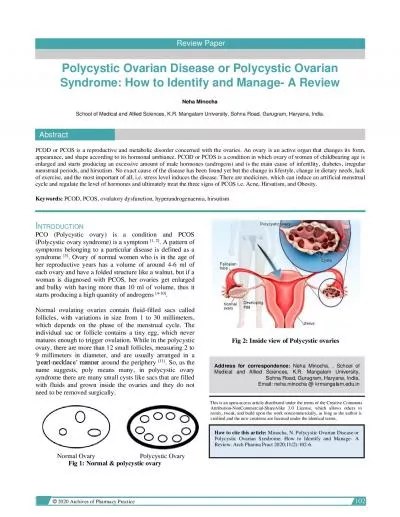
/
Ovarian Pathology POLYCYSTIC OVARIES (also called  Ovarian Pathology POLYCYSTIC OVARIES (also called 

Ovarian Pathology POLYCYSTIC OVARIES (also called  - PowerPoint Presentation

riley
riley . @riley
Follow
342 views
Uploaded On 2022-06-18

Ovarian Pathology POLYCYSTIC OVARIES (also called  - PPT Presentation

Stein Leventhal syndrome oligomenorrhea hirsutism infertility and obesity usually in girls after menarche secondary to excessive production of androgens by multiple cystic follicles in the ovaries unclear causes ID: 920425

ovarian tumors serous malignant tumors ovarian malignant serous cells benign borderline cell cancers cystic mucinous large germ gray unilateral

Share:

Link:

Embed:

Download Presentation from below link

Download Presentation The PPT/PDF document "Ovarian Pathology POLYCYSTIC OVARIES (al..." is the property of its rightful owner. Permission is granted to download and print the materials on this web site for personal, non-commercial use only, and to display it on your personal computer provided you do not modify the materials and that you retain all copyright notices contained in the materials. By downloading content from our website, you accept the terms of this agreement.


Presentation Transcript

Slide1

Ovarian Pathology

Slide2

POLYCYSTIC OVARIES (also called 

Stein-

Leventhal

syndrome).

oligomenorrhea

,

hirsutism

, infertility, and obesity

usually in girls after menarche

secondary to excessive production of androgens by multiple cystic follicles in the ovaries (unclear causes).

Pathogenesis: excessive production of androgens; high concentrations of LH, and low concentrations of FSH.

Slide3

The ovaries are enlarged, gray-white with a smooth outer cortex, and are studded with

subcortical

cysts 0.5 to 1.5 cm in diameter.

Histologically, there is a thickened, fibrotic outer surface, overlying cysts lined by granulosa cells with a hypertrophic and hyperplastic luteinized theca interna, with absence of corpora lutea

Slide4

5th

most common cancer in women.

5

th leading cause of cancer death in women. Ovarian Neoplastic Diseases

Slide5

three

cell types that make up the normal ovary:

1-The surface (

coelomic) covering epithelium 2- The germ cells 3- The sex cord/stromal cells. Each of these cell types gives rise to a variety of tumorsOrigin of ovarian tumors

Slide6

Ovarian Neoplasms

Slide7

Several risk factors for epithelial ovarian cancers have been recognized. Two of the most important are

nulliparity

and

family history.prolonged use of oral contraceptives may reduce the risk.Only 5%-10% of ovarian cancers are familial, the molecular pathogenesis of these cancers involve specific genes :mutations in the BRCA genes 1

 and 

2.

The average lifetime risk for ovarian cancer

30

% in 

BRCA1


carriers.

The risk in BRCA2 carriers is lower.

Pathogenesis-familial cases

Slide8

Mutations in 

BRCA

 genes mutations are seen in only 8% to 10% of sporadic ovarian cancers. Thus, there must be other molecular pathways for ovarian

neoplasms. HER2/NEU is overexpressed in 35% of ovarian cancers (poor prognosis). K-RAS protein is overexpressed in up to 30% of tumors, mostly mucinous cystadenocarcinomas.  

p53

 is mutated in about 50% of all ovarian cancers

Pathogenesis- sporadic cases

Slide9

derived

from

coelomic

mesothelium that covers the surface of the ovary. Benign lesions are usually cystic (cystadenoma) or can have an accompanying stromal component (cystadenofibroma). Malignant tumors may be cystic (cystadenocarcinoma

) or solid (carcinoma).

SURFACE EPITHELIAL TUMORS

Slide10

Serous

Mucinous

Endometrioid

Clear cell Brenner cystadenofibromaSURFACE EPITHELIAL TUMORS-types

Slide11

The surface epithelial tumors also have an intermediate,

borderline

category currently referred to as 

tumors of low malignant potential. These seem to be low-grade cancers with limited invasive potential. Thus, they have a better prognosis than the fully malignant ovarian carcinomas. borderline surface epithelial tumors

Slide12

the

most frequent ovarian tumors.

60% benign, 15% borderline, and 25% malignant.

Benign serous lesions 30 and 40 yearsmalignant serous tumors 45 and 65 years of age. Combined, borderline and malignant serous tumors are the most common malignant ovarian tumors and account for about 60% of all ovarian cancers.Mutations in BRAF and 

K-RAS

 are common in borderline tumors and low grade cancers.

High-grade serous carcinomas

mutations in 

p53

 and 

BRCA1.

1- Serous Tumors


Slide13

Benign serous tumors

:

large cystic structures, (30 to 40 cm in diameter

). May be bilateral.The serosa is smooth and glistening.usually filled with a clear serous fluidcharacterized

by a

single layer

of tall columnar epithelium. Some of the cells are

ciliated

.

Morphology

Slide14

Benign serous tumors:

Slide15

Borderline serous tumors:

more complex

architecture

mild cytologic atypia but no stromal invasion. However, they might be associated with peritoneal implants

Morphology

Slide16

Borderline serous tumors:

Slide17

Borderline

serous

tumors:

Slide18

Malignant serous carcinoma:

Anaplasia of the lining cells and invasion of the

stroma

. Note: Psammoma bodies (concentrically laminated calcified concretions) are common in the tips of papillae of all serous tumors in general.

Morphology

Slide19

Prognosis of serous tumors:

Benign and borderline

tumors:

excellent outcome (borderline tumors

100% survival, and even with peritoneal metastases it is nearly 75%, ).

Malignant invasive serous tumors

 prognosis

is poor and

depends on

the stage of the disease at the time of diagnosis.

Slide20

these tumors consists of

mucin

-

secreting cells.Only 10% of mucinous tumors are malignant(cystadenocarcinomas), while 10% are of low malignant potential (borderline

),

and

80% are

benign

.

2- Mucinous ovarian tumors

Slide21

Morphology

they are

large

and multilocular.psammoma bodies are not foundThe cysts are lined by mucin secreting cells with abundant vaculated cytoplasmDepending on the architectural complexity, these tumors are classified to benign,

borederline

or malignant

2- Mucinous ovarian tumors

Slide22

The

prognosis

of mucinous

cystadenocarcinoma is somewhat better than that for the serous counterpart, but the stage is the major determinant of treatment success.2- Mucinous ovarian tumors

Slide23

Mucinous ovarian tumors

Slide24

Microscopically they

are

similar to those of the endometrium, within the linings of cystic spaces.

endometrioid tumors are usually malignant. bilateral in about 30% of cases 15% to 30% of women with these ovarian tumors have a concomitant endometrial carcinoma. Many have mutations in the PTEN tumor suppressor

gene

3- Ovarian Endometrioid Carcinoma

Slide25

Germ cell tumors

Slide26

Benign (Mature) Cystic

Teratomas

:

differentiation of totipotential germ cells into mature tissues of all three germ cell layers Most are discovered in young women incidentally on abdominal scans90% are unilateral

Grossly: often filled with sebaceous secretion and

hair; bone

and

cartilage; epithelium

,

or teeth.

Germ cell tumors

Slide27

In 1% of cases

there is malignant transformation of one of the tissue elements.

May undergo torsion (10% to 15% of cases), producing an acute surgical emergency

Slide28

Benign (Mature)

Cystic

Teratomas

Slide29

Benign (Mature)

Cystic

Teratomas

Slide30

Other germ cell tumors

Slide31

Germ-Cell Origin

Peak

incidence


location


Morphology


Behavior


Dysgerminoma

Second to third decades Occur with

gonadal

dysgenesis

80% to 90% unilateral

Counterpart of testicular

seminoma

. Solid large to small gray masses. Sheets or cords of large cleared cells separated by scant fibrous strands.

Stroma

may contain lymphocytes and occasional

granuloma

.

All malignant but only one-third aggressive and spread; all radiosensitive with 80% cure.

Choriocarcinoma

First three decades of life

Unilateral

Identical to placental tumor. Often small, hemorrhagic focus with two types of epithelium;

cytotrophoblast

and

syncytiotrophoblast

.

Metastasizes early and widely. Primary focus may disintegrate, leaving only "

mets

." In contrast to placental tumors, ovarian primaries are resistant to chemotherapy.

Metastases to Ovary


Older ages

Mostly bilateral

Usually solid gray-white masses as large as 20cm in diameter.

Anaplastic

tumor cells, cords, glands, dispersed through fibrous background. Cells may be "signet-ring"

mucin

-secreting.

Primaries are gastrointestinal tract (

Krukenberg

tumors), breast, and lung.

Slide32

Sex cord-

stromal

tumors

Slide33

Sex Cord Tumors

Peak

incidence


location


Morphology


Behavior


Granulosa-thecal

cell

Most postmenopausal but at any

age

Unilateral

May be tiny or large, gray to yellow (with cystic spaces). Composed of mixture of

cuboidal

granulosa

cells in cords, sheets, or strands and spindled or plump lipid-laden

thecal

cells.

Granulosal

elements may recapitulate ovarian follicle as Call-

Exner

bodies.

May elaborate large amounts of estrogen (from

thecal

elements) and so may promote endometrial or breast carcinoma.

Granulosal

element may be malignant (5% to 25%).

Thecoma-fibroma

Any age

Unilateral

Solid gray fibrous cells to yellow (lipid-laden) plump thecal cells.

Most hormonally inactive. Few elaborate estrogens. About 40%, for obscure reasons, produce ascites and hydrothorax (

Meigs

syndrome). Rarely malignant.

Sertoli-Leydig

cell

All ages

Unilateral

Usually small, gray to yellow-brown, and solid. Recaps development of testis with tubules, or cords and plump pink

Sertoli

cells.

Many

masculinizing

or

defeminizing

. Rarely malignant.

Slide34


 All ovarian neoplasms

are clinical

challenges, because they produce no symptoms or signs until they are well advanced. The clinical presentation of all ovarian tumors is similar despite their great morphologic diversitythey cause local pressure symptoms (e.g., pain, gastrointestinal complaints, urinary frequency), sometimes they become twisted on their pedicles (torsion), producing severe abdominal pain mimicking an "acute abdomen." often cause ascites; espicially

Fibromas

and malignant serous

tumors.

Functioning ovarian tumors often come to attention because of the hormones they

induce (Estrogens or androgens).

Clinical Correlations for All Ovarian Tumors