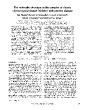
/
The molecular structure of the complex of Ascarischymotrypsinelastase The molecular structure of the complex of Ascarischymotrypsinelastase

The molecular structure of the complex of Ascarischymotrypsinelastase - PDF document

roberts
roberts . @roberts
Follow
342 views
Uploaded On 2022-09-07

The molecular structure of the complex of Ascarischymotrypsinelastase - PPT Presentation

680 Structure 1994 Vol 2 No 72830 squash seed trypsin inhibitor squash seedinhibitor family 31 serpins 3238 and hirudin3940 The inhibitors from these different familieshave a common ID: 952804

inhibitor bbo elastase structure bbo inhibitor structure elastase complex residues x97 x99 inhibitors hydrogen x01 crystal x399 disulfide protein

Share:

Link:

Embed:

Download Presentation from below link

Download Pdf The PPT/PDF document "The molecular structure of the complex o..." is the property of its rightful owner. Permission is granted to download and print the materials on this web site for personal, non-commercial use only, and to display it on your personal computer provided you do not modify the materials and that you retain all copyright notices contained in the materials. By downloading content from our website, you accept the terms of this agreement.


Presentation Transcript

The molecular structure of the complex of Ascarischymotrypsin/elastase inhibitor with porcine elastaseKui Huang1, Natalie CJ Strynadka1, Vincent D Bernard2,Robert J Peanasky2and Michael NG Jamesl*1Medical Research Council of Canada Group in Protein Structure and Function, Department ofBiochemistry, University of Alberta, Edmonton, Alberta T6G 2H7, Canada and 2Department of Biochemistryand Molecular Biology, University of South Dakota Medical School, Vermillion, SD 57069-2390, USABackground: The intestinal parasitic worm, Ascarissuum, produces a variety of protein inhibitors 680 Structure 1994, Vol 2 No 7[28-30], squash seed trypsin inhibitor (squash seedinhibitor family; [31]), serpins [32-38] and hirudin[39,40]. The inhibitors from these different familieshave a common conformation for the reactive-site loop,while displaying completely different overall structures(see [41,42] for reviews).An NMR study has reported the secondary structuralfeatures of one of the trypsin inhibitors from Ascaris(ATI) [43]. In it, two regions of 3-sheet were identified.In order to provide a complete tertiary structure and toestablish conclusively the disulfide bridge connections,we have undertaken the crystal structure determinationof one of the chymotrypsin/elastase inhibitors, desig-nated as the C/E-1 inhibitor. The C/E-1 inhibitor has63 amino acids and shares 40 % sequence identity withATI. It forms very tight complexes with chymotrypsinand pancreatic elastase, with dissociation constants of3.8 x 10-12M and 6.3 x 10⁻¹¹M, respectively [1]. Ithas been demonstrated that the reactive-site peptidebond (the scissile bond) is between Leu31 and Met32of the C/E-1 inhibitor [44].The C/E-1 inhibitor has been co-crystallized withporcine pancreatic elastase (PPE). Elastase is an im-portant enzyme in several regards [45]. Pancreatic elas-tase is of medical relevance, being implicated in pan-creatitis. The closely related human leukocyte elastase(which has 40 % sequence identity with PPE) has beenassociated with a number of inflammatory disordersincluding pulmonary emphysema, acute respiratory dis-tress syndrome, rheumatoid arthritis and cystic fibrosis.The crystal structure of PPE was among the first serineproteinase structures to be determined [46]. The diffi-culty we experienced in crystallizing the C/E-1 inhibitoralone was overcome by co-crystallizing it with PPE. Inaddition, the molecular replacement technique usingnative elastase [47] as the search model, greatly facili-tated the structure determination of the C/E-1 inhibitorand obviated the need for any heavy atom derivatives.Results and discussionOverall structure of the C/E-1 inhibitorThere are two copies of the enzyme-inhibitor com-plex, displaying nearly identical conformations, in theasymmetric unit of the crystals studied. Pairwise super-positions of the Ca atoms of the two elastase molecules(240 atoms) and the two inhibitor molecules (61 outof 63 atoms) give root mean square (rms) differencesof 0.35 A and 0.38 A, respectively. An rms difference of0.4A for all Ca atoms is obtained by superimposing thetwo copies of the entire complex, indicating that thebinding modes of the two inhibitors are virtually indis-tinguishable. Therefore, the following discussion of thestructure will not discriminate between each complexunless otherwise stated.The C/E-1 inhibitor folds into a wedge-shaped disc asillustrated in Figs la and lb. The diameter of the discis approximately 30 A and the thickness is about 15 Aon the thicker edge. The secondary structural elementsand the covalent pairings of the five disulfide bridgesare shown schematically in Fig. c. (In these figures andresulting discussion the inhibitor residues are labeledwith an I following the sequence number to distinguishthem from the residues of elastase.)The secondary structure assignment is based on thecriteria of Kabsch and Sander [48] as well as onthe main-chain hydrogen-bonding pattems. The criteriaused to define a hydrogen-bonding interaction were:the maximum donor to acceptor distance was 3.4Aand the maximum hydrogen to acceptor distance was2.4A; the angle defined by the donor atom-hydrogenatom-acceptor atom should be greater than 135°.Themajor secondary structures of the C/E-1 inhibitor aretwo 3-sheets. 3-sheet 1 comprises three strands, strand2 (Metl9I to Cys21I), strand 4 (Gly45I to Thr49I)and strand 5 (Lys53I to Ala57I). In fact, strands 4and 5 form a twisted 3-hairpin, while strand 2 runsparallel to strand 4. Only two hydrogen bonds areformed between the 3-hairpin and the otherwise iso-lated strand 2. The hydrogen-bonding pattern for this3-sheet is shown in Fig. 2. 3-sheet 2 consists of strand1 (VallOI to Thrl2 I) and strand 3 (Ser37I to Glu39 I).In total, four hydrogen bonds are formed between thetwo antiparallel P-strands. Immediately adjacent to 3-sheet 2, there is a single-residue -ladder (Thrl5 I andArg34 I), having two hydrogen bonds formed betweenthese two residues. Details of the hydrogen-bondinginteractions in 3-sheet 2 and the small 3-ladder areshown in Fig. 3.In contrast to most inhibitors of known structure, thereare no extensive helical regions in the C/E-1 inhibitor.Instead, only two single-tum 310-helices can be iden-tified. One such 310-helix (Ala57I to Gln59I) is lo-cated near the carboxyl terminus of the C/E-1

inhibitor.The other (Pro42 I to Arg44 I) is between -strands 3and 4. Interestingly, -structures are more commonlyobserved than a-helices among protein inhibitors ofserine proteinases. While there are no all-at structures,there are all-3 structures, such as Erythina trypsin in-hibitor [19], mung bean trypsin inhibitor [30], mu-cus proteinase inhibitor (SLPI) [27] and squash seedtrypsin inhibitor-1 (CMTI-I) [31].The crystallographically-determined hydrogen-bondingpatterns in the C/E-1 inhibitor agree in general withthose of ATI as observed by the NMR study [43]. Thesequence alignment of the C/E-1 inhibitor with ATI isshown in Fig. ld. The two hydrogen bonds definingthe two 310-helices are not reported for ATI by theNMR study, nor are the two hydrogen bonds definingthe small 3-ladder between Thrl 5I and Arg34I in theC/E-1 inhibitor (equivalent residues Gly15 and Lys34in ATI). In addition, the peptide oxygen of Asp49Iand the peptide nitrogen of Asn53I form a hydrogenbond in ATI, but the corresponding atoms from Thr49 Iand Lys53I diverge in the C/E-1 inhibitor. Nevertheless, Ascaris chymotrypsin/elastase inhibitor Huang et al. 681Fig. 1. (a) Schematic representation ofthe C/E-1 inhibitor. Arrows representPJ-strands. Disulfide bridges are shownby yellow bonds. The side chains ofthe P1and P1' residues Leu311 andMet321 are represented in green. Thepeptide bond Leu31 I-Met32 I is the scis-sile bond. (Diagram generated usingMOLSCRIPT [601.) (b) An all-atom rep-resentation of the C/E-1 inhibitor, fromapproximately the same view as (a).Main-chain atoms are shown in thicklines, side-chain atoms are shown inthin lines. Every fifth residue is labeled.The carboxy-terminal residues, Glu621and His631 have poor electron den-sity and are shown here in an arbitraryconformation. (c) Sequence, secondarystructure and disulfide bridges of theCIE-1 inhibitor. Residues in -strandsare enclosed in arrow-shaped boxes.The two single-turn 310-helices (Pro42 Ito Arg441 and Ala571 to Gln591) aredenoted by the helical symbol abovethe sequence letters of the residuesinvolved. The vertical arrow indicatesthe scissile peptide bond. (d) Sequencealignment of the C/E-1 inhibitor with ATI(a trypsin inhibitor from Ascaris). Con-served residues are boxed.it seems that the molecular scaffold of the C/E-1 in-hibitor is already established in its free form in solu-tion, and is largely retained in the crystal lattice of theenzyme-inhibitor complex.The other major secondary structural features presentin the C/E-1 inhibitor are reverse turns. Three turns arepresent including two type I turns and one type II turn.A type I turn (Glu2 I to Cys5 I) and a type II turn (Glu6 Ito Glu9 I) are located at the amino terminus; the othertype I turn (Thr49I to Gly52 I) connects -strands 4and 5.Solvent accessible surface calculations fail to identifya hydrophobic core in the C/E-1 inhibitor. The ab-sence of a hydrophobic core is understandable giventhe fact that only 25 % of the residues of the C/E-1inhibitor are hydrophobic residues, and half of theseare prolines. The relatively large number of disulfidebridges thus serves as an alternative to a hydrophobiccore for maintaining a well-defined tertiary structure.The pairing of the 10 cysteine residues is shown inFig. c. Interestingly, except for the disulfide bridgeformed by Cysl7 I and Cys29 I, which is proximal to theamino-terminal side of the reactive site peptide bond(Leu31 I-Met32 I), the other four disulfide bridges areall involved in cross-linking potentially flexible loops tothe relatively well-defined 13-sheet scaffold (Fig. la). Forexample, Cys38 I from strand 3 forms a disulfide bridgewith Cys5 I from a type I turn near the amino terminus,thereby imposing constraints on the conformation ofthe amino terminus. Similarly, the single-turn 310-helixnear the carboxyl terminus is clamped to strand 2 bythe disulfide bridge Cys21 I-Cys60I. The stabilizing ef-fects of this disulfide bridge on the conformation ofthe carboxyl terminus can be appreciated by the factthat Pro61l I, the residue immediately following Cys60 I, 682 Structure 1994, Vol 2 No 7Fig. 2. (a) Hydrogen-bonding pattern in P-sheet 1. Main-chain atoms are shown in thick lines, side-chain atoms are shown in thin lines.Dashed lines represent possible hydrogen bonds. (b) Schematic representation of the inter-strand hydrogen bonds of P-sheet 1.Fig. 3. (a) Hydrogen-bonding pattern in P-sheet 2 and the small P-ladder. Main-chain atoms are shown in thick lines, side-chain atomsare shown in thin lines. Dashed lines represent possible hydrogen bonds. (b) Schematic representation of inter-strand hydrogen bondsin P-sheet 2 and the p-ladder.has only weak electron density and the two carboxy-ter-minal residues, Glu62 I and His63 I, have no recogniz-able electron density associated with them. The disul-fide bridge Cys40 I-Cys54 I attaches the loop betweenGlu39I and Gly45I to strand 5. The disulfide bridgeconnecting Cysl4 Ito Cys33 I brings Thr51 I and Arg34 Iof the small -ladder close in space, so as to resume theinter-strand hydrogen bonding interrupted by Pro36Ifollowing P-strand 3.Such disulfide bridge-enhanced structures are observedin the squash seed trypsin inhibitor [31] and the mucusproteinase inhibitor [27] in addition to C/E-1 inhibitor.However, the disposition of th

e disulfide bridges arecompletely different in these three inhibitor families, asare the overall tertiary structures.In addition to the interconnections between secondarystructural elements introduced by the disulfide bridges,electrostatic and hydrogen-bonding interactions involv-ing side chains also contribute substantially to main-tain the integrity of the overall structure. The hydrogen-bonding potential of the side chains is well satisfied formost residues in the C/E-1 inhibitor. Prominent interac-tions are made by Arg48I (Fig. 4) and Arg44 I (Fig. 5).Projecting out of strand 4, the side chain of Arg48Iforms hydrogen bonds and/or salt bridges with thepeptide oxygen of Gly52 I and the carboxylate oxygenatoms of Glu9 I and Glu18 I. These glutamate residuesI IO=C NHEtE EDHN C=OI IC=O HN NHO=C NH NHHN C=O C=ONH O=C C=OO-CHN(a) (b) NH O=CGt l[--'C=O _ HNHN C=OO=C NHI INH -O=CC=O _ HNHC -- C=OO=C NHI INH O-CO-C NHI INn -u C=O HN1-1 L,I Ascaris chymotrypsin/elastase inhibitor Huang et al. 683also form hydrogen bonds to the peptide nitrogenatoms of Gly6 I and Cys40 I, respectively. Through thiselectrostatic/hydrogen-bond network, connections areestablished between the P-sheets and the three loops,i.e. the loop preceding strand 1, the loop betweenstrand 1 and strand 2 and the loop linking strand 3and strand 4. On the obverse face of the disc, Arg44 Ilinks a reverse turn (Ser41 I to Arg44 I) to strand 3 bysalt bridges or hydrogen bonds to Glu39I and Ser41 I.Geometry of the reactive-site loopA close-up view of part of the reactive-site loop ofthe C/E-1 inhibitor and the active site region of elas-tase is shown in Fig. 6, superimposed onto the elec-tron density map computed with coefficients derivedfrom SIGMAA [49]. As expected, the reactive-site loop(residues Asn26 I to Cys33 I) of the C/E-1 inhibitor ex-hibits an extended conformation,. closely resemblingthose of other protein inhibitors. Fig. 7 shows thesuperposition of the reactive-site loops from severalprotein inhibitors of serine proteinases. The rms dif-ferences in main-chain atoms from residues P3to P2'(nomenclature of Schechter and Berger [50]) are sum-marized in Table 1.The distance from the carbonyl carbon of Leu31 I (P1residue) to OY of Serl95 is 2.9A in one C/E-1 inhibitorand 3.1 A in the other C/E-1 inhibitor in the asymmet-ric unit. These distances are 0.2-0.4A longer than theunusually short (2.7 + 0.1 A) non-bonded contact dis-tances observed in other serine proteinase-inhibitorcomplexes (see, for examples, [10,12-14,26]). Theselonger distances in the C/E-1 complexes may reflect aless than ideal complementarity between the reactive-site loop of the C/E-1 inhibitor and the active site ofelastase or the relatively limited resolution (2.4A) ofthe present determination.Enzyme-inhibitor complexA unique feature of the C/E-1 inhibitor and elastasecomplex is their mutual penetration. This is charac-terized by the penetration of the P1residue, Leu31 I,into the S1 substrate specificity pocket, and by Arg217 A(Arg217A is an insertion in the elastase structure rel-ative to the sequence of chymotrypsin) from elastasepenetrating through a pore in the C/E-1 inhibitor (Fig.8). The latter penetration has not been observed in anyother protein inhibitor-proteinase complex. The porein the C/E-1 inhibitor is about 5A in diameter, encir-Fig. 4. The salt bridge/hydrogen-bondnetwork centered on Arg481. The sidechain of Arg481, and the peptide ni-trogen atoms of Gly61 and Cys401 areshown in blue. The side chain of Glu9 I,the side chain of Glu181 and the pep-tide oxygen of Gly52 I are shown in red.Dashed lines represent hydrogen bondsor salt bridges within 3.2 A.Fig. 5. The salt bridge/hydrogen-bondnetwork centered on Arg441. The sidechains of Arg44 I, Glu39 I and Ser41 I areshown in blue, red and green, respec-tively. Dashed lines represent possiblehydrogen bonds or salt bridges within3.3 A. ��Vol 2 No 7 Table 1. Comparison of residues ��C/E-1 from &#x/BBo;&#xx [2;.1;֙ ;̕.;妙&#x 224;&#x.399;&#x 322;&#x.800; ]0;&#x/BBo;&#xx [2;.1;֙ ;̕.;妙&#x 224;&#x.399;&#x 322;&#x.800; ]0;P3 to &#x/BBo;&#xx [2;7.1; 3;.8;Δ ;Ʌ.;冒&#x 323;&#x.519; ]0;&#x/BBo;&#xx [2;7.1; 3;.8;Δ ;Ʌ.;冒&#x 323;&#x.519; ]0;P2' of the reactive-site loop with equivalent residues in a selection of protein inhibitors of serine proteinases. Inhibitor Residue position &#x/BBo;&#xx [2;.8;ޒ ;ɵ.;瘁&#x 219;&#x.839; 28;.04; ];&#x/BBo;&#xx [2;.8;ޒ ;ɵ.;瘁&#x 219;&#x.839; 28;.04; ];Rms difference of the &#x/BBo;&#xx [1;.0;ޖ ;ɤ.;⎙&#x 112;&#x.079; 27;.44; ];&#x/BBo;&#xx [1;.0;ޖ ;ɤ.;⎙&#x 112;&#x.079; 27;.44; ];P3 &#x/BBo;&#xx [1;%.7;֔ ;ɤ.;&#x 131;&#x.759;&#x 271;&#x.199; ]0;&#x/BBo;&#xx [1;%.7;֔ ;ɤ.;&#x 131;&#x.759;&#x 271;&#x.199; ]0;P2 &#x/BBo;&#xx [1;E.1;গ ;ɤ.;⎙&#x 150;&#x.959; 27;.44; ];&#x/BBo;&#xx [1;E.1;গ ;ɤ.;⎙&#x 150;&#x.959; 27;.44; ];P1 &#x/BBo;&#xx [1;d.1;֙ ;ɤ.;⎙&#x 172;&#x.319; 27;.91;“ ];&#x/BBo;&#xx [1;d.1;֙ ;ɤ.;⎙&#x 172;&#x.319;&#

x5 27;.91;“ ];PI' &#x/BBo;&#xx [1;„.3;Ƙ ;ɤ.;⎙&#x 192;&#x.479; 27;.91;“ ];&#x/BBo;&#xx [1;„.3;Ƙ ;ɤ.;⎙&#x 192;&#x.479; 27;.91;“ ];P2' 20 main-chain atoms &#x/BBo;&#xx [6;.51;’ 2;D.0; 84;&#x.959; 25;�.07;– ];&#x/BBo;&#xx [6;.51;’ 2;D.0; 84;&#x.959; 25;�.07;– ];C/E-la C P &#x/BBo;&#xx [1;C.5;ƙ ;Ʉ.;㆕&#x 153;&#x.599; 25;�.55;— ];&#x/BBo;&#xx [1;C.5;ƙ ;Ʉ.;㆕&#x 153;&#x.599; 25;�.55;— ];L31' M C &#x/BBo;&#xx [6;.27;— 2;4.4;ࠁ ;.1;Ƙ ;ɀ.;閘&#x ]00;&#x/BBo;&#xx [6;.27;— 2;4.4;ࠁ ;.1;Ƙ ;ɀ.;閘&#x ]00;BPTI~ P c &#x/BBo;&#xx [1;C.0;Θ ;ȴ.;䠁&#x 153;&#x.839; 24;�.47;— ];&#x/BBo;&#xx [1;C.0;Θ ;ȴ.;䠁&#x 153;&#x.839; 24;�.47;— ];~lsl A R &#x/BBo;&#xx [2;1.5;ক ;ȴ ;Ʉ.;ՙ ;ȹ.;( ];&#x/BBo;&#xx [2;1.5;ক ;ȴ ;Ʉ.;ՙ ;ȹ.;( ];0.87 A &#x/BBo;&#xx [6;.27;— 2;$.4; ;–.2;Α ;ȩ.;栂&#x ]00;&#x/BBo;&#xx [6;.27;— 2;$.4; ;–.2;Α ;ȩ.;栂&#x ]00;OMTKY3= C T &#x/BBo;&#xx [1;C.2;ޓ ;ȣ.;ᦓ&#x 153;&#x.359; 22; .20; ];&#x/BBo;&#xx [1;C.2;ޓ ;ȣ.;ᦓ&#x 153;&#x.359; 22; .20; ];Ll8l E &#x/BBo;&#xx [1;†.2;Α ;Ȥ.;写&#x 190;&#x.319; 22; .55;“ ];&#x/BBo;&#xx [1;†.2;Α ;Ȥ.;写&#x 190;&#x.319; 22; .55;“ ];Y 0.89 &#x/BBo;&#xx [2;F.2;Ι ;Ȥ.;写&#x 250;&#x.799; 23;�.76; ];&#x/BBo;&#xx [2;F.2;Ι ;Ȥ.;写&#x 250;&#x.799; 23;�.76; ];d &#x/BBo;&#xx [6;.27;— 2;.4;Ι ;„.4;ޔ ;ȡ.;ᘃ&#x ]00;&#x/BBo;&#xx [6;.27;— 2;.4;Ι ;„.4;ޔ ;ȡ.;ᘃ&#x ]00;PCI-ld &#x/BBo;&#xx [1;.7;঒ ;ȕ.;䀁&#x 111;&#x.359; 22;�.91;— ];&#x/BBo;&#xx [1;.7;঒ ;ȕ.;䀁&#x 111;&#x.359; 22;�.91;— ];C P &#x/BBo;&#xx [1;C.2;ޓ ;Ȓ.;首&#x 153;&#x.359; 21; .23;™ ];&#x/BBo;&#xx [1;C.2;ޓ ;Ȓ.;首&#x 153;&#x.359; 21; .23;™ ];L38' N C 0.76 &#x/BBo;&#xx [2;F.2;Ι ;Ȕ.;㈂&#x 250;&#x.799; 22;�.55;“ ];&#x/BBo;&#xx [2;F.2;Ι ;Ȕ.;㈂&#x 250;&#x.799; 22;�.55;“ ];d &#x/BBo;&#xx [6;.27;— 2;.3; 83;&#x.759; 20; .87;• ];&#x/BBo;&#xx [6;.27;— 2;.3; 83;&#x.759; 20; .87;• ];EG-Ce V T &#x/BBo;&#xx [1;C.2;ޓ ;Ȃ.;醗&#x 153;&#x.359; 20; .15;™ ];&#x/BBo;&#xx [1;C.2;ޓ ;Ȃ.;醗&#x 153;&#x.359; 20; .15;™ ];L591 D L 0.74 &#x/BBo;&#xx [2;E.9;ও ;Ȅ.;␂&#x 250;&#x.559; 21;�.47;” ];&#x/BBo;&#xx [2;E.9;ও ;Ȅ.;␂&#x 250;&#x.559; 21;�.47;” ];A &#x/BBo;&#xx [6;.27;— 1;”.5;ƕ ;„.4;ޔ ;ȁ.;⎙&#x ]00;&#x/BBo;&#xx [6;.27;— 1;”.5;ƕ ;„.4;ޔ ;ȁ.;⎙&#x ]00;B-Birkf C T &#x/BBo;&#xx [1;C.0;Θ ;Ɠ.;ࠃ&#x 153;&#x.839; 19; .31;• ];&#x/BBo;&#xx [1;C.0;Θ ;Ɠ.;ࠃ&#x 153;&#x.839; 19; .31;• ];K261 S M 0.73 &#x/BBo;&#xx [2;F.2;Ι ;Ɣ.;螙&#x 250;&#x.799; 20;.12; ];&#x/BBo;&#xx [2;F.2;Ι ;Ɣ.;螙&#x 250;&#x.799; 20;.12; ];d &#x/BBo;&#xx [6;.27;— 1;r.5;؁ ;y.4;Ι ;Ÿ.;㈂&#x ]00;&#x/BBo;&#xx [6;.27;— 1;r.5;؁ ;y.4;Ι ;Ÿ.;㈂&#x ]00;aThe &#x/BBo;&#xx [8;.23;™ 1;r.3;ƕ ;™.5;ঙ ;Ÿ.;㈂&#x ]00;&#x/BBo;&#xx [8;.23;™ 1;r.3;ƕ ;™.5;ঙ ;Ÿ.;㈂&#x ]00;CIE-1 inhibitor from &#x/BBo;&#xx [8;.23;™ 1;r.3;ƕ ;™.5;ঙ ;Ÿ.;㈂&#x ]00;&#x/BBo;&#xx [8;.23;™ 1;r.3;ƕ ;™.5;ঙ ;Ÿ.;㈂&#x ]00;Ascaris &#x/BBo;&#xx [1;€.9;֑ ;Ų.;㆕&#x 199;&#x.679; 17;.39;˜ ];&#x/BBo;&#xx [1;€.9;֑ ;Ų.;㆕&#x 199;&#x.679; 17;.39;˜ ];suurn. &#x/BBo;&#xx [2;.7;ƒ ;Ų.;㆕&#x 229;&#x.439; 17;.80; ];&#x/BBo;&#xx [2;.7;ƒ ;Ų.;㆕&#x 229;&#x.439; 17;.80; ];bBovine pancreatic trypsir &#x/BBo;&#xx [2;.7;ƒ ;Ų.;㆕&#x 229;&#x.439; 17;.80; ];&#x/BBo;&#xx [2;.7;ƒ ;Ų.;㆕&#x 229;&#x.439; 17;.80; ];1101. &#x/BBo;&#xx [1;.0;ޒ ;Ţ.;䠁&#x 152;&#x.879; 16;.24; ];&#x/BBo;&#xx [1;.0;ޒ ;Ţ.;䠁&#x 152;&#x.879; 16;.24; ];COvomucoid inhibitor third domain from turkey 1121 &#x/BBo;&#xx [6;.27;— 1;P.9;֘ ;Ē.;㆘&#x 159;&#x.599; ]0;&#x/BBo;&#xx [6;.27;— 1;P.9;֘ ;Ē.;㆘&#x 159;&#x.599; ]0;dChymotrypsin inhibitor-1 from potato &#x/BBo;&#xx [1;.5;Y 1;R.1;֕ ;Ȃ.;㆘&#x 158;&#x.399; ]0;&#x/BBo;&#xx [1;.5;Y 1;R.1;֕ ;Ȃ.;㆘&#x 158;&#x.399; ]0;[261. &#x/BBo;&#xx [2;.9;֘ ;Œ.;䀁&#x 226;&#x.559;&#x 158;&#x.160; ]0;&#x/BBo;&#xx [2;.9;֘ ;Œ.;䀁&#x 226;&#x.559;&#x 158;&#x.160;&

#x3 ]0;eLeech inhibitor eglin-c &#x/BBo;&#xx [2;.0;Α ;ő.;’ 2;‘.3;֖ ;Ř.;ᘃ&#x ]00;&#x/BBo;&#xx [2;.0;Α ;ő.;’ 2;‘.3;֖ ;Ř.;ᘃ&#x ]00;1221 &#x/BBo;&#xx [6;.27;— 1;B.8; ;Ĉ.;閑&#x 149;&#x.519; ]0;&#x/BBo;&#xx [6;.27;— 1;B.8; ;Ĉ.;閑&#x 149;&#x.519; ]0;fBowmarA3irk inhibitor from &#x/BBo;&#xx [6;.27;— 1;B.8; ;Ĉ.;閑&#x 149;&#x.519; ]0;&#x/BBo;&#xx [6;.27;— 1;B.8; ;Ĉ.;閑&#x 149;&#x.519; ]0;[291. cled by the long &#x/BBo;&#xx [6;.27;— 1;B.8; ;Ĉ.;閑&#x 149;&#x.519; ]0;&#x/BBo;&#xx [6;.27;— 1;B.8; ;Ĉ.;閑&#x 149;&#x.519; ]0;Cysl7 &#x/BBo;&#xx [8;.31;‘ 1;.0;Ђ ;Ġ.;閕&#x 110;&#x.399; ]0;&#x/BBo;&#xx [8;.31;‘ 1;.0;Ђ ;Ġ.;閕&#x 110;&#x.399; ]0;I-Cys29 I. The side chain &#x/BBo;&#xx [8;.31;‘ 1;.0;Ђ ;Ġ.;閕&#x 110;&#x.399; ]0;&#x/BBo;&#xx [8;.31;‘ 1;.0;Ђ ;Ġ.;閕&#x 110;&#x.399; ]0;17 A inserts into the pore, the pore. The &#x/BBo;&#xx [8;.31;‘ 1;.0;Ђ ;Ġ.;閕&#x 110;&#x.399; ]0;&#x/BBo;&#xx [8;.31;‘ 1;.0;Ђ ;Ġ.;閕&#x 110;&#x.399; ]0;Arg217A are better ordered in as they display sig- B-factors in the complex. Electron density active site complex, showing &#x/BBo;&#xx [8;.31;‘ 1;.0;Ђ ;Ġ.;閕&#x 110;&#x.399; ]0;&#x/BBo;&#xx [8;.31;‘ 1;.0;Ђ ;Ġ.;閕&#x 110;&#x.399; ]0;His57, &#x/BBo;&#xx [4;Q.6;ޘ ;ؑ.;➓&#x 477;&#x.119; 61; .19;“ ];&#x/BBo;&#xx [4;Q.6;ޘ ;ؑ.;➓&#x 477;&#x.119; 61; .19;“ ];Asp102 and &#x/BBo;&#xx [4;—.7;֘ ;ؓ.;䐃&#x 521;&#x.039; 61; .43;™ ];&#x/BBo;&#xx [4;—.7;֘ ;ؓ.;䐃&#x 521;&#x.039; 61; .43;™ ];Ser195 of elas- &#x/BBo;&#xx [4;—.7;֘ ;ؓ.;䐃&#x 521;&#x.039; 61; .43;™ ];&#x/BBo;&#xx [4;—.7;֘ ;ؓ.;䐃&#x 521;&#x.039; 61; .43;™ ];Asn26 &#x/BBo;&#xx [4;™.1;™ 6;.5;؁ ;Ԁ.;ᖒ&#x 610;&#x.320; ]0;&#x/BBo;&#xx [4;™.1;™ 6;.5;؁ ;Ԁ.;ᖒ&#x 610;&#x.320; ]0;1 to &#x/BBo;&#xx [5;.9;ও ;؂.;ᖕ&#x 536;&#x.399; 61;�.32; ];&#x/BBo;&#xx [5;.9;ও ;؂.;ᖕ&#x 536;&#x.399; 61;�.32; ];Cys33 1 of the reactive-site loop of the &#x/BBo;&#xx [5;.8;ޙ ;֔.;熖&#x 536;&#x.159; 60;.67;” ];&#x/BBo;&#xx [5;.8;ޙ ;֔.;熖&#x 536;&#x.159; 60;.67;” ];C/E-1 in- hibitor. The map was calculated with coefficients &#x/BBo;&#xx [4;R.1;֙ ;յ.;➓&#x 478;&#x.319; 58;.40; ];&#x/BBo;&#xx [4;R.1;֙ ;յ.;➓&#x 478;&#x.319; 58;.40; ];2m(Fd- &#x/BBo;&#xx [4;€.2;Ι ;յ.;➓&#x 492;&#x.719; 58;.40; ];&#x/BBo;&#xx [4;€.2;Ι ;յ.;➓&#x 492;&#x.719; 58;.40; ];dlFd and calculated &#x/BBo;&#xx [4;€.2;Ι ;յ.;➓&#x 492;&#x.719; 58;.40; ];&#x/BBo;&#xx [4;€.2;Ι ;յ.;➓&#x 492;&#x.719; 58;.40; ];[491, contoured at the 2.00 level and was displayed on a Silicon Graph- using a locally modified version of &#x/BBo;&#xx [4;€.2;Ι ;յ.;➓&#x 492;&#x.719; 58;.40; ];&#x/BBo;&#xx [4;€.2;Ι ;յ.;➓&#x 492;&#x.719; 58;.40; ];FRODO &#x/BBo;&#xx [5;&.3;Ƙ ;Հ.;阂&#x 539;&#x.279; 54;.67;” ];&#x/BBo;&#xx [5;&.3;Ƙ ;Հ.;阂&#x 539;&#x.279; 54;.67;” ];[611. &#x/BBo;&#xx [4;.2;Ι ;у.;Ђ&#x 419;&#x.999; 45;.83;— ];&#x/BBo;&#xx [4;.2;Ι ;у.;Ђ&#x 419;&#x.999; 45;.83;— ];Fiq 7. Superposition of &#x/BBo;&#xx [4;.2;Ι ;у.;Ђ&#x 419;&#x.999; 45;.83;— ];&#x/BBo;&#xx [4;.2;Ι ;у.;Ђ&#x 419;&#x.999; 45;.83;— ];Pj to &#x/BBo;&#xx [4;.4;ޔ ;з.;搄&#x 414;&#x.959; 44;.55;“ ];&#x/BBo;&#xx [4;.4;ޔ ;з.;搄&#x 414;&#x.959; 44;.55;“ ];PZ of the reactive-site loops selection of &#x/BBo;&#xx [4;.4;ޔ ;з.;搄&#x 414;&#x.959; 44;.55;“ ];&#x/BBo;&#xx [4;.4;ޔ ;з.;搄&#x 414;&#x.959; 44;.55;“ ];ser- ine proteinases. The &#x/BBo;&#xx [4;”.8;ޒ ;Р.;␂&#x 512;&#x.639; 42;.20; ];&#x/BBo;&#xx [4;”.8;ޒ ;Р.;␂&#x 512;&#x.639; 42;.20; ];CIE-1 inhibitor is shown in green, bovine pancreatic &#x/BBo;&#xx [4;”.8;ޒ ;Р.;␂&#x 512;&#x.639; 42;.20; ];&#x/BBo;&#xx [4;”.8;ޒ ;Р.;␂&#x 512;&#x.639; 42;.20; ];(BPTI) in yellow, &#x/BBo;&#xx [5;4.2;Ι ;Ђ.;閘&#x 550;&#x.08 ;Ї.;➗&#x ]00;&#x/BBo;&#xx [5;4.2;Ι ;Ђ.;閘&#x 550;&#x.08 ;Ї.;➗&#x ]00;ovo- mucoid inhibitor third domain from &#x/BBo;&#xx [5;4.2;Ι ;Ђ.;閘&#x 550;&#x.08 ;Ї.;➗&#x ]00;&#x/BBo;&#xx [5;4.2;Ι ;Ђ.;閘&#x 550;&#x.08 ;Ї.;➗&#x ]00;(OMTKY3) in red, chymotrypsin inhibitor-I from potato &#x/BBo;&#xx

[5;4.2;Ι ;Ђ.;閘&#x 550;&#x.08 ;Ї.;➗&#x ]00;&#x/BBo;&#xx [5;4.2;Ι ;Ђ.;閘&#x 550;&#x.08 ;Ї.;➗&#x ]00;(PCI-1) in cyan, leech inhibitor eglin-c in purple and Bowman-Birk inhibitor from beans in pink. is probably preformed, since the free &#x/BBo;&#xx [4;‚.8;ޙ ;̤.;䠁&#x 506;&#x.159; 33;.59;™ ];&#x/BBo;&#xx [4;‚.8;ޙ ;̤.;䠁&#x 506;&#x.159; 33;.59;™ ];C/E-1 inhibitor, as shown by the &#x/BBo;&#xx [3;.9;֕ ;̓.;’ 4;.0;ޖ ;̠.;螙&#x ]00;&#x/BBo;&#xx [3;.9;֕ ;̓.;’ 4;.0;ޖ ;̠.;螙&#x ]00;NMR study on the related trypsin &#x/BBo;&#xx [3;.9;֕ ;̓.;’ 4;.0;ޖ ;̠.;螙&#x ]00;&#x/BBo;&#xx [3;.9;֕ ;̓.;’ 4;.0;ޖ ;̠.;螙&#x ]00;(ATI) &#x/BBo;&#xx [3;w.5;ƙ ;̀.;閕&#x 397;&#x.199; 31;�.79;™ ];&#x/BBo;&#xx [3;w.5;ƙ ;̀.;閕&#x 397;&#x.199; 31;�.79;™ ];[43], displays a very similar (albeit &#x/BBo;&#xx [5;4.4;ޔ ;̂.;搄&#x 550;&#x.08 ;̐.;ࠃ&#x ]00;&#x/BBo;&#xx [5;4.4;ޔ ;̂.;搄&#x 550;&#x.08 ;̐.;ࠃ&#x ]00;lirn- ited) secondary structure to the molecular complexes in this crystal structure. On the other hand, induce conformational inhibitor-2 from &#x/BBo;&#xx [5;4.4;ޔ ;̂.;搄&#x 550;&#x.08 ;̐.;ࠃ&#x ]00;&#x/BBo;&#xx [5;4.4;ޔ ;̂.;搄&#x 550;&#x.08 ;̐.;ࠃ&#x ]00;[21]. Since chymotrypsin does not residue corresponding to &#x/BBo;&#xx [5;4.4;ޔ ;̂.;搄&#x 550;&#x.08 ;̐.;ࠃ&#x ]00;&#x/BBo;&#xx [5;4.4;ޔ ;̂.;搄&#x 550;&#x.08 ;̐.;ࠃ&#x ]00;Arg2l7 A, the structure of the complex of chymotrypsin with the &#x/BBo;&#xx [3;.3;™ 2; .7;֘ ;̷.;枑&#x 218;&#x.879; ]0;&#x/BBo;&#xx [3;.3;™ 2; .7;֘ ;̷.;枑&#x 218;&#x.879; ]0;C/E-1 inhibitor should help to &#x/BBo;&#xx [4;P.7;Ɩ ;Ȉ.;禖&#x 476;&#x.159; 21;.64;&#x ]00;&#x/BBo;&#xx [4;P.7;Ɩ ;Ȉ.;禖&#x 476;&#x.159; 21;.64;&#x ]00;clanfy this point. The intermolecular interactions between the &#x/BBo;&#xx [5;.6;ޔ ;ƒ.;閕&#x 534;&#x.72 ;Ȃ.;ࠃ&#x ]00;&#x/BBo;&#xx [5;.6;ޔ ;ƒ.;閕&#x 534;&#x.72 ;Ȃ.;ࠃ&#x ]00;C/E-1 in- hibitor and elastase are similar to those observed protein inhibitors &#x/BBo;&#xx [5;.6;ޔ ;ƒ.;閕&#x 534;&#x.72 ;Ȃ.;ࠃ&#x ]00;&#x/BBo;&#xx [5;.6;ޔ ;ƒ.;閕&#x 534;&#x.72 ;Ȃ.;ࠃ&#x ]00;C/E-1 inhibitor has secondary &#x/BBo;&#xx [5;.6;ޔ ;ƒ.;閕&#x 534;&#x.72 ;Ȃ.;ࠃ&#x ]00;&#x/BBo;&#xx [5;.6;ޔ ;ƒ.;閕&#x 534;&#x.72 ;Ȃ.;ࠃ&#x ]00;subsites elongated on both sides of the &#x/BBo;&#xx [5;2.0; 14;.08; 5;I.8;Δ ;ŕ.;➗&#x ]00;&#x/BBo;&#xx [5;2.0; 14;.08; 5;I.8;Δ ;ŕ.;➗&#x ]00;scis- &#x/BBo;&#xx [3;.6;Ζ ;Ķ.;醓&#x 328;&#x.319; 14;.59;™ ];&#x/BBo;&#xx [3;.6;Ζ ;Ķ.;醓&#x 328;&#x.319; 14;.59;™ ];sile bond. In total, 105 contacts (within 4A) are &#x/BBo;&#xx [3;.6;Ζ ;Ķ.;醓&#x 328;&#x.319; 14;.59;™ ];&#x/BBo;&#xx [3;.6;Ζ ;Ķ.;醓&#x 328;&#x.319; 14;.59;™ ];C/E-1 inhibitor and residues (20 from &#x/BBo;&#xx [3;.6;Ζ ;Ķ.;醓&#x 328;&#x.319; 14;.59;™ ];&#x/BBo;&#xx [3;.6;Ζ ;Ķ.;醓&#x 328;&#x.319; 14;.59;™ ];C/E-1 inhibitor) are involved in these &#x/BBo;&#xx [3;V.6;Γ ;‰.2;ޓ ;в.;⎕&#x 99.;ᆘ&#x ]00;&#x/BBo;&#xx [3;V.6;Γ ;‰.2;ޓ ;в.;⎕&#x 99.;ᆘ&#x ]00;enzymeinhibitor interactions. By compari- son, only 11 residues of ovomucoid inhibitor third do- main from turkey &#x/BBo;&#xx [3;–.2;Ε ;g.4;Ζ ;у.;馓&#x 76.;禙&#x ]00;&#x/BBo;&#xx [3;–.2;Ε ;g.4;Ζ ;у.;馓&#x 76.;禙&#x ]00;(OMTKY3) are involved in interac- tions in &#x/BBo;&#xx [3;–.2;Ε ;g.4;Ζ ;у.;馓&#x 76.;禙&#x ]00;&#x/BBo;&#xx [3;–.2;Ε ;g.4;Ζ ;у.;馓&#x 76.;禙&#x ]00;OMTKY3-Streptomyces &#x/BBo;&#xx [4;p.3;ঔ ;T.9;؂ ;ԁ.;6 6;.08;&#x ]00;&#x/BBo;&#xx [4;p.3;ঔ ;T.9;؂ ;ԁ.;6 6;.08;&#x ]00;griseus &#x/BBo;&#xx [5;.5;ƙ ;U.1;গ ;Հ ;c.3;؄ ;&#x]000;&#x/BBo;&#xx [5;.5;ƙ ;U.1;গ ;Հ ;c.3;؄ ;&#x]000;protease B (SGPB) complex &#x/BBo;&#xx [3;–.4;y 4;.40; 4;.6;ޔ ;T.2;Ε ;&#x]000;&#x/BBo;&#xx [3;–.4;y 4;.40; 4;.6;ޔ ;T.2;Ε ;&#x]000;[I &#x/BBo;&#xx [4;.8;ޒ ;D.1;֕ ;Е.;醓&#x 54 ;&#x]000;&#x/BBo;&#xx [4;.8;ޒ ;D.1;֕ ;Е.;醓&#x 54 ;&#x]000;21, and 10 residues Ascaris chymotrypsin/elastase inhibitor Huang et al. 685P7, P18, P8' and P13' of the C/E-1 inhibitor are alsoinvolved in intermolecular contacts in the complex.These intermolecular interactions were not observed inany of the previously determined complex structures.Fig. 8. A schematic representation of the penetration of Arg217 Aof el

astase through the pore of the C/E-1 inhibitor. The van derWaals surface of elastase is shown in white. Arg217A, whose vander Waals surface is finger-shaped, projects from the main bodyof elastase. The backbone of the C/E-1 inhibitor is depicted as ared tube. The five disulfide bridges are shown in yellow. The sidechains of Leu311 and Met32 I are shown in green and orange,respectively. (Diagram was generated using GRASP [62].)are involved in the OMTKY3-chymotrypsin complex[13]. Also, 10 residues of polypeptide chymotrypsininhibitor-1 (PCI-1) are involved in the PCI-1-SGPBcomplex [26]. Apart from those residues interactingsolely with Arg217 A from elastase, residues at positionsComparison of elastase in the free and complexed statesThe structure of elastase in the complex agrees closelywith that of elastase in the native, uncomplexed state[47]. Pairwise superpositions of the native elastasestructure with this complexed elastase show that 212out of the total 240 Ct atoms fit within 1.0 A, while 28CaG atoms deviate by more than 1.0A. The rms devi-ations for the 212 Ca atoms are 0.562A and 0.553A,respectively for the two copies of elastase in the com-plex. The two copies of elastase in the asymmetric unitare more similar, having an rms deviation of only 0.35 Afor all main-chain atoms. Almost all the 28 residuesexhibiting deviations greater than 1.0 A are located onsurface loops, except for two of the residues on oneof the loops in the substrate-binding pocket. Most ofthese displacements may be attributed to the differ-ence in crystal packing environment between the na-tive crystal and the complex crystal, and to the bindingof the inhibitor to elastase. A significant change takesplace in the calcium-binding loop of elastase (Asn74to Gly78). Consequently, the expected six-coordinationgeometry for Ca2+ binding is disrupted. In the com-plex, there is a five-coordinated water molecule occu-pying the expected calcium-binding site of native elas-tase. The corresponding loop (Ser75 to Lys79) in thechymotrypsin-OMTKY3 complex also undergoes con-Table 2. Interactionsa within 4 A made between the C/E-1 inhibitor (listed vertically) and porcine pancreatic elastase (listed horizontally).Residues T4 H57 R61 N148 W172 T175 G190 C191 Q192 G193 D194 5195 T213 S214 F215 V216 S217 R217A L218 K224 SumP18 C141 1 1P1S C171 (1) 1P14E18 I (1) 1P13M191 1 1 2P7E251 1 1 2P6N261 9(1) 5(2) 14P5T271 2 6 (2) 8P4P281 1 2 3(1) 6P3C291 1 4 8(2) 1 14P2P301 2 2 1 5P1L311 2 4 3 4(1) 1 9(2) 1 3(1) 2 29P1' M321 3 1 1 1 6P2' C331 2 2P3' R341 1 1P4' R351 3 (1) 3P8' E391 1 1Pl1' S411 2 2P11' P421 2 2P12' G431 2 2P13' R441 3 3Sum 3 3 3 6 9 5 2 4 9 4 1 10 2 5 6 12 4 14 2 1 105aThe interactions listed in this table are observed in one copy of the complex. Both copies of the complex exhibit minor differences. The number ofinteractions may change slightly when refinement with higher resolution data is completed. The numbers in parentheses are the numbers of hydrogenbonds included in the total. 686 Structure 1994, Vol 2 No 7siderable shifts relative to native chymotrypsin. There isa minor conformational readjustment in the polypep-tide chain bordering the elastase active site (Cys191to GlnI92). Residues Cys191 and Gln192 have movedby an average of 1.2A relative to the native elastase,presumably to allow favorable contacts with the boundC/E-1 inhibitor at Leu31 I.Chemical determination of disulfide bond pairingTo learn as much as possible about the structureof Ascaris serine proteinase inhibitors, Bernard andPeanasky in South Dakota began a study to assignthe pairing of the 10 half-cystine residues by chemicalmethods [51]. Native inhibitors were found to be re-sistant to both chemical and en ymatic fragmentation.Collaborations with laboratories in Edmonton (X-raycrystallography) and at the National Institutes of Health(NMR spectroscopy) were initiated. Samples of nativeAscaris suum serine proteinase inhibitors were madeavailable; the same preparations were sent to both lab-oratories. In our laboratory we studied the C/E-1 in-hibitor; the NMR group studied Ascaris trypsin inhibitor(ATI) [43].Treatment of methionine-containing inhibitors (C/E-inhibitors) with cyanogen bromide inactivated the in-hibitors, cleaved the molecules at expected peptidebonds, but did not release cystine-containing peptides.A variety of proteinases failed to release cystine-contain-ing peptides even though they were able to hydroly esome peptide bonds. Cleavage with cyanogen bromidewas then combined with hydrolysis by glycyl endopep-tidase (Gly-C) from the latex of Carica papaya andfollowed by hydrolysis by staphylococcal serine pro-teinase (Glu-C). All three steps, employed in sequence,were required to release cystine-containing peptides.The peptides were resolved by HPLC, repurified, ana-ly ed for amino acid composition and then sequencedusing a manual double coupling method [52]. Whenthis study was completed, it was submitted for publi-cation. Within weeks after the chemical study was pub-lished, the X-ray laboratory solved the C/E-1 structureand made an assignment of the disulfide bonds. Regret-tably, the published disulfide bond assignment madeusing chemical methods supports neither the structuredetermined by X-ray, nor disulfide bond assignmentsmade by calculations from the NMR spectra o

f the Ascars trypsin inhibitor (M Clore, personal communica-tion). The disulfide bond assignments by X-ray analysisand by calculations from the NMR spectra agree witheach other.The experiments and data on which the chemically de-termined disulfide bond assignments were based werereviewed. It appears that the fragmentation data ob-tained permit the assignments reported by Bernard andPeanasky [51]. There is no clear explanation for thediscrepancy. It is possible that the inactivation withcyanogen bromide and the unique arrangement of thefive disulfide bonds in native inhibitors may have per-mitted the disulfides to rearrange either to some inter-mediate folding form or a dead-end product which wasunwittingly determined.Biological implicationsIt has been proposed [8] that most protein in-hibitors of serine proteinases act by a commonmechanism; they bind very tightly to their cog-nate enzymes, but are hydrolyzed very slowly.This tight binding has been explained by crystal-lographic studies, which have revealed extensivesurface complementarity between the reactive-site loops of the inhibitors and the active-siteclefts of the enzymes. Thus, the protein inhibitorsrecognize and bind to their cognate enzymes inthe manner of a good substrate. Presumably, theexcellent fit throughout the enzyme-inhibitor in-terface results in a remarkably deep minimum inthe free-energy profile, thereby presenting a veryhigh activation energy barrier to hydrolysis and todissociation. In the complex of elastase with thechymotrypsin/elastase (C/E-1) inhibitor, the com-plementarity is extremely good throughout thereactive-site loop as well as for more peripheralregions. For example, the side chain of Arg217Aof elastase fits neatly into a preformed pore in theinhibitor, without any collision. The reactive-siteloop of the C/E-1 inhibitor is similar in conforma-tion to those of most typical proteinase inhibitors,indicating that the C/E-1 inhibitor probably con-forms to the standard inhibitory mechanism.The structure of the C/E-1 inhibitor presentedhere confirms that this inhibitor indeed differsfrom the other known inhibitor families in tertiarystructure and topography of disulfide bridges, andthus represents a novel family of inhibitors. Onthe basis of the high sequence identity and theconserved positions of cysteine residues, it islikely that the other seven known serine pro-teinase inhibitors of the Ascaris family will alsoadopt a structure similar to that of the C/E-1 in-hibitor.Hydrophobic interactions are believed to be themain driving forces in the protein folding pro-cess. The absence of a hydrophobic core in theC/E-1 inhibitor is an exception to the generalrule, thereby challenging the current folding al-gorithms. The five disulfide bridges, as well as theelaborate network of electrostatic and hydrogen-bond interactions, appear to be the major stabiliz-ing forces in the C/E-1 inhibitor. Mutagenesis ofeach disulfide pair would readily reveal its signif- Ascaris chymotrypsin/elastase inhibitor Huang et al. 687icance in the folding process, and in maintainingthe structure and function of the C/E-1 inhibitor.An analogous observation was made with an acid-stable human mucus proteinase inhibitor (SLPI)[27], which is also rich in disulfides and lacksextensive secondary structure. Buried chargedresidues, other than hydrophobic residues, com-prise the molecular core of SLPI. These small pro-teins may provide valuable model systems withwhich to study how the primary sequence of aprotein dictates its tertiary structure.Materials and methodsCrystallization of the complexPorcine pancreatic elastase (Lot 31013C; Serva FeinbiochemicaGmbH and Co.) was used without further purification. The C/E-1 inhibitor was purified as described previously [1]. The com-plex was prepared by mixing the C/E-1 inhibitor with elastasein a 1:1.1 molar ratio of en yme to inhibitor. Crystals of thecomplex were grown from solutions of 10 mg ml- protein and12 % polyethylene glycol 6000, buffered with 50 mM sodium cit-rate at pH 6.5. Two crystal forms were observed from growth atdifferent temperatures. Extremely thin plate-shaped crystals (di-mensions 0.6 x 0.2 x 0.001 mm3) were obtained at room tem-perature; they belong to space group P21212, with unit cell pa-rameters a = 70.45A, b = 114.33A c = 74.55Ak Growth at 4°Cyielded large chunky crystals (dimensions 0.4 x 0.3 x 0.2 mm3)which belong to space group P3221, with a = b = 84.02Ac = 190.93k Both crystal forms have values of Vm [53] thatsuggest two sets of molecular complexes per asymmetric unit.Data collectionThe trigonal crystal form was chosen for the present structuredetermination because of its better diffracting ability. Crystalswere mounted into glass capillaries at 4°C. Diffraction data wererecorded at room temperature, with a Siemens area detector,X-1000, mounted on an 18kW Siemens rotating anode X-raygenerator. Graphite monochromati ed Cu K, radiation was usedfor data collection. Three data sets were collected from threecrystals; each set is nearly complete to 2.4k. All three data setswere merged and used for the structure solution and refinement.The data processing and merging were performed with the Xen-gen package [54]. Data collection statistics are given in Table 3.Structure determinationThe initial phases were det

ermined by the molecular replace-ment method, using the native porcine pancreatic elastase [47]as the search model. Reflections between 20-3.5A were usedfor the rotation function search that was conducted with theprogram ROTING [55]. The highest peak given by the Nava arotation function search was 8.5cy above the mean; the secondhighest peak was significantly lower (4.2o). This suggested thatthe two sets of complex molecules were in nearly the sameorientation in the asymmetric unit. The translation search wasdone using BRUTE [56], with the two elastase molecules in anidentical orientation in the unit cell. Itgave two peaks, one foreach elastase molecule. Rigid-body refinement in X-PLOR [57]was used to optimi e the molecular replacement solution. TheR-factor that resulted was 41.8 % and the correlation coefficientwas 54.3 %, with two elastase molecules in one asymmetric unit.An initial 2.4 A electron density map computed after the rigid-body refinement showed very good electron density for the twoelastase molecules. So the remaining task was to build the C/E-1inhibitor into the complex. The presence of two sets of molec-ular complexes per asymmetric unit made electron density aver-aging feasible. The DEMON package was used for this purpose[58]. A local correlation map was calculated to generate theinitial molecular mask embracing a single copy of the complex.This defined the region where averaging should take place. Afterseveral cycles of two-fold averaging and solvent flattening, 85 %of the residues of the C/E-1 inhibitor could be built into theresulting electron density map. The molecular envelope was thenadjusted accordingly for further averaging cycles. Eventually 61residues of the C/E-1 inhibitor could be built into the densityunambiguously. The last two residues at the carboxyl terminus,Glu62I and His63 I, were not well defined in this or subsequentelectron density maps.RefinementThe complex model was subjected to energy minimi ation andsimulated annealing molecular dynamics refinement using theX-PLOR package. Reflections between 10-2.4A with intensitiesgreater than 3c(I) were used in the X-PLOR refinement. Themodel was then refined with the program TNT [59], using allreflections between 20-2.4 A with no a cut-off. The initial B-fac-tors of the elastase residues in the complex were taken from thenative elastase structure [47]; the initial B-factors of the C/E-1inhibitor residues were assigned as 15.0A2. The positional pa-rameters and the B-factors were refined at the same time whilethe occupancy factors were kept at 1.0 for all atoms. About 50ordered solvent molecules have been added to each complex.The current model contains two copies of elastase, two copiesof the C/E-1 inhibitor (from which the last two residues aremissing), and 100 solvent molecules. The present quality of thecomplex structure is summari ed in Table 3.The coordinates of the elastase-C/E-1 inhibitor complex havebeen deposited with the Brookhaven Protein Data Bank.Table 3. Data collection and refinement.Space group P3221Cell dimensions a = b = 84.02 A, c = 190.93 AData collectionMaximum resolution 2.4 ANo. of crystals used 3Total observations 192 689Unique reflections 29 261Average redundancy 6.58Completeness of data 20 A-2.5 A: 99 %/o2.5 A-2.4 A: 71 O/%Rmergea7.00 %Refinement statisticsNo. of protein atoms 4538No. of solvent atoms 100Missing residues Glu62 I, His63 IReflections used 29 261Resolution range 20 A-2.4 AR-factor 19.1%Rms deviation from idealBond distance 0.021 ABond angle 3.8°Planar groups 0.026 AaRmerge = ZhkI [(iIli -�I)/ili] 688 Structure 1994, Vol 2 No 7Note added in proofThe three-dimensional structure of the Ascars trypsin inhibitordetermined at two pHs by NMR spectroscopy has been solvedindependently (BL Grasberger, GM Clore and AM Gronenbom,Structure 1994, 2:669-678).Acknowledgements. We thank Marc Allaire and Norma Duke for as-sistance in the data collection. We thank Anita Sielecki for helpfuldiscussions and Mae Wylie for help in preparing the manuscript. Thiswork was funded by the grants from the Medical Research Councilof Canada, the Alberta Heritage Foundation for Medical Research andthe National Institute of Allergy and Infectious Diseases (RJP). KH issupported by the Medical Research Council of Canada.References1. Peanasky, RJ., Bent, Y., Paulson, B., Graham, D.L. & Babin, D.R.(1984). The isoinhibitors of chymotrypsin/elastase from Ascarislumbricoides: isolation by affinity chromatography and associa-tion with the en ymes. Arch. Biochem Biophys 232, 127-134.2. Goodman, RB. & Peanasky, RJ. (1982). Isolation of the trypsininhibitors in Ascaris lumbricoides var suum using affinity chro-matography. Anal Biochem. 120, 387-393.3. Abuereish, G.M. & Peanasky, RJ. (1974). Pepsin inhibitors formAscaris lumbricoides isolation, purification and some properties.J Biot Chem 249, 1558-1565.4. Homandberg, GA & Peanasky, RJ. (1976). Characteri ation ofproteins from Ascaris lumbricoides which bind specifically tocarboxy peptidase. J Biol Chemr 251, 2226-2233.5. Mart en, M.R., Geise, G.L., Hogan, B. & Peanasky, RJ. (1985).Ascaris suum: locali ation by immunochemical and fluorescentprobes of host proteases and parasite proteinase inhibitors incross-sections. Expl Parasitl. 60, 139-149.6. Peanasky, R.J., Mart en, M.R., Homandb

erg, G.A, Cash, J.M.,Babin, D.R. & Litweiler, B. (1987). Proteinase inhibitors fromintestinal parasitic helminths: structure and indications of somepossible functions. In Molecular Paradigms for EradicatingHelminthic Parasites (Maclnnis, AJ., ed), pp. 349-366, DPL, NewYork7. Babin, D.R., Peanasky, RJ. & Goss, S.M. (1984). The isoinhibitorsof chymotrypsin/elastase from Ascaris lumbricoides- the primarystructure. Arch. Biochem. Biophy& 232, 143-161.8. Laskowski, M. Jr. & Kato, I. (1980). Protein inhibitors of pro-teinases. Annu Rev. Biochem 49, 593-626.9. Bigler, T.L., et at, & Laskowski, M. (1993). Binding of aminoacid side chains to preformed cavities: interaction of serine pro-teinases with turkey ovomucoid third domains with coded andnoncoded P1 residues. Protein Sci 2, 786-799.10. Marquart, M., Walter, J., Deisenhofer, J., Bode, W. & Huber, R.(1983). The geometry of the reactive site and of the peptidegroups in trypsin, trypsinogen and its complex with inhibitors.Acta Crystallogr. B 39, 480-490.11. Wlodawer, A, Deisenhofer, J. & Huber, R. (1987). Comparisonof two highly refined structures of bovine pancreatic trypsin in-hibitor. J. Mol Biol 193, 145-156.12. Read, RJ., Fujinaga, M., Sielecki, AR & James, M.N.G. (1983).Structure of the complex of Streptomyces griseus protease Band the third domain of the turkey ovomucoid inhibitor at 1.8Aresolution. Biochemistry 22, 4420-4433.13., Fujinaga, M., Sielecki, AR., Read, RJ., Ardelt, W., Laskowski, M.Jr. & James, M.N.G. (1987). The crystal and molecular structuresof the complex of turkey ovomucoid inhibitor third domain witha-chymotrypsin at 1.8A resolution. J. Mol Biol 195, 397-418.14. Bode, W., Wei, AZ., Huber, R., Meyer, E., Travis, J. & Neumann,S. (1986). X-ray crystallographic structure of the complex ofhuman leukocyte elastase (PMN elastase) and the 3rd domainof the turkey ovomucoid inhibitor. EMBO J. 5, 2453-2458.15. Papamokos, E., et at, & Laskowski, M. Jr. (1982). Crystallographicrefinement of Japanese quail ovomucoid, a Ka al-type inhibitor,and model building studies of complexes with serine proteases.J Mol Biol 158, 515-537.16. Bode, W., Epp, O., Huber, R., Laskowski, M. Jr. & Ardelt, W.(1985). The crystal and molecular structure of the 3rd domainof silver pheasant ovomucoid (OMSVP3). Eur. J Biochem. 147,387-395.17. Hirono, S., Akagawa, H., Mitsui, Y. & litaka, Y. (1984). Crystalstructure at 2.6A resolution of the complex of subtilisin BPN'with Streptomyces subtilisin inhibitor. J. Mot Biol 178, 389-413.18. Sweet, RM., Wright, H.T., Janin, J., Chothia, C.H. & Blow, D.M.(1974). Crystal structure of the complex of porcine trypsin withsoybean trypsin inhibitor (Kunit ) at 2.6A resolution. Biochem-istry 13, 4212-4228.19. Onesti, S., Brick, P. & Blow, D.M. (1991). Crystal structure of aKunit -type trypsin inhibitor from Erythina caffra seeds. J. MotBiol 217, 153-176.20. McPhalen, CA, Svendsen, I., Jonassen, I. & James, M.N.G. (1985).Crystal and molecular structure of the inhibitor CI-2 from barleyseeds in complex with subtilisin Novo. Proc. Natl. Acad Sci.USA 82, 7242-7246.21. McPhalen, CA & James, M.N.G. (1987). Crystal and molecu-lar structure of the serine proteinase inhibitor CI-2 from barleyseeds. Biochemistry 26, 261-269.22. McPhalen, CA, Schnebli, H.P. & James, M.N.G. (1985). Crystaland molecular structure of the inhibitor eglin from leeches incomplex with subtilisin Carlsberg. FEBS Lett. 188, 55-58.23. McPhalen, C.A. & James, M.N.G. (1988). Structural compari-son of two serine proteinase-protein inhibitor complexes: eglin-c-subtilisin Carlsberg and CI-2-subtilisin Novao. Biochemistry 27,6582-6598.24. Bolognesi, M., et al, & Ascen i, P. (1990). X-ray crystal structureof the bovine at-chymotrypsin/eglin c complex at 2.6A resolution.J Mol Recogn 3, 163-168.25. Dauter, Z. & Bet el, C. (1991). Complex between the subtilisinfrom a mesophilic bacterium and the leech inhibitor eglin-c. ActaCrystallogr. B 47, 707-730.26. Greenblatt, H.M., Ryan, CA & James, M.N.G. (1989). Structure ofthe complex of Streptomyces griseus proteinase B and polypep-tide chymotrypsin inhibitor-1 from Russet Burbank potato tubersat 2.1k J. Mol Biol 205, 201-228.27. Grutter, M.G., Fendrich, G., Huber, R & Bode, W. (1988). The2.5A X-ray crystal structure of the acid-stable proteinase inhibitorfrom human mucous secretions analy ed in complex with bovinea-chymotrypsin. EMBO J 7, 345-351.28. Chen, P., Rose, J., Love, R., Wei, C.H. & Wang, B.C. (1992).Reactive sites of an anticarcinogenic Bowman-Birk proteinaseinhibitor are similar to other trypsin inhibitors. J- Biol Chem,267, 1990-1994.29. Tshuogae, Y., et al, & Takahashi, K. (1986). Structure ofthe trypsin binding domain of Bowman-Birk type proteinaseinhibitor and its interaction with trypsin. J. Biochem 100,1637-1646.30. Lin, G., Bode, W., Huber, R., Chi, C. & Engh, RA (1993). The0.25-nm X-ray structure of the Bowman-Birk type inhibitor frommung bean in ternary complex with porcine trypsin. Eur. J.Biochen 212, 549-555.31. Bode, W., Greyling, HJ., Huber, R., Otlewski, J. & Wilus, T.(1989). The refined 2.OA X-ray crystal structure of the complexformed between bovine -trypsin and CMTI-I, a trypsin inhibitorfrom squash seeds (Curcurbita maxima): topological similarityof the squash seed inhibitors with the carboxypeptidase A in-hibitor from pota

toes. FEBS Lett 242, 285-292.32. Lobermann, H., Tokuoka, R., Deisenhofer, J. & Huber, R. (1984).Human cl-proteinase inhibitor: crystal structure analysis of twocrystal modifications, molecular model and preliminary analysisof the implications for function. J. Mol Biol 177, 531-556.33. Delarue, M., Samama, J.P. & Moras, D. (1990). Crystal structureof bovine antithrombin III. Acta Crystallogr. B 46, 550-556.34. Baumann, U., Huber, R., Bode, W., Grosse, D., Lesjak, M. &Laurell, C.B. (1991). Crystal structure of cleaved human -l-anti-chymotrypsin at 2.7A resolution and its comparison with otherserpins. J Mol Biol 218, 595-606.35. Wright, H.T., Qian, H.X. & Huber, R (1990). Crystal structureof plakalbumin, a proteolytically nicked form of ovalbumin. Itsrelationship to the structure of cleaved al-proteinase inhibitor.J, Mol Biol. 213, 513-528.36. Stein, P.E., Leslie, AG.W., Finch, J.T., Turnell, W.G., McLaughlin,PJ. & Carrell, RW. (1990). Crystal structure of ovalbumin as amodel for the reactive centre of serpins. Nature 347, 99-102.37. Schreuder, HA, et al, & Hol, W.G.J. (1994). The intactand cleaved human antithrombin III complex as a model forserpin-proteinase interactions. Nature Struct Biol 1, 48-54. Ascaris chymotrypsin/elastase inhibitor Huang et al. 68938. Carrell, RW., Stein, P.E., Fermi, G. & Wardell, M. (1994). Bio-logical implications of a 3A structure of dimeric antithrombin.Structure 2, 257-270.39. Rydel, TJ., et al, & Fenton, J.W. (1990). The structure of acomplex of recombinant hirudin and human -thrombin. Science249, 277-280.40. Gritter, M.G., et al, & Stone, S.R. (1990). Crystal structure of thethrombin-hirudin complex: a novel mode of serine proteinaseinhibition. EMBO J. 9, 2361-2365.41. Read, RIJ. & James, M.N.G. (1986). Introduction to the protein in-hibitors: X-ray crystallography. In Proteinase Inhibitors (Barrett,AJ. & Salvesen, G., eds), pp. 301-336, Elsevier, Amsterdam.42. Bode, W. & Huber, R (1992). Natural protein proteinase in-hibitors and their interaction with proteinases. Eur. J. Biochem.204, 433-451.43. Gronenbom, AM., Nilges, M., Peanasky, RJ. & Clore, G.M.(1990). Sequential resonance assignment and secondary struc-ture determination of the Ascaris trypsin inhibitor, a member ofa novel class of proteinase inhibitors. Biochemistry 29, 183-189.44. Peanasky, R.J., Bent, Y., Homandberg, GA, Minor, S.T. & Babin,D.R. (1984). The inhibitors of chymotrypsin/elastase from As-caris lumbricoides the reactive site. Arch. Biocbem. Biophys 232,135-142.45. Bode, W., Meyer, E. & Powers, J. (1989). Human leukocyte andporcine pancreatic elastase: X-ray crystal structures, mechanism,substrate specificity and mechanism-based inhibitors. Biochem-istry 28, 1951-1963.46. Shotton, D.M. & Watson, H.C. (1970). Three-dimensional struc-ture of tosyl-elastase. Nature 225, 811-816.47. Meyer, E., Cole, G. & Radharkrishnan, R. (1988). Structure ofnative porcine pancreatic elastase at 1.65 A resolution. Acta Crystallogr. B 44, 26-38.48. Kabsch, W. & Sander, C. (1983). Dictionary of protein secondarystructure: pattern recognition of hydrogen bonded and geomet-rical features. Biopolymers 22, 2577-2637.49. Read, R.J. (1986). Improved Fourier coefficients for maps usingphases from partial structures with errors. Acta Crystallogr. A42, 140-149.50. Schechter, I. & Berger, A (1967). On the si e of the activesite in proteases. I. Papain. Biochem. Biophys Res Commun. 27,157-162.51. Bemard, V.D. & Peanasky, RJ. (1993). The serine protease in-hibitor family from Ascaris suunm chemical determination of thefive disulfide bridges. Arch. Biochem. Biophys 303, 367-376.52. Chang, J.Y. (1981). N-terminal sequence analysis of polypeptidesat the picomole level. Biochem J 199, 557-564.53. Matthews, B.W. (1968). Solvent content of protein crystals. JMol. Biol 33, 491-497.54. Howard, AJ., Gilliland, G.L., Fin el, B.C. & Poulos, T.L. (1987).The use of an imaging proportional counter in macromolecularcrystallography. J Appl Crystallogr. 20, 383-387.55. Nava a, J. (1990). Accurate computation of the rotation matrices.Acta Crystallogr. A 46, 619-620.56. Fujinaga, M. & Read, RJ. (1987). Experiences with a newtranslation-function program. j Appl Crystallogr. 20, 517-521.57. Brunger, AT., Kuriyan, J. & Karplus, M. (1987). Crystallographic Rfactor refinement by molecular dynamics. Science 235, 458-460.58. Vellieux, F.M.D., et al, & Hol, W.GJ. (1993). Structure of gly-cosomal glyceraldehyde-3-phosphate dehydrogenase from Trypanosoma brucei determined from Laue data. Proc Natl AcadSci USA 90, 2355-2359.59. Tronrud, D.E. (1992). Conjugate-direction minimi ation. An im-proved method for the refinement of macromolecules. Acta Cry&tallogr. A 48, 912-916.60. Kraulis, PJ. (1991). MOLSCRIPT: a program to produce bothdetailed and schematic plots of protein structures. J Appl Crystallogr. 24, 946-950.61. Jones, TA (1985). Interactive computer graphics: FRODO. Withmodifications for TOM UQV/CIT v.2.8.0 by Cambillau, C., Israel,M., Griffin, G. and Chirino, A Methods Enzymol 115, 157-171.62. Nicholls, A, Sharp, KA & Honig, B. (1991). Protein foldingand association: insights from the interfacial and thermodynamicproperties of hydrocarbons. Proteins 11, 281-296.Received: 13 May 1994; revisions requested: 3 Jun 1994;revisions received: 9 Jun 1994. Accepted: 9 Jun 199