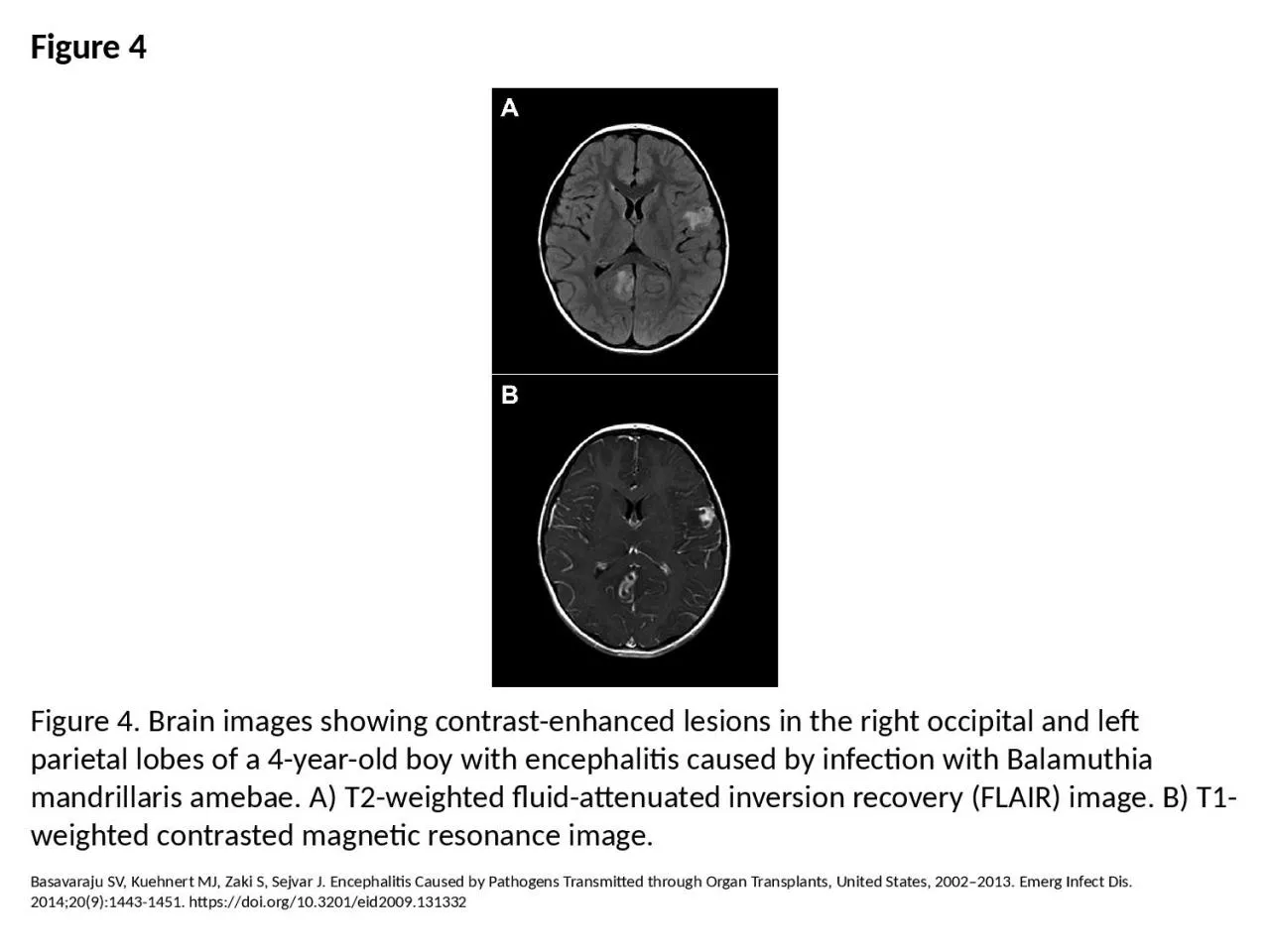
PPT-Figure 4 Figure 4. Brain images showing contrast-enhanced lesions in the right occipital

Author : rose | Published Date : 2024-01-03

Basavaraju SV Kuehnert MJ Zaki S Sejvar J Encephalitis Caused by Pathogens Transmitted through Organ Transplants United States 20022013 Emerg Infect Dis 201420914431451

Presentation Embed Code

Download Presentation

Download Presentation The PPT/PDF document "Figure 4 Figure 4. Brain images showing ..." is the property of its rightful owner. Permission is granted to download and print the materials on this website for personal, non-commercial use only, and to display it on your personal computer provided you do not modify the materials and that you retain all copyright notices contained in the materials. By downloading content from our website, you accept the terms of this agreement.

Figure 4 Figure 4. Brain images showing contrast-enhanced lesions in the right occipital: Transcript


Basavaraju SV Kuehnert MJ Zaki S Sejvar J Encephalitis Caused by Pathogens Transmitted through Organ Transplants United States 20022013 Emerg Infect Dis 201420914431451 httpsdoiorg103201eid2009131332.  . Dr.Kalpana Devi K.R., Dr. Francis G., Dr. Murali K., Dr. Madan R., Dr. . Subhapradha. , Dr . Geetha. S Nair,.  . MIOT hospitals, . Manapakkam. , Chennai. .. .  .  .  . ABSTRACT. ENERGY CONTRAST ENHANCED SPECTRAL . MAMMOGRAM : . AS A PROBLEM SOLVING TOOL IN EQUIVOCAL . CASES. Abstract . ID . IRIA-1121. B. reast . cancer incidence has increased by more than . 20% ,mortality . Abstract Id. : IRIA - 1052. Introduction. Among the widely used . neuro. imaging techniques . ie. computed topography and MRI, numerous etiologies may present as ring enhancing lesions. On non contrast images these lesions usually appear as . What is the cerebral cortex?. The cerebral cortex is the newest part of the brain (evolutionarily speaking. ). It also consists of two main layers:. Gray matter. White matter. . . Gray . matter. Entering the Debate on Cerebral Asymmetry. Paul Greenberg, 2015. Anoka Ramsey Community College. The Left vs. Right Controversy. No, the other controversy.. http://cercor.oxfordjournals.org/content/11/9/868.full. 11/20/2015. Elaine Smith . TJUH Emergency Medicine. Class of 2018. Contrast enhanced ultrasound. Detects. blood pooling/slow flow . by . using contrast to amplify . signal from blood . flow. Displays parenchymal . A. . clause. is a collection of words that has a subject that is actively doing a verb. The following are examples of clauses:. Clauses . of contrast : . although , even if, for all , in spite of , however, much as.. Hua Li and David . Mould. UNC Wilmington and Carleton University. Presented by Ling Xu . lihua@uncw.edu. . 1. The Problem of Tone . R. eduction. It is difficult to obtain a . clear segmentation without user . Diogo. Torres Marques, MD. Luiz Tenório de Brito Siqueira, MD. Regis Otaviano Franca Bezerra, MD. Marcos Roberto Menezes, MD. Manoel de Souza Rocha, MD, PhD. Giovanni Guido Cerri, MD, PhD. Address correspondence to. Adrenal glands can be affected by a variety of lesions. Adrenal lesions can either be primary, of adrenal origin, or secondary to other pathologies. Primary adrenal lesions can further be either of cortical or medullary origin. Functioning adrenal lesions can also give clues to the histologic diagnosis and direct workup. Over the years, various imaging techniques have been developed that have increased diagnostic accuracy and helped in better characterization of adrenal lesions non-invasively. In the first part of the two part series, we review adrenal imaging techniques and adrenal cortical tumors such as adenomas, adrenocortical tumors, adrenal hyperplasia and oncocytomas. Mubemba B, Chanove E, Mätz-Rensing K, Gogarten JF, Düx A, Merkel K, et al. Yaws Disease Caused by Treponema pallidum subspecies pertenue in Wild Chimpanzee, Guinea, 2019. Emerg Infect Dis. 2020;26(6):1283-1286. https://doi.org/10.3201/eid2606.191713. Song D, Kang B, Lee C, Saif LJ, Ha G, Kang D, et al. Transmission of Avian Influenza Virus (H3N2) to Dogs. Emerg Infect Dis. 2008;14(5):741-746. https://doi.org/10.3201/eid1405.071471. Shirwadkar CG, Samant R, Sankhe M, Deshpande R, Yagi S, Schuster FL, et al. Acanthamoeba Encephalitis in Patient with Systemic Lupus, India. Emerg Infect Dis. 2006;12(6):984-986. https://doi.org/10.3201/eid1206.060087. Wolfs TF, Wagenaar JA, Niesters H, Osterhaus A. Rat-to-Human Transmission of Cowpox Infection. Emerg Infect Dis. 2002;8(12):1495-1496. https://doi.org/10.3201/eid0812.020089.

Download Document

Here is the link to download the presentation.
"Figure 4 Figure 4. Brain images showing contrast-enhanced lesions in the right occipital"The content belongs to its owner. You may download and print it for personal use, without modification, and keep all copyright notices. By downloading, you agree to these terms.

Related Documents