/
ANATOMY-EAR(DOG) Types Of Ear Affections ANATOMY-EAR(DOG) Types Of Ear Affections

ANATOMY-EAR(DOG) Types Of Ear Affections - PowerPoint Presentation

ruby
ruby . @ruby
Follow
69 views
Uploaded On 2023-05-23

ANATOMY-EAR(DOG) Types Of Ear Affections - PPT Presentation

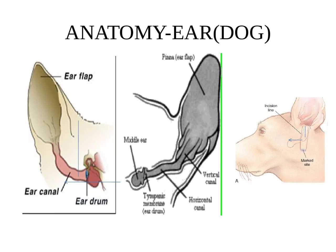
Otitis externa Inflammation of epithelium of the vertical or horizontal ear canal or both and surrounding structure Otitis media Inflammation of tympanic cavity ampmembrane Otitis ID: 999268

canal ear skin vertical ear canal vertical skin hematoma suture surface incision externa bulla tympanic cavity otitis drain left

Share:

Link:

Embed:

Download Presentation from below link

Download Presentation The PPT/PDF document "ANATOMY-EAR(DOG) Types Of Ear Affections" is the property of its rightful owner. Permission is granted to download and print the materials on this web site for personal, non-commercial use only, and to display it on your personal computer provided you do not modify the materials and that you retain all copyright notices contained in the materials. By downloading content from our website, you accept the terms of this agreement.


Presentation Transcript

1. ANATOMY-EAR(DOG)

2.

3. Types Of Ear AffectionsOtitis externa-Inflammation of epithelium of the vertical or horizontal ear canal or both and surrounding structureOtitis media -Inflammation of tympanic cavity &membraneOtitis interna -Inflammation of inner ear that causes vestibular diseases in dogsPresbycusis:Age related hearing loss.Swimmer’s Ear: Otitis externa develop after swimming or Bathing

4. Factors-predisposed To Ear Infections Long floppy earsAbnormal ear conformation or anatomyWater or hair in the earsAllergiesTraumaTumorsForeign material in the earsParasitesAutoimmune diseaseGeneralized skin diseaseEar infections can occur in dogs of any age breed or sex.

5. Clinical SignsScratching or rubbing the earsHead shaking Abnormal odor or discharge from the earPain on opening the mouthRedness and swelling of the external ear canalLack of appetiteHead tilt, circlingleaning to one sideRollingStumblingVomitingSide to side involuntary continuous eye movement (nystagmus)

6. DiagnosisClinical signsOtoscopic examinationRadiography of skullAntibiotic sensitivity – cultures from ear discharges collected asepticallyCT SCANMRI SCAN

7. Otoscopy of EarNormal Ear canalInflamed Ear canal and ruptured Tympanic membrane

8. Radiography of skullCalcification of Left ear canal (Arrow) and normal right ear canal (star)

9. CT- SCANFluid filled bulla and dense petrosal part of temporal bone-Left Ear

10. Flushing Solutions-earIncase of ruptured tympanic membraneDo Use-Tris-EDTA,ciprofloxacin,clotrimazole, miconazole,nystatin,ofloxacin,silver sulphadiazine, ticarcillin,tolnaftateDon’t usechlorhexidine 0.2%,nondiluted iodine/iodophores,ethanol,benzalkonium, aminoglycosides

11. Surgical CorrectionsIts mainly to improve drainage and facilitate applying medicationsOtitis externaLateral ear canal resection(Zepp method)Vertical ear canal ablationTotal ear canal ablationOtitis media &internaLateral bulla osteotomyVentral bulla osteotomy

12. Lateral Ear Canal Resection(Zepp Procedure)Lacroix procedure

13. Zepp Drain BoardLateral recumbency, head elevatedStand vertical aspect of headVertical ear canal distance measured with forcepMake the site one and half times the length of vertical canalTwo parallel incision from tragus,connect ventrally,reflect skin flapCut the vertical ear canal,upto exposing horozontal canalMake a drain board then suture by simple interrupted

14. Vertical Ear Canal AblationWhen entire vertical canal diseased and horizontal canal normal

15. Total Ear Canal Abalation (TECA)Chronic Otitis Externa with severe calcification and ossification of ear cartilageSevere epithelial hyperplasia extend beyond pinna or vertical canal Calcification of Left ear canal (Arrow) and normal right ear canal (star)

16. Lateral Bulla OsteotomyTo expose tympanic cavity for drainage of middle and inner ear secretion/exudate

17. Ventral Bulla OsteotomyBetter exposure of tympanic cavity without TECA

18. Pinna LacerationSkin of both side sutured with simple interrupted suture pattern (Fig. Top) ORVertical mattress pattern to appose the skin and cartilage on one side and simple interrupted pattern to appose the skin on the opposite site (Fig. Bottom)

19. Aural HematomaAn aural (Auricular) hematoma is a collection of blood within the cartilage plate of the ear characterized by fluctuant, fluid -filled swellings on concave surface of the pinna.It occurs due to bleeding of the branches of caudal auricular artery within the fractured auricular cartilage rather than between the skin and cartilageHead shaking may cause sinusoidal wave motion in the ear, resulting in fracture of the cartilageHistory-Violent head shaking and acute or chronic otitis externa may be noted.In Dog it mainly occurs due to bacterial infection in ear and O. cyanotis in cat.The affected dog tries to shake their head and scratch at the ear due to irritation or pain associated with otitis externa that may predisposed to this condition

20. Surgical CorrectionThe goals of the surgery are to remove the hematoma, prevent recurrence, and retain the natural appearance of the ear (i.e., minimize thickening of the ear and scarring)After aseptic preparation make an S-shaped incision on the concave surface of skin underlying hematoma covering full length of fluid filled area to remove the hematoma, its contents and irrigate the cavity.S- shaped incision prevent twisting of the ear during healing, as longitudinal contraction less likely to occur.

21. Place simple mattress suture incorporating cartilage between dorsal and ventral surface of skin and put final knot on dorsal surface of skin.Place ample number of stitches so that no pockets are left in which fluid can accumulate.Sutures should placed parallel to the line of incision and major vessels of ear.

22. Suture Materials: Non-absorbable Silk, Nylon, Polypropylene Absorbable: Polydioxanone, Polyglyconate Incorrect method of suture placementCorrect method of suture placementBlood vessels pattern of ear

23. Sutureless Technique For Aural HematomaPossible disadvantage of suture technique:-Ear pinna thickeningWrinklingCauliflower like appearanceThese complication avoided by sutureless technique.After aseptic preparation elliptical incision made on concave side to expose the hematoma cavity from end to end and lavage

24. Tape placed on rostral and caudal border on convex surface of earLong tape placed on rostral and caudal border on concave surface of earPinna reflected over large roll of cast padding to secure ear.

25. If minimal fibrin is present then teat cannula or fenestrated latex drain can be used.

26. Laser surgery: Carbon dioxide laser also used to drain hematoma then multiple incision made on hematoma to promote healing. Major disadvantage : Prolong inflammation and post surgery ear swellingSurgical Glue (Cyanoacrylate): Tissue glue also injected into hematoma to close the edges but formation of granuloma has been reported, so using surgical glue is unnecessary

27. Thanks