/
The Bangkok Medical Journal Vol 6 September 2013 The Bangkok Medical Journal Vol 6 September 2013

The Bangkok Medical Journal Vol 6 September 2013 - PDF document

ruby
ruby . @ruby
Follow
342 views
Uploaded On 2022-08-16

The Bangkok Medical Journal Vol 6 September 2013 - PPT Presentation

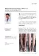
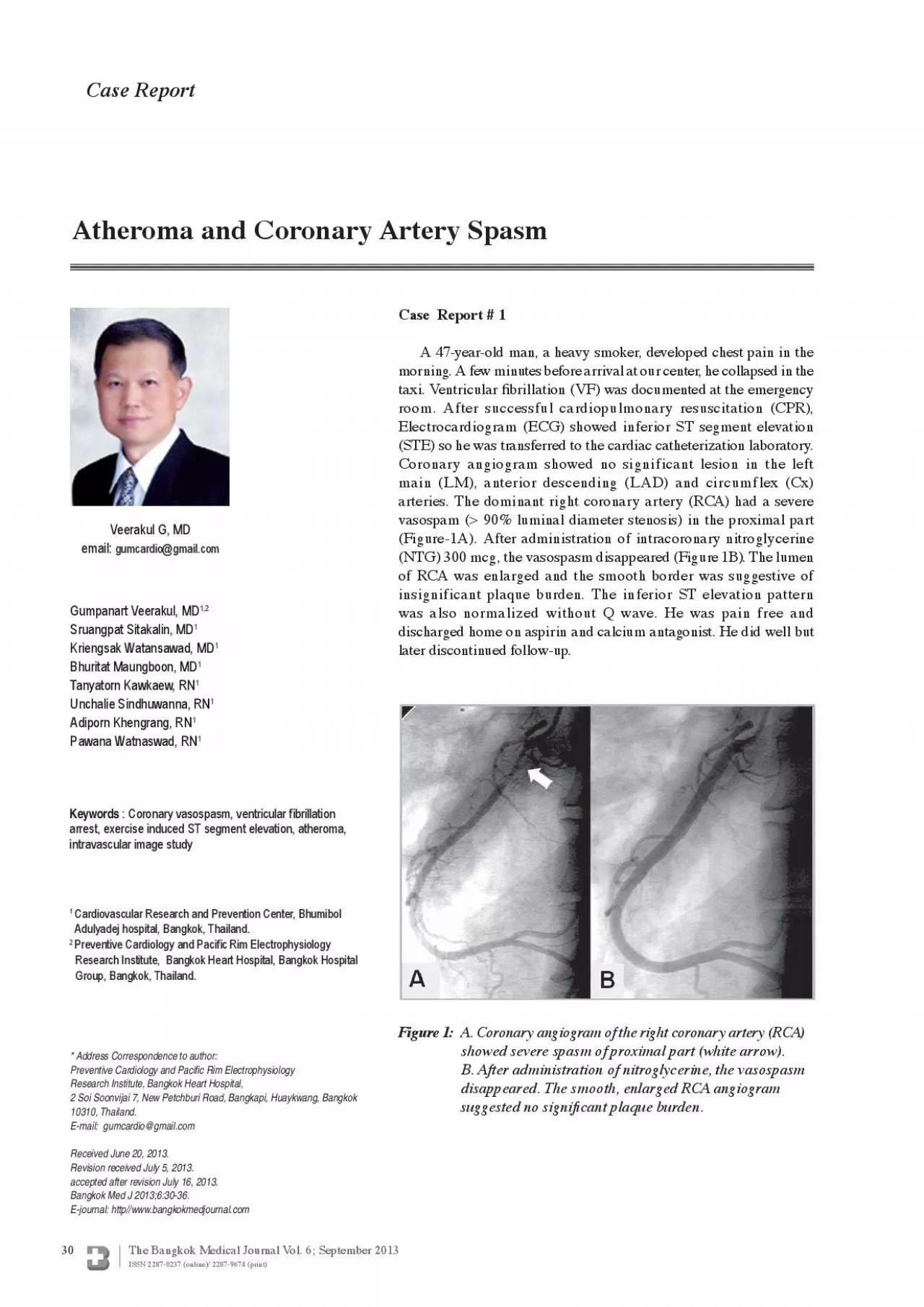
30 Case ReportGumpanart Veerakul MDKriengsak Watansawad MDTanyatorn Kawkaew RNPawana Watnaswad RNVeerakul G MDAtheroma and Coronary Artery Spasm Case Report 1 A 47yearold man a heavy smoker ID: 936491

artery coronary angina spasm coronary artery spasm angina vasospasm myocardial lad segment figure lesion stenosis atheroma normal patients angiogram

Share:

Link:

Embed:

Download Presentation from below link

Download Pdf The PPT/PDF document "The Bangkok Medical Journal Vol 6 Septem..." is the property of its rightful owner. Permission is granted to download and print the materials on this web site for personal, non-commercial use only, and to display it on your personal computer provided you do not modify the materials and that you retain all copyright notices contained in the materials. By downloading content from our website, you accept the terms of this agreement.


Presentation Transcript

30 The Bangkok Medical Journal Vol. 6; September 2013 Case ReportGumpanart Veerakul, MDKriengsak Watansawad, MDTanyatorn Kawkaew, RNPawana Watnaswad, RNVeerakul G, MDAtheroma and Coronary Artery Spasm Case Report # 1 A 47-year-old man, a heavy smoker, developed chest pain in the morning. A few minutes before arrival at our center, he collapsed in the taxi. Ventricular brillation (VF) was documented at the emergency room. After successful cardiopulmonary resuscitation (CPR),Electrocardiogram (ECG) showed inferior ST segment elevation(STE) so he was transferred to the cardiac catheterization laboratory.Coronary angiogram showed no significant lesion in the left main (LM), anterior descending (LAD) and circumflex (Cx)arteries. The dominant right coronary artery (RCA) had a severe�vasospam ( 90% luminal diameter stenosis) in the proximal part(NTG) 300 mcg, the vasospasm disappeared (Figure 1B). The lumenof RCA was enlarged and the smooth border was suggestive ofinsignificant plaque burden. The inferior ST elevation patternwas also normalized without Q wave. He was pain free anddischarged home on aspirin and calcium antagonist. He did well butlater discontinued follow-up.Cardiovascular Research and Prevention Center, Bhumibol Adulyadej hospital, Bangkok, Thailand. c Rim Electrophysiology Group, Bangkok, Thailand.* Address Correspondence to author: c Rim ElectrophysiologyE-journal: http//www.bangkokmedjournal.com brillation arrest, exercise induced ST segment elevation, atheroma, Figure 1: A. Coronary angiogram of the right coronary artery (RCA) showed severe spasm of proximal part (white arrow). B. After administration of nitroglycerine, the vasospasm disappeared. The smooth, enlarged RCA angiogram suggested no signi cant plaque burden. 31 The Bangkok Medical Journal Vol. 6; September 2013 Case Report # 2 A 54-year-old man experienced crescendo angina for a month. Chest pain started every morning, right after minimal exertion, and lasted three minutes. His known coronary risk factors included impaired fasting glucose, untreated dyslipidemia (LDL-cholesterol of 170 mg/dl) and hypertension. He had stopped smoking cigarettes 10 years before and did not use any illicit drugs. The physical examination was unremarkable, BP was 122/88 mmHg, HR was 72 beats per minutes. Baseline ECG showed a normal sinus rhythm without ST-T changes. The echocardiogram revealed mild concentric left ventricu-lar hypertrophy with well-preserved systolic function, ejec-tion fraction of 0.55. Trace mitral regurgitation and mild diastolic dysfunction (grade 1) were also observed. The Bruce protocol exercise stress test was performed. After walking three minutes, chest pain occurred. It was associated with 2-3 mm ST segment elevation (STE) in leads V1-3, aVR and 2 mm ST depression in leads II,III,F (Figure 2). These ndings suggested a critical stenosis in at least one or more major coronary arteries. A coronary angiography was then performed and showed no signi cant lesion in the LM trunk, Cx and RCA. There was a modest lesion (50-60% luminal stenosis) in the mid part of the LAD artery at the origin

of an unobstructed diagonal branch. The lumen of mid-distal LAD artery was rather small (Figure 3A). Since this moderate lesion could not entirely explain an ischemic exercise response at low workload, we decided to assess the functional status of this lesion. A pressure wire (St. Jude Medical Company) was passed across the lesion into the distal LAD artery. After obtaining maximal hyperemia by an intracoronary injection of Adenosine 60 cc,the measured fractional ow reserved (FFR) was in the borderline normal zone, 0.76. However, after intracoronary administration of NTG 400 mcg, FFR increased to thewas deferred. The repeat angiogram showed a signi cantlyenlarged luminal diameter of the whole LAD artery(Figure 3B-C) suggestive of coronary vasospasm. The mid LAD lesion persisted in the range of 50% luminal stenosis. To study the patho-anatomy of atheromatous plaque, we examined this lesion with intravascular ultrasound (IVUS) catheter (Eagle Eye Gold, Volcano Cooperation, US). Despite the normal appearing angiogram, crescentic plaques were noted in the left main (Figure 4A) and proximal LAD (Figure 4B) arteries with an area stenosis of 30% and 54.9% respectively. The mid LAD lesion had an elliptical lumen surrounded by bro-lipid plaque causing an area stenosis of 55.6-67% (Figure 4C-E). There was no signi cant atheroma observed in the distal LAD segment (Figure 4F). All of these ndings suggested a non-hemody-namic signi cant plaque burden so medical treatment was administered with verapamil SR 240 mg, aspirin (300 mg/day), clopidogrel (75 mg/day) and simvastatin (40 mg/day). After treatment, the patient had no more pain and was able to walk around as usual. The repeat exercise test after two weeks of medication showed no inducible ischemia. He walked through 9 minutes (10 mets) on standard Bruce protocol with a maximal heart rate of 130 bpm (80% of age predicted maximal heart rate), a maximal BP of 160/79 mmHg. There was no signi cant ST elevation observed as shown in Figure 5.Figure 2: ST segment elevation in leads V1-3, aVL , aVR and ST depression in leads II, III, aVF, V4-6 were documented during chest pain after exercise for 3 minutes. The maximal heart rate was only 105 bpm. His angina and ST deviation disappeared within 4 minutes. Atheroma and Coronary Artery Spasm 32 The Bangkok Medical Journal Vol. 6; September 2013 Figure 3: A: The left coronary angiogram revealed a moderate lesion localized in the mid LAD segment (white arrow, A-C), close to the origin of an unobstructed diagonal branch (DG). The mid LAD segment had diffuse severe stenosis (black arrow). B-C: After administration of intra-coronary nitroglycerine, the whole LAD diameter was enlarged but the mid LAD lesion (black arrow) remained in 50-60% diameter stenosis (white arrow B,C). FFR, performed after intracoronary administration of adenosine 60 cc and 400 microgram of NTG, was in the normal range, 0.89-0.95, indicative of a non-hemodynamic signi cant lesion, so coronary intervention was deferred.Figure 4: Intravascular ultrasound imaging showed an angiographic silent atheroma from 2 to 12 o’clock in the left main,

(A) and proximal LAD artery (B). At the mid LAD segment, the lumen (arrow sign) shape was elliptical since it was surrounded by an eccentric bro-lipid atheroma, causing an area stenosis of 55.6% and 60% (C &D). A similar lesion was observed along the vaso-spastic segment (E). However, there was no signi cant plaque burden in the distal LAD (F).Veerakul G, et al. Lumen 2.8x2.5, Area 5.1 mmVessel 3.9x3.9, Area 11.3 mmLumen 3.1x2.2, Area 5.2 mmVessel 3.9x3.8, Area 11.7 mmLumen 2.5x1.9, Area 3.6 mmVessel 3.8x3.1, Area 9.1 mmLumen 2.2x2.1, Area 3.5 mmVessel 3.4x3.2, Area 8.3 mm cant atheroma 33 The Bangkok Medical Journal Vol. 6; September 2013 After administration of slow release verapamil 240 mg for two weeks, the repeat exercise stress test showed no reproducible chest pain after walking 9 minutes on Bruce protocol (10 mets achieved). There was mild STE in aVR and J point ST depression in leads V4-6 and II, aVF but the patient had no symptoms.Spectrum of coronary artery spasm Coronary artery spasm or coronary vasospasm is ned as a transient abnormal vasoconstriction of one or more epicardial coronary arteries which results in compromising coronary blood flow and myocardial ischemia. The clinical spectrum largely depends on the degree of coronary spasm, for example in complete myocardial infarction (MI) and ST segment elevation If occlusion were incomplete, ischemia and display ST depression. Historically, rst group who linked In 1962, the angiographic evidence of reversible coronary spasm In or after exposure to various active substances such as cocaine, marijuana, amphetamine, alcohol, and, on rare occasions, has led to cardiogenic Currently, spontaneous coronary vasospasm is 11,12 silent myocardial ischemia advancedAV block and sudden cardiac death. Although the underlying mechanism remains unclear, a recent study suggested that hyper-reactivity of the smooth muscle cell The role of post-receptor alterations, gene mutations, extensively reviewed by Lanza GA and colleagues.The prevalence of coronary vasospasm in the Japanese population is higher than in westerners and genetic factors are involved. For example, polymorphism of the gene associated with endothelium nitric oxide Clinical presentation and risk pro Like formerly reported cases, the first patient presented with VF arrest on arrival. After successful defibrillation, transient inferior STE was documented before catheterization. Spasm of proximal RCA disappeared after intra-coronary administration of NTG and the RCA angiogram was completely normal (Figure 1B-C). Thus spontaneous coronary vasospasm was likely the cause of ischemic VF arrest in this par-ticular case. In addition, smoking was the only risk he McKenna et al studied 10 cases of young (40 years) myocardial infarction (MI) victims Interestingly, they found only one associated risk factor, heavy cigarette smoking. Sugiishi and Fumimaro compared all risk factors of 175 proven coronary spasm cases who had Atheroma and Coronary Artery Spasm 34 The Bangkok Medical Journal Vol. 6; September 2013 near normal angiogram (25% diameter stenosis) with . Again, cigarette smo

king cant associated risk factor with the odd ratio (by multivariate logistic regression analysis) 0.05).How smoking cant coronary stenosis was not entirely clear. Several vasoactive sub-stances in cigarettes, such as nicotine and carbon monoxide, potentially produce lipid peroxidation products causing low grade inflammation, pro-thrombotic states and In fact, high levels of ammatory marker, C-reactive protein, has been therefore, smoking cessation is mandatory. In contrast, the second patient presented with unstableangina (increased frequency of attack on minimal exertion) which was also the common manifestation in Although he had quit smokingcoronary risk profiles: impaired fasting glucose, hypertension and a high LDL cholesterol level. Thus, it was not surprising that he had diffuse atheroma with area stenosis of 30-60%, starting from the left main to ndings were observed in McKenna’s report: 30 young MI cases with obstructive coronary angiograms also had multiple risk Vasospasm and lesion severity Recent studies have focused on the hypersensitive While the vascular smooth muscle it was better preserved in mild to moderate atherosclerotic lession as ected by mild to moderate () near normal angiograms. Therefore, it was likely possible might be the main site of the vasospasm. There was some necropsy studies to support this hypothesis. Firstly, victims with a normal angiographic study, necropsy or both) increased from 4-7% in the general population to almost four times in younger It suggested that either coronary spasm or other non-atherosclerotic disease could be the cause of MI in the young. Second, Ong and colleagues studied had a non-obstructive coronary angiogram. Nearly half of this particular group had abnormal vasoconstriction Third, in a necropsy study of 10 fatal myocardial infarction (MI) cases by Elliot et al, had only mild to moderate (50%) stenotic lesions. In addition, McKenna et al described two fatal MI casesfrom coronary thrombosis in whom no atheromatous Fourth, the link between coronary spasm and subsequent thrombosis was reported in one Recently, Reynolds et al studied multi-modalities of cardiac imaging in MI women who had non-obstructive lesions (50% diameter stenosis by angiogram). Half of the patients had either normal (30%) or minimal lesions (median diameter stenosis of only 20%) on angiogram. By intravascular ultrasound study (IVUS) imaging, plaque disruption was detected Although our rst patient did not have an IVUS examination, it was less likely that cant atheroma as evidenced by his segment of our second case, which also contributed to vasospasm (Figure 3A), was free of atheroma by IVUS, (Figure 4F). All of this evidence suggests that severe fatal coronary spasm required an active muscle cell located in the non-obstructive lesion where the Vasospasm and more advanced atheroma To study lesion characteristics requires more sophisticated ects only the silhouette lling lumen, so-called luminogram. In fact,the angiogram provides no detail of the arterial wall and its wall component. Thus, in angiographically normal segments, like in the LM and proximal LAD artery

of the second case, the silent atheroma was depicted by stages of atheroma had been shown at the site of the In contrast to minimal lesions, more advanced 4D-E), the more advanced atherosclerotic plaques after bifurcation were mostly eccentric in distribution In necropsy cases, the disease-free segment (opposite to the plaque) was observed between 2.3-32% and vascular media in this segment Since the media behind the advanced atherosclerotic wall was thin or absent, it has been postulated that this spared segment might be the 29, 30How the plaque severity contributes to various degrees of vasospasm remains unknown at the present time and further study is mandatory. Another way to assess functional severity of the stenotic ow within the coronary artery by Fractional Flow Reserve (FFR) technique. FFR refers to ow through the stenotic ow during maximal Since the resistant vessels were maximally dilated, the flow and pressure were well-correlated in a linear curve. This technique had been clinically validated In a DEFER study, Veerakul G, et al. 35 The Bangkok Medical Journal Vol. 6; September 2013 Owing The prevention of angina attacks with calcium antago- and both patients responded well to slow release Verapamil. After two minutes (10 mets) without chest pain or STE. Smoking Lowering cholesterol with statin has been Aspirin must be continued to reduce thromboxane A2 production from activated De ciency of magnesium and vitamin Ehad been reported in vasospasm cases and replacement of both agents were recommended in Japanese guidelines.Infrequently, alcohol could induce vasospastic angina but It is postulated thatthis particular case, alcohol restriction is mandatory. In and coronary bypass graft surgery has beenperformed with favorable outcomes. In aborted VF victims, brillator had effectively We reported two coronary vasospasm cases with different clinical manifestations, one with VF arrest and another with unstable angina. The correlative pathology and vasospasm were discussed. Severe intense spasm a normal coronary artery, like the rst case. The relative ease- free segment might be the site of the vasoactive spasm. This latter group, as represented by our second responded well to a long-acting calcium antagonist. To date, it remains unclear how the different types of atheromatous plaque contribute to vasospasm. Thus, further study is mandatory.References 1. Guidelines for Diagnosis and Treatment of Patients with Vasospastic Angina (Coronary Spastic Angina). 2008;72:1239-52. 2. Prinzmetal M, Kennamer R, Merliss R, et al. Angina pectoris. The variant form of angina pectoris. Am J Med 1959;27:375-88. 3. Gensini GG, Di Giorgi S, Murad-Netto S, et al. Arteriographic demonstration of coronary artery spasm and its release after the use of a vasodilator in a case of angina pectoris and in the experimental animal. Angiology 1962;13:550-53. 4. El Menyar AA. Drug-induced myocardial infarction secondary to coronary artery spasm in teenagers and young adults. J Postgrad Med 2006;52:51-8. 5. Fernandez D, Rosenthal JE, Cohen LS, et al. Alcohol- induced Prinzmetal variant angina. Am J Cardio

l 1973; 32:238-39. 6. Wasson S, Jayam VK. Coronary vasospasm and myocardial infarction induced by oral sumatriptan. Clin Neuropharmacol 2004;27:198-200. 7. Sestito A, Sgueglia GA, Pozzo C, et al. Coronary artery spasm induced by capecitabine. J Cardiovasc Med 2006; 7:136-8. 8. Bathina JD, Yusuf SW. 5-Fluorouracil-induced coronary vasospasm. J Cardiovasc Med 2010;11:281-84. 9. Shlomo S, Bajes de Luna A. Coronary Artery Spasm, A 2009 update. Circulation 2009;119:2531-34. 10. Wong A, Cheng A, Chan C, et al. Cardiogenic shock caused by severe coronary artery spasm immediately after stenting. Tex Heart Inst J 2005;32:78-80. 11. Maseri A, Mimmo R, Chierchia S, et al. Coronary spasm as a cause of acute myocardial ischemia in man. Chest 1975;68:625-33. 12. Ong P, Athanasiadis A, Hill S, et al. Coronary artery spasm as a frequent cause of acute coronary syndrome: the CASPAR (Coronary Artery Spasm in Patients with Acute Coronary Syndrome) study. J Am Coll Cardiol 2008;52: 523-27. 13. Specchia G, De Servi S, Falcone C, et al. Coronary arterial spasm as a cause of exercise-induced ST-segment elevation in patients with variant angina. Circulation 1979;59:948-54. 14. Myerburg RJ, Kessler KM, Mallon SM, et al. Life- threatening ventricular arrhythmias in patients with silent myocardial ischemia due to coronary-artery spasm. N Engl J Med 1992;326:1451–55. 15. Sanna T, Lanza GA, Niccoli G, et al. Coronary artery vasospasm causing ventricular brillation. An external loop recording. Resuscitation 2009;80:393–94. 16. Roberts WC, Curry RC Jr, Isner JM, et al. Sudden death in Prinzmetal’s angina with CAS documented by angiography. Analysis of three necropsy patients. Am J Cardiol 1982;50:203–10.Atheroma and Coronary Artery Spasm 36 The Bangkok Medical Journal Vol. 6; September 2013 17. Kaski JC, Maseri A, Vejar M, et al. Spontaneous coronary artery spasm in variant angina results from a local hyperreactivity to a generalized constrictor stimulus. J Am Coll Cardiol 1989;14:1456. 18. Lanza GA, Careri G, Cres F. Mechanism of coronary artery spasm. Circulation 2011;124:1774-82. 19. McKenna WJ, Chew CY, Oakley CM. Myocardial infarction with normal angiogram: Possible mechanism of smoking risk in coronary artery disease. Br Heart J 1982;43:493-98. 20. Sugiishi M, Takatsu F. Cigarette smoking is a major risk factor for CAS. Circulation 1993;87:76-9. 21. Takaoka K. Comparison of the risk factors for coronary artery spasm with those for organic stenosis in a Japanese population: role of cigarette smoking. Int J Cardiol 2000;72:121-26. 22. Morrow JD, Frai B, Longmire AW, et al. Increase in circulating products of lipid peroxidation (F2-isopros- tanes) in smokers as a cause of oxidative damage. N Engl J Med 1995; 332:1198-03. 23. Katayama N, Nakao K, Horiuchi K, et al. Disease activities and serum C-reactive protein levels in patients with vasospastic angina pectoris. J Cardiol 2005;46:63-70. 24. Waller BF. Atherosclerotic and nonatherosclerotic coronary artery factors in acute myocardial infarction. In: Pepine CJ, ed. Acute

Myocardial Infarction. Phila- delphia, PA: FA Davis;1989:29-104. 25. Eliot RS, Baroldi G, Leone A. Necropsy Studies in Myocardial Infarction with Minimal or No Coronary Luminal Reduction Due to Atherosclerosis. Circulation 1974; 49:1127-31. 26. Cheitlin MD, McAllister HA, deCastro CM. Myocardial infarction without atherosclerosis. JAMA 1975;231:951-9. 27. Maseri A, L’Abbate A, Baroldi G, et al. Coronary vasospasm as a possible cause of myocardial infarction. A conclusion derived from the study of “preinfarction angina”. New Engl J Med 1978;299:1271-7. 28. Reynolds HR, Srichai MB, Igbal SN et al. (2011). Mechanisms of myocardial infarction in women with out angiographically obstructive coronary artery disease. Circulation 2001;124:1414-25. 29. Isner JM, Donaldson RF, Katsas GC. Spasm at autopsy: a prospective study [abstract]. Circulation 1983;68:III- 1028. 30. Waller BF. The eccentric coronary atherosclerotic plaque: morphologic observations and clinical relevance. Clin Cardiol 1988;12:14-20. 31. Pijls NH, De Bruyne B, Peels K, et al. Measurement of fractional ow reserve to assess the functional severity of coronary-artery stenoses. N Engl J Med 1996;334:1703-8. 32. Watkins S, McGeoch R, Lyne J, et al. Validation of magnetic resonance myocardial perfusion imaging with fractional ow reserve for the detection of signi cant coronary heart disease. Circulation 2009;120:2207-13. 33. Yasue H, Mizuno Y, Harada E, et al for the SCAST (Statin and Coronary Artery Spasm Trial) Investigators. Effects of a 3-hydroxy-3-methylglutaryl coenzyme A reductase inhibitor, Fluvastatin, on coronary spasm after withdrawal of calcium-channel blockers.J Am CollCardiol 2008;51:1742-48. 34. Tada M, Kuzuya T, Inoue M, et al. Elevation of throm- boxane B2 levels in patients with classic and variant angina pectoris. Circulation 1981;64:1107. 35. Goto K, Yasue H, Okumura K, et al. Magnesium de ciency detected by intravenous loading test in variant angina pectoris. Am J Cardiol 1990;65:709-712. 36. Miwa K, Miyagi Y, Igawa A, et al. Vitamin E de ciency in variant angina. Circulation 1996;94:14-8. 37. Miwa K, Igawa A, Nakagawa Ket, al. Consumption of vitamin E in coronary circulation in patients with variant angina. Cardiovasc Res 1999;41:291-8. 38. Motoyama T, Kawano H, Kugiyama K, et al. Vitamin E administration improves impairment of endothelium dependent vasodilation in patients with coronary spastic angina. J Am Coll Cardiol 1998;32:1672-79. 39. Sueda S, Suzuki J, Watanabe K, et al. Comparative results of coronary intervention in patients with variant angina versus those with non-variant angina. Jpn Heart J2001;42:657-7. 40. Ono T, Ohashi T, Asakura T, et al. Internal mammary revascularization in patients with variant angina and normal coronary arteries. Interact Cardiovasc Thorac Surg2005;4:426-8. 41. Al-Sayegh A, Shukkur AM, Akbar M. Automatic implantable cardioverter de brillator for the treatment of ventricular brillation following coronary artery spasm: a case report. Angiology 2007;58:122-5.Veerakul G,