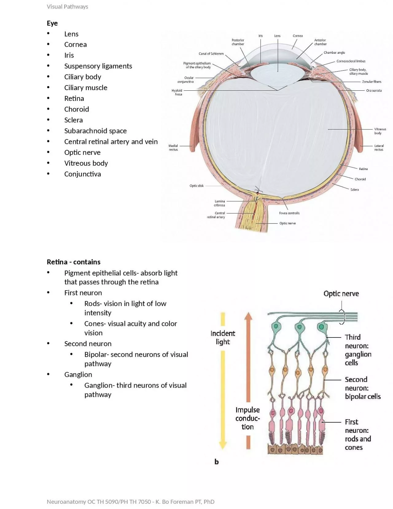
/
Retina - contains Pigment epithelial cells- absorb light that passes through the retina Retina - contains Pigment epithelial cells- absorb light that passes through the retina

Retina - contains Pigment epithelial cells- absorb light that passes through the retina - PowerPoint Presentation

samantha
samantha . @samantha
Follow
64 views
Uploaded On 2024-01-29

Retina - contains Pigment epithelial cells- absorb light that passes through the retina - PPT Presentation

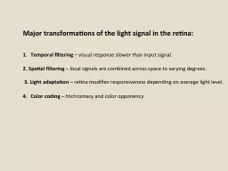
First neuron Rods vision in light of low intensity Cones visual acuity and color vision Second neuron Bipolar second neurons of visual pathway Ganglion Ganglion third neurons of visual pathway ID: 1042468

optic visual 7050 foreman visual optic foreman 7050 5090 neuroanatomy left cells hemianopsia homonymous pupil retina occipital sympathetic superior

Share:

Link:

Embed:

Download Presentation from below link

Download Presentation The PPT/PDF document "Retina - contains Pigment epithelial cel..." is the property of its rightful owner. Permission is granted to download and print the materials on this web site for personal, non-commercial use only, and to display it on your personal computer provided you do not modify the materials and that you retain all copyright notices contained in the materials. By downloading content from our website, you accept the terms of this agreement.


Presentation Transcript

1. Retina - containsPigment epithelial cells- absorb light that passes through the retinaFirst neuronRods- vision in light of low intensityCones- visual acuity and color visionSecond neuronBipolar- second neurons of visual pathwayGanglionGanglion- third neurons of visual pathwayVisual PathwaysNeuroanatomy OC TH 5090/PH TH 7050 - K. Bo Foreman PT, PhDEyeLensCorneaIrisSuspensory ligamentsCiliary bodyCiliary muscleRetinaChoroidScleraSubarachnoid spaceCentral retinal artery and veinOptic nerveVitreous bodyConjunctiva

2. Visual Pathway-Light travels through retina from internal to external exciting the rods and conesImpulse is transferred from rods and cones to bipolar cells Bipolar cells synapse on the dendrites of the ganglion cells Neuroanatomy OC TH 5090/PH TH 7050 - K. Bo Foreman PT, PhDAdditional areas:Optic disc- Area optic nerve exits retina (blind spot- no receptors)Macula lutea- central region of the retinaFovea centralis- area of the central part of the retina that is indented and contains mainly cones for visual acuity

3. Visual Pathway- forth order neuronsCell body located in the lateral geniculate bodyAxon travels posteriorly (optic radiations)Terminates in the visual cortex in the occipital lobeVisual Pathway-Ganglion cells form the optic nerve at the level of the optic discOptic nerves combine to form optic chiasmEnters lateral geniculate body to synapse

4. Neuroanatomy OC TH 5090/PH TH 7050 - K. Bo Foreman PT, PhD1. Right eye blindness - trauma, optic neuritis2. Bitemporal hemianopsia - pituitary tumors3. Right nasal hemianopsia - pressure by aneurysm of internal carotid artery4. Left homonymous hemianopsia - abscess or tumor of temporal lobe5. Left homonymous hemianopsia - anterior choroidal artery dysfunction6. Left homonymous superior quadratic anopsia - temporal or occipital lobe dysfunction7. Left homonymous inferior quadratic anopsia - parietal or occipital lobe tumor8. Left homonymous hemianopsia with macular preservation - posterior cerebral artery dysfunctions, tumors, trauma1.2.3.4.5.6.7.8.

5. Neuroanatomy OC TH 5090/PH TH 7050 - K. Bo Foreman PT, PhDVisual ReflexesLight reflex (constriction- parasympathetic): when light enters the pupil the pupil constrictsNeuronal elements of the retinaOptic nerveOptic chiasmOptic tractEnter the brachium of the superior colliculus (superior brachium)Pretectal regionOculomotor nuclear complex (Edinger-Westphal nucleus)Preganglionic parasympathetic axons to the ciliary ganglion behind eyeShort ciliary nervesTerminate on the constrictor muscle of the irisLecture 14 - Visual

6. Neuroanatomy OC TH 5090/PH TH 7050 - K. Bo Foreman PT, PhDVisual ReflexesLight reflex (dilation- sympathetic): when the pupil widens. Occurs passively when parasympathetic tone decreases and actively when sympathetic tone increasesHypothalamusSpinal cord (T1)Ventral rootWhite communicating ramiAscend sympathetic trunkSynapse in superior cervical ganglion with post-synaptic sympathetic ganglionTravel in the carotid plexusTerminate on the dilator muscle of the irisLecture 14 - Visual

7. Neuroanatomy OC TH 5090/PH TH 7050 - K. Bo Foreman PT, PhDVisual ReflexesAccommodation reflex: Process in which a clear visual image is maintained as gaze is shifted from a distant to a near point3 componentsConvergence of the eyesPupillary constrictionThickening of the lensInitiated by occipital cortexImpulses go to:Edinger-Westphal nucleus for changes in the lens and pupil (parasympathetic)Somatic nuclei: for convergence of the eyes (medial rectus