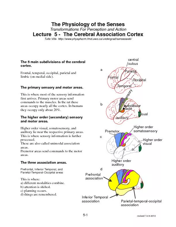
/
FrontalFrontal FrontalFrontal

FrontalFrontal - PDF document

sherrill-nordquist
sherrill-nordquist . @sherrill-nordquist
Follow
373 views
Uploaded On 2016-10-09

FrontalFrontal - PPT Presentation

ParietalParietal OccipitalOccipital TemporalTemporal a motormotor visualvisual auditoryauditory vestibularvestibular tastetaste b Higher order visual Premotor Higher ordersomatosensory Higher orderaud ID: 473569

ParietalParietal OccipitalOccipital TemporalTemporal a motormotor visualvisual auditoryauditory vestibularvestibular tastetaste b Higher order visual Premotor Higher ordersomatosensory Higher orderaud

Share:

Link:

Embed:

Download Presentation from below link

Download Pdf The PPT/PDF document "FrontalFrontal" is the property of its rightful owner. Permission is granted to download and print the materials on this web site for personal, non-commercial use only, and to display it on your personal computer provided you do not modify the materials and that you retain all copyright notices contained in the materials. By downloading content from our website, you accept the terms of this agreement.


Presentation Transcript

FrontalFrontal ParietalParietal OccipitalOccipital TemporalTemporal a motormotor visualvisual auditoryauditory vestibularvestibular tastetaste b Higher order visual Premotor Higher ordersomatosensory Higher orderauditory c Inferior Temporalassociation Parietal-temporal-occipitalassociation Prefrontalassociation d The Physiology of the SensesTransformations For Perception and ActionLecture 5 - The Cerebral Association CortexTutis Vilis http://www.physpharm.fmd.uwo.ca/undergrad/sensesweb/ The 5 main subdivisions of the cerebral cortex.Frontal, temporal, occipital, parietal and limbic (on medial side).The primary sensory and motor areas.This is where most of the sensory information first arrives. Primary motor areas send The higher order (secondary) sensory and motor areas.Higher order visual, somatosensory, and auditory lie near the respective primary areas. The three association areas.Prefrontal, Inferior Temporal, and Parietal-Temporal-Occipital areasThis is where:a) different modalities combine.b) attention is shifted. revised 14-8-2010 centralsulcus 5-1 somatosensorysomatosensory How are the different areas of cortex interconnected? 5-2 Long loop reflexes, such as writing down the name of a seen object, require the complex The neurons in all areas of the cortex are confined to a thin sheet called the gray matter. The sheet is extensively folded to maximize its All adjacent areas of cortex are extensively interconnected as are selected distant areas. These extensive interconnections predisposes the cortex to epilepsy. A locus of abnormal activity in one area quickly spreads to These interconnections are, as we will see later, where our memory is. Our grey matter contains an Grey matter consists of six, anatomically distinct, layers. Information arrives in layer 4, Layer 4 receives input from the thalamus and other cortical regions. It is thickest in primary The striate cortex (primary visual) is so-called because of its thick layer 4.Layers 3 and 5 send output to other cortical regions, the brain stem, and spinal cord. These layers are thickest in primary motor cortex. Primary SecondaryHigher order Association Sensory Premotor motor sensory Short loop reflexes long loop reflexes Inferior Temporallong term memory Prefrontalworking memory P-T-Oattention 5-3 What are the functions of the different association areas?a) Are the functions of different cortical areas unique?There are two opposing theories: Lashley's equipotential theory: Information on a particular function is spread out over the entire cortex.Evidence for: The loss of a few cells from a small lesion in one part results in minimal impairment.Evidence against: The cortex is not uniform. Different regions serve different functions. Grandmother cell theory: Simple cells connect to complex cells, complex cells connect to hyper-complex cells, and so on, until finally there is one unique cell that fires when you see your grandmother. If you loose that cell, you can no longer recognize your grandmother but have no problems with grandfather.Evidence for: Some lesions do impair the recognition of faces selectively. Some cells are activated only by a particular face.Evidence against: Brain cell death is common, yet the memory loss observed is a general fuzziness in remembering faces, not an absolute loss of one face and not of another.Truth probably lies somewhere between these two extremes. b) Is the function of a particular cortical area identical in different people?No. The cortex is very plastic, particularly in early life.If a particular sensory input is missing.That area which previously received this input will now receive a different sensory input. This is similar to the competition for cortical representation between the two eyes. If one eye is This may explain the very fine acuity of these senses in the blind.If a particular cortical area is damaged.The same function becomes organized in a new cortical area. For example language is usually represented on the left. If the left is damaged early in life, the right side develops How and why do the two sides of the cortex differ in function?a) Each hemisphere excels in different tasksDominant (usually left) - sequential or serial taskse.g. language (reading writing speaking signing), analytic (math A=B, B=C, therefore A=C)Non-dominant (usually right)- tasks requiring parallel processinge.g. spatial tasks, intuitive (C resembles O as I resembles L), geometry, music. 5-4 b) Patients with a section of the corpus callosum.The corpus callosum is the large fiber tract that interconnects the two hemispheres.When a patient with a sectioned corpus callosum is shown an apple on the left, the patient cannot name The subject could visually recognize an apple and pick it out from a group of other objects with his Two independent brains function in one person (e.g. a patient would hug wife with one arm and c) What are the advantages and disadvantages of lateralization?Advantage: Shorter paths to interconnect related regions.. Redundancy is good if one part breaks down. Front to left arm motor area left arm corpus callosum Inferior Temporal Association AreasThese are large area located on the underside of the cortex.The inferior and medial portions of the temporal lobe are involved in long term memory.The right side is more involved with pictorial memory (e.g. faces) and the left side, in verbal memory (e.g. names of people). We will look at memory in detail in the last session. 5-6 Parietal- Occipital-Temporal (PTO): Polimodal convergence of senses and attention.e.g. locating objects in space by touch, sight, sound etc.e.g. language: sound of words, written words (sight), or Braille (touch)Attention allows us to focus in on specific stimuli and neglect others, for example a specific voice in Parietal-temporal-occipitalassociation Prefrontal association area: Two other very important functions of the prefrontal association area are decision making and planning.After lesions here, patients are calmer and less frustrated. If one has no plans, one has no expectations and is less likely to be frustrated if things go wrong.Because this has a calming effect, frontal lobotomies used to be a popular cure for aggression. Unfortunately it also destroyed initiative.Neurologist António Egas Moniz won the Nobel Prize for medicine in 1949 for inventing this technique.Dr. Walter Freeman used an ice pick hammered through the back of the eye socket into the brain. He performed thousands of lobotomies, each in minutes from his office. 5-5 Prefrontal association area: The prefrontal cortex has become larger, as a percentage of total brain size, over the course Lesions of the prefrontal association area produce deficits in tasks that are spatial and This is called working memory, a form of short term memory. Prefrontalassociation Depletion of dopamine in the prefrontal cortex impairs working memory.The prefrontal cortex is interconnected with the basal ganglia. Parkinson patients have difficulty in initiating movements to targets in working memory. They need some external stimulus to initiate movement. Children prior to the age of 1 yr have not developed this working memory.Neurons in the prefrontal cortex showIf a toy is covered by one of two covers, the child cannot find it. Out of sight is out of mind.a) activity which starts when a stimulus appears (T on) in a particular locationb) unlike neurons in V1, here activity continues even when the stimulus disappears (T off).This tonic activity holds the object location in .Different cells hold the memory of objects in different target locations. toy cover X X stimuluson stimulusoff T on T off noactivity The spot light of attentionResearchers at Western Washington University questioned students who walked while talking into their cell phonesThey were twice as likely of not noticing the clown than those without cell phones. In attention True whether you are walking, driving a car or listening to this lecture.An analogy that is simple and useful in beginning to understanding attention is that of a flash light that selectively Attention acts like a bottle neck. It limits what enters the brain. The retina and visual cortex see both the person and the bike but attention limits what gets further down into the what We are blind to what does not get through the bottleneck.Visual perception is a two stage process Stage 1) an early stage that performs rapid low level , awareness and consciousness.Visual objects compete for your attention. While attention is processing a visual object one is blind to the presence of other This is known as the attentional blink, that is we behave as if our Attention can be drawn from below by things that pop out from the background. In early visual areas, like or similar ferent objects are accentuated and thus drawing attention to themselves. Or attention can be directed voluntarily from above by areas in the parietal dorsal stream.Finding Waldo takes time. This occurs for 3 reasons.1) Because Waldo does not automatically draw your attention to him as does a horizontal line among a background of Waldo to another.3) At each location, it takes time to process the image and decide if it is Waldo or not. 5-7 X a X b X c X Attention allows us to focus in on a specific location. This is illustrated by a simple experiment developed by a) Subject is asked to fixate on the Xb) Subject cued to expect an stimulus on the A test stimulus is presented to the left or rightThe subject has better sensitivity to the cued side because attention was shifted to it. Most people, take longer to respond on the trial cue is to one side and the test is to the . This is because on these trials the cue shifts attention to the wrong place. In trials This is an example of spatially directed attention.Posner Attention is not the same as arousal.Arousal is general while attention is specific. Arousal is mediated by one of several diffuse systems arising from the brainstem. One of these is the locus They act like a volume control to increase one’s level of alertness. 5-8 Attending to different features causes activity in different cortical regions A subject is shown an image A and then, after a delay of .5 sec, image B and then CInstruction to the subject:the same color.”ii)“Indicate when two successive objects have the same shape.”iii) “Indicate when two successive objects have the same direction of motion.”Attention can be viewed as a flash light that is directed at different aspects of an objectBut the big question is: “What directs the flash light?” A B C ii) enhancedactivity iii) enhancedactivity i) enhancedactivityin early visual areas 5-9 Attention to a particular color or orientation Find the line that is different.That was easy because, as we saw in session 3, your early visual areas automatically cause Now find the line that is different in a more complex pattern.This takes a bit longer. Now the odd line no longer pops out. You need to serially scan the image to find the Now try it again but this time focus your The same thing happens if one focuses one's attention to just . 5-10 We have seen 2 types of attention: 1. spatial: you can voluntarily direct the 2. feature-based: you can voluntarily focus your attention onto a particular attribute such , etc.rdIn addition there is a 3 type of attentionObject-based: Objects that stand out from the background automatically attract attention.This can bea vertical line that pops out when seen in a 5-11 NeglectWe saw that the PTO is involved in to the spatial location of objects in space and to a particular object.A lack of attention leads to lack of awareness. A lesion of the right PTO causes neglect of things on the left. The patient is unaware that half the world is gone.focussing our attention 5-12 In a right V1 lesion, one is blind to everything to the left of where one's eyes look.In many right PTO lesions, the left side of the face is neglected independent of whether you look (X) to the right This is because what is left and right depends on how the brain frames the object.In objects like faces, the frame is drawn around the face and patients neglect the left side of the frame independent of its XX XX You might suppose that a lesion of the left PTO would result in neglect of things on the right. Strangely it does not. One suggested FrontFront normal LL RR RR Thus after a lesion on the left, the right side still attends to things on the right (as well as the left). After a lesion on the right, the representation of things on the left is lost. FrontFront right lesion Front left lesion 5-13 Coordinate FramesThere are many ways of stating an object’s location. Two ways are:i) Allocentric coordinates: State its location with respect to some other object, e.g., a table in terms of its location in a room, or a feature of a face in terms of its location on the face. you. (Note you can be your body, your head or your eye).ference between these is to consider what happens when you move (e.g. to the right). A useful model: Take two transparencies. On one draw an x indicating your location. On the other draw the location of a few objects. Place the two on top of each other. For allocentric, move the sheet with you on it.The ruler is on the room. See how you re-code For egocentric, move the sheet with the objects on it. The ruler is on you.See how you re-code the location of all the 5-14 Allocentric & egocentric coordinates may have nested frames. An example of a nested allocentric frame:Frame 1. The location of the apple may be specified with respect to the table.Frame 2. The location of the table may be specified with respect to the room. i) allocentric object 1 object 2 me room X ii) egocentric object 1 object 2 me X 5-15 Coordinate FramesAreas in the "where" stream tend to encode locations in egocentric coordinates.Areas in the "what" stream tend to encode locations in allocentric (object centered) If the task is to recognize a face, the "what" stream is involved. The frame of attention is centered on the face. The features of the face are coded in an allocentric frame and If you are standing at one end of a square and the task is to walk to restaurant on your

Related Contents


Next Show more