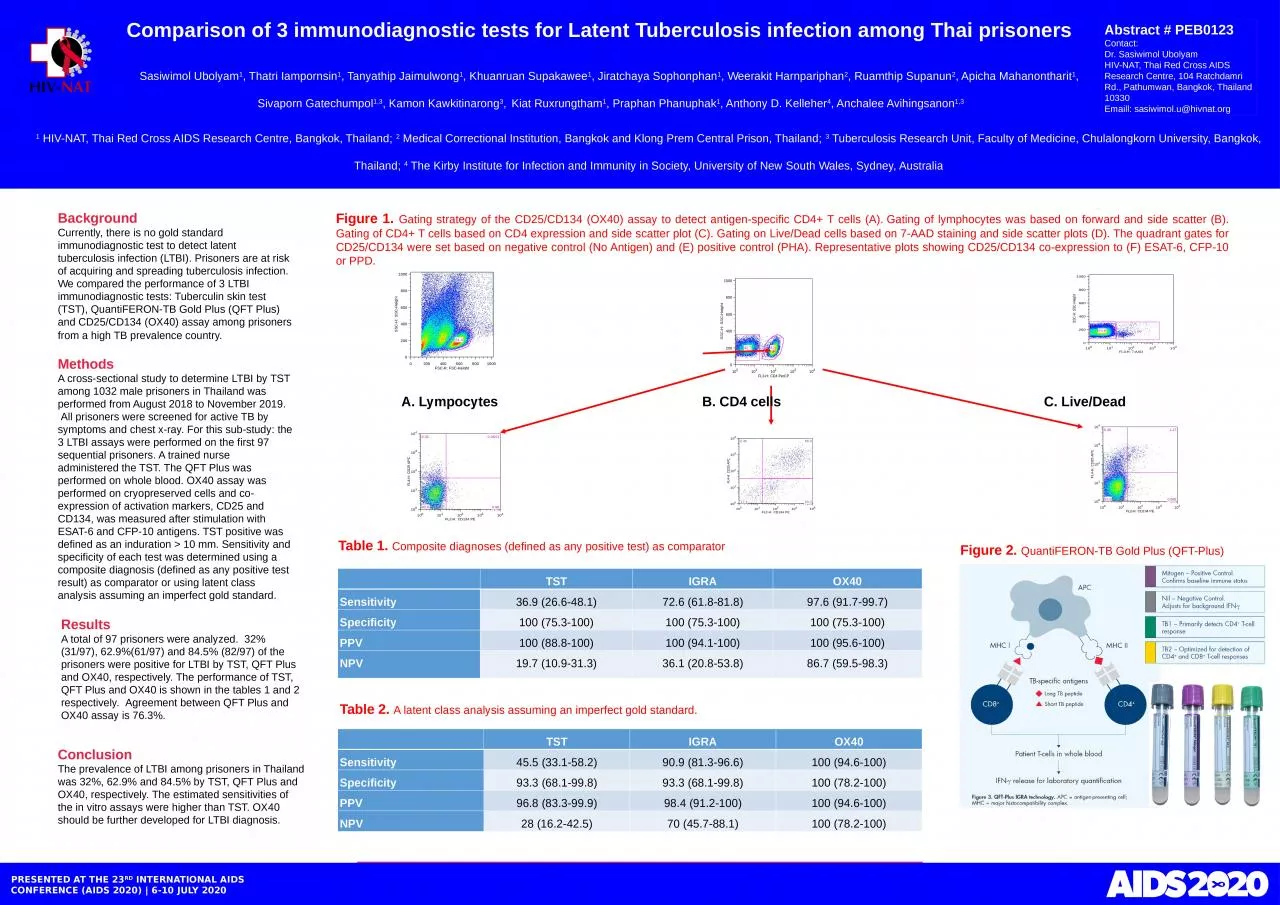
/
Background Currently, there is no gold standard immunodiagnostic test to detect latent Background Currently, there is no gold standard immunodiagnostic test to detect latent

Background Currently, there is no gold standard immunodiagnostic test to detect latent - PowerPoint Presentation

singh
singh . @singh
Follow
27 views
Uploaded On 2024-02-09

Background Currently, there is no gold standard immunodiagnostic test to detect latent - PPT Presentation

QuantiFERON TB Gold Plus QFT Plus and CD25CD134 OX40 assay among prisoners from a high TB prevalence country   Methods A crosssectional study to determine LTBI by TST among 1032 male prisoners in Thailand was performed from August 2018 to November 2019  All prisoners were screened for ID: 1045832

prisoners 100 tst ox40 100 prisoners ox40 tst qft ltbi thailand positive test cells gold cd25 cd134 cd4 performed

Share:

Link:

Embed:

Download Presentation from below link

Download Presentation The PPT/PDF document "Background Currently, there is no gold s..." is the property of its rightful owner. Permission is granted to download and print the materials on this web site for personal, non-commercial use only, and to display it on your personal computer provided you do not modify the materials and that you retain all copyright notices contained in the materials. By downloading content from our website, you accept the terms of this agreement.


Presentation Transcript

1. BackgroundCurrently, there is no gold standard immunodiagnostic test to detect latent tuberculosis infection (LTBI). Prisoners are at risk of acquiring and spreading tuberculosis infection. We compared the performance of 3 LTBI immunodiagnostic tests: Tuberculin skin test (TST), QuantiFERON-TB Gold Plus (QFT Plus) and CD25/CD134 (OX40) assay among prisoners from a high TB prevalence country. MethodsA cross-sectional study to determine LTBI by TST among 1032 male prisoners in Thailand was performed from August 2018 to November 2019.  All prisoners were screened for active TB by symptoms and chest x-ray. For this sub-study: the 3 LTBI assays were performed on the first 97 sequential prisoners. A trained nurse administered the TST. The QFT Plus was performed on whole blood. OX40 assay was performed on cryopreserved cells and co-expression of activation markers, CD25 and CD134, was measured after stimulation with ESAT-6 and CFP-10 antigens. TST positive was defined as an induration > 10 mm. Sensitivity and specificity of each test was determined using a composite diagnosis (defined as any positive test result) as comparator or using latent class analysis assuming an imperfect gold standard.  Comparison of 3 immunodiagnostic tests for Latent Tuberculosis infection among Thai prisoners 1 HIV-NAT, Thai Red Cross AIDS Research Centre, Bangkok, Thailand; 2 Medical Correctional Institution, Bangkok and Klong Prem Central Prison, Thailand; 3 Tuberculosis Research Unit, Faculty of Medicine, Chulalongkorn University, Bangkok, Thailand; 4 The Kirby Institute for Infection and Immunity in Society, University of New South Wales, Sydney, AustraliaPRESENTED AT THE 23RD INTERNATIONAL AIDS CONFERENCE (AIDS 2020) | 6-10 JULY 2020ResultsA total of 97 prisoners were analyzed.  32% (31/97), 62.9%(61/97) and 84.5% (82/97) of the prisoners were positive for LTBI by TST, QFT Plus and OX40, respectively. The performance of TST, QFT Plus and OX40 is shown in the tables 1 and 2 respectively.  Agreement between QFT Plus and OX40 assay is 76.3%.ConclusionThe prevalence of LTBI among prisoners in Thailand was 32%, 62.9% and 84.5% by TST, QFT Plus and OX40, respectively. The estimated sensitivities of the in vitro assays were higher than TST. OX40 should be further developed for LTBI diagnosis. Abstract # PEB0123Contact:Dr. Sasiwimol UbolyamHIV-NAT, Thai Red Cross AIDS Research Centre, 104 Ratchdamri Rd., Pathumwan, Bangkok, Thailand 10330Emaill: sasiwimol.u@hivnat.orgSasiwimol Ubolyam1, Thatri Iampornsin1, Tanyathip Jaimulwong1, Khuanruan Supakawee1, Jiratchaya Sophonphan1, Weerakit Harnpariphan2, Ruamthip Supanun2, Apicha Mahanontharit1, Sivaporn Gatechumpol1,3, Kamon Kawkitinarong3, Kiat Ruxrungtham1, Praphan Phanuphak1, Anthony D. Kelleher4, Anchalee Avihingsanon1,3Figure 1. Gating strategy of the CD25/CD134 (OX40) assay to detect antigen-specific CD4+ T cells (A). Gating of lymphocytes was based on forward and side scatter (B). Gating of CD4+ T cells based on CD4 expression and side scatter plot (C). Gating on Live/Dead cells based on 7-AAD staining and side scatter plots (D). The quadrant gates for CD25/CD134 were set based on negative control (No Antigen) and (E) positive control (PHA). Representative plots showing CD25/CD134 co-expression to (F) ESAT-6, CFP-10 or PPD.A. Lympocytes B. CD4 cells C. Live/DeadTable 1. Composite diagnoses (defined as any positive test) as comparator TSTIGRAOX40Sensitivity36.9 (26.6-48.1)72.6 (61.8-81.8)97.6 (91.7-99.7)Specificity100 (75.3-100)100 (75.3-100)100 (75.3-100)PPV100 (88.8-100)100 (94.1-100)100 (95.6-100)NPV19.7 (10.9-31.3)36.1 (20.8-53.8)86.7 (59.5-98.3)Table 2. A latent class analysis assuming an imperfect gold standard. TSTIGRAOX40Sensitivity45.5 (33.1-58.2)90.9 (81.3-96.6)100 (94.6-100)Specificity93.3 (68.1-99.8)93.3 (68.1-99.8)100 (78.2-100)PPV96.8 (83.3-99.9)98.4 (91.2-100)100 (94.6-100)NPV28 (16.2-42.5)70 (45.7-88.1)100 (78.2-100)Figure 2. QuantiFERON-TB Gold Plus (QFT-Plus)