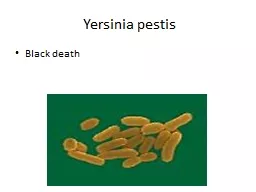
/
Yersinia  pestis Black death Yersinia  pestis Black death

Yersinia pestis Black death - PowerPoint Presentation

sistertive
sistertive . @sistertive
Follow
343 views
Uploaded On 2020-06-17

Yersinia pestis Black death - PPT Presentation

taxonomy Member of the Enterobacteriaceae family yersinia is gram nagative coccobacilli 4 Yersinia   General characteristics Yersinia ID: 780489

plague yersinia disease bubonic yersinia plague bubonic disease animal cases organisms test diagnostic staining virulence bacteria pestis black human

Share:

Link:

Embed:

Download Presentation from below link

Download The PPT/PDF document "Yersinia pestis Black death" is the property of its rightful owner. Permission is granted to download and print the materials on this web site for personal, non-commercial use only, and to display it on your personal computer provided you do not modify the materials and that you retain all copyright notices contained in the materials. By downloading content from our website, you accept the terms of this agreement.


Presentation Transcript

Slide1

Yersinia pestis

Black death

Slide2

taxonomy

Member of the

Enterobacteriaceae

family

yersinia

is gram

nagative

coccobacilli

Slide3

4-Yersinia


General

characteristics

:- Yersinia pestis is the causative agents of plague (the Black Death), and another two less important species: Yersinia enterocoltitica & Y. pseudo tuberculosis.- It is a small Gram –ve bacilli, with bipolar staining with clear central area.- Freshly isolated organisms possess a capsule composed of polysaccharide-protein complex, where it is considered as virulence factor, where its loss leads to loss virulence.- It is considered as the virulent most bacteria known, where only 1-10 bacteria are capable of causing the disease.- Wild rodents are the animal reservoir, where it is endemic now in western U.S.A and in South East Asia.

Pathogenesis

:

-

The

vector is the wild rodent’s fleas.

Dogs could as the main reservoir, and human is the accidental host

.

-

The

organisms inoculated by the flea bite are spread to the regional lymph nodes which become swollen and tender (buboes), bubonic plague

.

-

Organisms

reach high concentration in the blood lead to abscesses in different organs, and the endotoxins relayed symptoms, such as disseminated intravascular coagulation, and Cutaneous hemorrhage gives the name of Black Death.

Slide4

The virulence factors include

:

*

The

capsular antigens F-1* The cell wall endotoxic* An exotoxin (its action is unknown)* V & W antigens (protect organisms from intracellular killing and digestion)* Yops (Yersinia Outer Proteins), experimentally shown to inhibit phagocytosis and cytokine production, as well as inhibit tumor necrosis factor production.Laboratory diagnosis:* Smear and culture from the blood or bubo is the best diagnostic test.* Great care must be taken during samplings.* Giemsa stain shows safety pin cell appearance.* For further diagnosis, fluorescent-antibody staining is used. Treatment:Combination of erythromycin and tetracycline is used without waiting the result to appear.

Slide5

Target tissues

This disease direct effects the lymph node which can be found in the groin , neck , and armpits cause them to enlarge and suppurate

Slide6

Symptoms

Bubonic Plague

bacteria infect lymph nodes

Bubos

Fever

HeadacheVomiting Blood

Slide7

Diagnostic Tests

Take smear from blood or feces for bubonic plague

> bacteria has “safety pin” appearance

Can also use FA (fluorescent-antibody) test

All plague bacilli have unique diagnostic envelope glycoprotein called the Fraction 1 (F1) antigen

Slide8

Epidemiology: Transmission

Bubonic

Infected Rodent

 Fleas  Humans

Can also enter through breaks in skin when handling infected animal

Slide9

Other

Yersinia

cause disease.

Yersinia

enterocolitica

Typically, only a small number of human cases of Yersiniosis are recognized. Symptoms are like that of appendicitis and out breaks are often detected by a sudden increase in appendectomies in a particular region. The Center for Disease Control & Prevention estimates that about 17,000 cases occur each year in the United States.

Slide10

Mortality

Bubonic Plague

Untreated 50- 60% mortality rate

Treated 5 – 20% mortality rate

Killed one third of the world’s population during the 14th century Latest reports As of 15 March 2001, World Health Organization has reported a total of 436 suspected cases, including 11 deaths in Nyanje area in Zambia. As of 27 May 2002, the Malawian Ministry of Health has reported a total of 71 cases of bubonic plague in Malawi.

Slide11

WHO reports 1,000 to 3,000 cases of plague every year!

Slide12

EVOLUTION: A single gene change in a relatively benign recent ancestor of the bacterium that causes bubonic plague played a key role in the evolution of the deadly disease from a germ that causes a mild human stomach illness acquired via contaminated food or water to the flea-borne agent of the "Black Death.”

GENETICS: Research on three genes,

hemin

storage

(hms) genes, in Y. pestis that change it from a harmless, long-term inhabitant in the flea midgut to one that amasses in its foregut. PREVENTION: Current prevention measures include dusting family pets with insecticides to prevent the spread of the Yersinia pestis organism from the native prairie dog populations

Slide13

5-Francisella


Francisella

tularensis, the causative agent of Tularemia.Small Gram –ve bacilli with a single serologic type.  Pathogenesis: - It is enzootic, isolated from more than 100 different species of wild animals, mainly rabbits, deer, and a variety of rodents.- The vectors are: ticks, mites and lice, where ticks can pass it to their offspring.- Human acquire the infection by a tick bite or due a direct contact with the animal during removal of the hide.- No person to person transmission.- Rarely transmitted by ingestion or inhalation causing gastrointestinal or pneumonic tularemia respectively.- Clinical findings, vary from sudden influenza- like syndrome to prolonged onset of a low grade fever and adenopathy, and may be other gastrointestinal and typhoid symptoms.Disease usually confers a lifelong immunity.

Laboratory diagnosis

:

It is very risky dealing with this organism.

Media should contain

cystcine

.

The best diagnostic tests are

:

1-

agglutination

test and

2-

fluorescent- antibody staining test of the infected tissues.

Slide14

6-Pasteurella

*

Pasteurella

multocida causes wound infections associated with cat and dogs bites.* It is short Gram –ve, encapsulated with bipolar staining.* 25% of these animal bites will transmit these organisms with other anaerobic and facultative anaerobes present in the mouth.* The capsule and the endotoxins are considered as the virulence factors* The incubation period is less than 24 hrs.* Rapidly spreading cellulites at the site of the animal bite is an indicative of the P.multocida.* Osteomyelitis may complicate cat bite.* Laboratory diagnosis is binding the organism in culture of wound sample.

*

Penicillin

G

is the drug of choice.

Slide15

Thank you