/
Clinical Enzymology ☼ Clinical Enzymology ☼

Clinical Enzymology ☼ - PowerPoint Presentation

sophia
sophia . @sophia
Follow
66 views
Uploaded On 2023-08-30

Clinical Enzymology ☼ - PPT Presentation

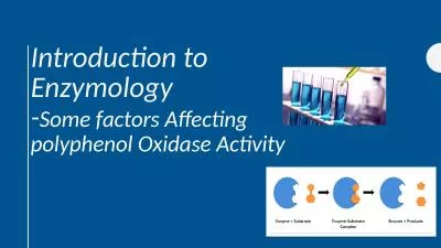
Enzymes are catalysts that increase the rate of physiologic reactions Each and every reaction in our body catalyzed by enzyme In general most enzymes are present in cells at much higher concentrations than in plasma ID: 1014836

enzymes plasma ldh levels plasma enzymes levels ldh muscle tissue disorders activity enzyme liver serum elevated myocardial ast isoenzyme

Share:

Link:

Embed:

Download Presentation from below link

Download Presentation The PPT/PDF document "Clinical Enzymology ☼" is the property of its rightful owner. Permission is granted to download and print the materials on this web site for personal, non-commercial use only, and to display it on your personal computer provided you do not modify the materials and that you retain all copyright notices contained in the materials. By downloading content from our website, you accept the terms of this agreement.


Presentation Transcript

1. Clinical Enzymology

2. ☼ Enzymes are catalysts that increase the rate of physiologic reactions. Each and every reaction in our body catalyzed by enzyme.☼ In general, most enzymes are present in cells at much higher concentrations than in plasma.☼ Measurement of their levels in plasma indicates whether their tissue of origin is damaged leading to the release of intracellular components into the blood.☼ This forms the basis of clinical enzymology.Thus clinical enzymology refers to measurement of enzyme activity for the diagnosis and treatment of diseases.Since the tight control of enzyme activity is essential for homeostasis, any malfunction of a single critical enzyme (mutation, overproduction, underproduction or deletion) can lead to a genetic disease - commonly called inborn errors of metabolism.One example is the most common type of phenylketonuria caused by a mutation of a single amino acid in the enzyme phenylalanine hydroxylase.

3. DIAGNOSTIC CLINICAL ENZYMOLOGY:☼ One major item of the diagnostic clinical enzymology is the investigation of changes in the level of enzymes and their correlation to the differential diagnosis of diseases and to establish cut-off levels for; normal, benign and malignant diseases.☼ These enzymes changes could be followed up in plasma, serum, urine, blood cells or tissue biopsies.☼ Thus, detection of the plasma level of an enzyme immunologically (for its protein amount) or colorimetrically (for its activity, preferred) have the following applications:

4. Diagnosis: As example, high serum creatine phosphokinase (CPK2) on the day of a suspected case of myocardial infarction strengthen the diagnosis if ECG changes are doubtful.Differential diagnosis: e.g., chest pain associates myocardial infarction and pulmonary embolism. Elevated serum glutamate-oxaloacetate transaminase (GOT) and lactate dehydrogenase (LDH) characterizes myocardial infarction, whereas, elevated serum LDH only characterizes pulmonary embolism.

5. PLASMA ENZYMES☼ Enzymes present in plasma can be classified into 2 types, they areFunctional Plasma enzymes and Non-functional plasma enzymes.·Plasma-derived enzymes (functional plasma enzymes):They are normally occurring functional plasma enzymes.Mostly synthesized by the liver.Usually decreased in disease conditions.Their field of activity is plasma components and their activity is higher in plasma than in cells, e.g., coagulation and lipoprotein-metabolizing enzymes.Their clinical importance is limited to diseases related to their own synthesis and function; i.e., abnormalities of metabolism of plasma lipoproteins and blood clotting, and the organ function of their synthesizing tissues, e.g., thromboplastin as a liver function test.

6. Cell-Derived enzymes (Non-functional plasma enzymes):Normally they locate to intracellular compartments; i.e., they are non-functional plasma enzymes.A very low plasma level normally exists due to normal wear and tear and diffusion through undamaged cell membranes.The amount and nature of the plasma enzyme(s) reflects the extent and nature of the damaged tissue.Measurement of these enzymes in plasma can be used to assess cell damage and proliferation i.e. diagnosis of disease.

7. They are further subdivided into; secretory and metabolic non-functional plasma enzymes:Secretory: They are synthesized and secreted by specialized glands into body lumens mainly for digestion.Metabolic: They are intracellular metabolic enzymes and their appearance in the plasma is mainly due to cellular damage among other factors.INCREASED non-functional plasma enzymes could be due to increased release and/or impaired clearance.

8. ISOENZYMES:☼ Isoenzymes (also known as isozymes) are enzymes that differ in amino acid sequence but catalyze the same chemical reaction☼ Believed to be originating from closely linked genes or from multiple gene loci.☼ Evolution from a single form possibly due to long-term mutations☼ They vary with respect to their kinetic parameters, electrophoretic mobility, and localization.☼ They all have independent action E.g. Lactate dehydrogenase have 5 isoenzymes (LDH1, LDH2, LDH3, LDH4 & LDH5)☼ They can be used to identify the specific affected tissues.☼ They can be differentiated from each other and can be clinically quantified in the lab.

9. CREATINE KINASECK is an enzyme that is generally associated with ATP regeneration in contractile or transport systems.☼ Its predominant physiologic function occurs in muscle cells, where it is involved in the storage of high-energy creatine phosphate.☼ Every contraction cycle of muscle results in creatine phosphate use, with the production of ATP.☼ This results in relatively constant levels of muscle ATP. The reversible reaction catalyzed by CK is shown in the following Equation.

10. Tissue SourceCK is widely distributed in tissue, with highest activities found in skeletal muscle, heart muscle, and brain tissue.CK is present in much smaller quantities in other tissue sources, including the bladder, placenta, gastrointestinal tract, thyroid, uterus, kidney, lung, prostate, spleen, liver, and pancreas.Diagnostic SignificanceBecause of the high concentrations of CK in muscle tissue, CK levels are frequently elevated in disorders of cardiac and skeletal muscle.The CK level is considered a sensitive indicator of acute myocardial infarction (AMI) and muscular dystrophy.Total CK levels are not entirely specific indicators in as much as CK elevation is found in various other abnormalities of cardiac and skeletal muscle.Levels of CK also vary with muscle mass and, therefore, may depend on;Gender, race, degree of physical conditioning, and age.Elevated CK levels are also occasionally seen in central nervous system disorders such as cerebrovascular accident, seizures, nerve degeneration, and central nervous system shock.Damage to the blood–brain barrier must occur to allow enzyme release to the peripheral circulation.

11. CK Isoenzyme:Because enzyme elevation is found in numerous disorders, the separation of total CK into its various ISOENZYME fractions is considered a more specific indicator of various disorders than total levels.Typically, the clinical relevance of CK activity depends more on isoenzyme fractionation than on total levels.CK occurs as a dimer consisting of two subunits that can be separated readily into three distinct molecular forms.The three isoenzymes have been designated as CK-BB (brain type), CK-MB (hybrid type), and CK-MM (muscle type).Hypothyroidism results in CK-MM elevations because of the involvement of muscle tissue (increased membrane permeability), the effect of thyroid hormone on enzyme activity, and, possibly, the slower clearance of CK as a result of slower metabolism.

12.

13. It has been observed that CK-BB may be significantly elevated in patients with carcinoma of various organs.These findings indicate that CK-BB may be a useful tumor-associated marker.The most common causes of CK-BB elevations are central nervous system damage, tumors, childbirth, and the presence of macro-CK, an enzyme–immunoglobulin complex.

14. The value of CK isoenzyme separation can be found principally in detection of myocardial damage.Cardiac tissue contains significant quantities of CK-MB, approximately 20% of all CK-MB.Whereas CK-MB is found in small quantities in other tissue, myocardium is essentially the only tissue from which CK-MB enters the serum in significant quantities.Demonstration of elevated levels of CK-MB, greater than or equal to 6% of the totalCK, is considered a good indicator of myocardial damage, particularly AMI.The typical time course of CK-MB elevation following AMI is not found in other conditions.

15. Reference Range Male, 15–160 U/L (37°C).Female, 15–130 U/L (37°C).The higher values in males are attributed to increased muscle mass.

16. LACTATE DEHYDROGENASELDH is an enzyme that catalyzes the interconversion of lactic and pyruvic acids. Tissue SourceLDH is widely distributed in the body. High activities are found in the heart, liver, skeletal muscle, kidney, and erythrocytes; lesser amounts are found in the lung, smooth muscle, and brain.

17. Because of its widespread activity in numerous body tissues, LDH is elevated in a variety of disorders.Increased levels are found in cardiac, hepatic, skeletal muscle, and renal diseases, as well as in several hematologic and neoplastic disorders.Liver disorders, such as viral hepatitis and cirrhosis, show slight elevations of two to three times.AMI and pulmonary infarct also show slight elevations of approximately the same degree.In AMI, LDH levels begin to rise within 12 to 24 hours, reach peak levels within 48 to 72 hours, and may remain elevated for 10 days.Skeletal muscle disorders and some leukemias contribute to increased LDH levels. Marked elevations can be observed in most patients with acute lymphoblastic leukemia in particular.Because of the many conditions that contribute to increased activity, an elevated total LDH value is a rather nonspecific finding.

18. LDH IsoenzymesLDH assays, therefore, assume more clinical significance when separated into isoenzyme fractions.The enzyme can be separated into five major fractions, each comprising four subunits. It has a molecular weight of 128,000 daltons.Each isoenzyme comprises four polypeptide chains with a molecular weight of 32,000 daltons each. Two different polypeptide chains, designated H (heart) and M (muscle), combine in five arrangements to yield the five major isoenzyme fractions.In the sera of healthy individuals, the major isoenzyme fraction is LDH-2, followed by LDH-1, LDH-3, LDH-4, and LDH-5.However, cardiac tissue and red blood cells contain a higher concentration of LDH- 1, Therefore, in conditions involving cardiac necrosis (AMI) and intravascular hemolysis.

19.

20. However, LDH is not specific to cardiac tissue and is not a preferred marker of diagnosis of AMI.LDH-1/LDH-2 ratios greater than 1 also may be observed in hemolyzed serum samples.Elevations of LDH-3 occur most frequently with pulmonary involvement and are also observed in patients with various carcinomas.The LDH-4 and LDH-5 isoenzymes are found primarily in liver and skeletal muscle tissue, with LDH-5 being the predominant fraction in these tissues.LDH-5 levels have greatest clinical significance in the detection of hepatic disorders, particularly intrahepatic disorders.Disorders of skeletal muscle will reveal elevated LDH-5 levels, as depicted in the muscular dystrophies.Reference RangeLDH, 100–225 U/L (37°C).

21. ASPARTATEAMINOTRANSFERASE☼ Aspartate aminotransferase (AST) is an enzyme belonging to the class of transferases. It is commonly referred to as a transaminase and is involved in the transfer of an amino group between aspartate and α-keto acids.☼ The older terminology, serum glutamic-oxaloacetic transaminase (SGOT, or GOT), may also be used.

22. Tissue SourceAST is widely distributed in human tissue. The highest concentrations are found in cardiac tissue, liver, and skeletal muscle, with smaller amounts found in the kidney, pancreas, and erythrocytes.Diagnostic SignificanceThe clinical use of AST is limited mainly to the evaluation of hepatocellular disorders and skeletal muscle involvement.AST elevations are frequently seen in pulmonary embolism.In AMI, AST levels begin to rise within 6 to 8 hours, peak at 24 hours, and generally return to normal within 5 days.

23. AST levels also may be increased, probably reflecting liver involvement as a result of inadequate blood supply to that organ.AST levels are highest in acute hepatocellular disorders.In viral hepatitis, levels may reach 100 times.In cirrhosis, only moderate levels—approximately four times—are detected Skeletal muscle disorders, such as the muscular dystrophies, and inflammatory conditions also cause increases in AST levels.The intracellular concentration of AST may be 7,000 times higher than the extracellular concentration.AST exists as two isoenzyme fractions located in the cell cytoplasm and mitochondria.The cytoplasmic isoenzyme is the predominant form occurring in serum.In disorders producing cellular necrosis, the mitochondrial form may be significantly increased.Reference RangeAST, 5 to 30 U/L (37°C).

24. ALANINEAMINOTRANSFERASE☼ Alanine aminotransferase (ALT) is a transferase with enzymatic activity similar to that of AST. Specifically, it catalyzes the transfer of an amino group from alanine to α- ketoglutarate with the formation of glutamate and pyruvate.☼ The older terminology was serum glutamic-pyruvic transaminase (SGPT, or GPT).Tissue Source☼ ALT is distributed in many tissues, with comparatively high concentrations in the liver. It is considered the more liver-specific enzyme of the transferases.

25. Diagnostic SignificanceClinical applications of ALT assays are confined mainly to evaluation of hepatic disorders. Higher elevations are found in hepatocellular disorders than in extrahepatic or intrahepatic obstructive disorders.In acute inflammatory conditions of the liver, ALT elevations are frequently higher than those of AST and tend to remain elevated longer as a result of the longer half-life of ALT in serum (16 and 24 hours, respectfully).Cardiac tissue contains a small amount of ALT activity, but the serum level usually remains normal in AMI unless subsequent liver damage has occurred.ALT levels have historically been compared with levels of AST to help determine the source of an elevated AST level and to detect liver involvement concurrent with myocardial injury.Reference RangeALT, 6–37 U/L (37°C).

26. ENZYMES & DISEASE DIGAGNOSIS

27. PANCREATIC ENZYMES:Acute pancreatitisIt is an inflammatory process in which pancreatic enzymes are activated and cause auto- digestion of the gland.It is a result of anatomical changes that arise from two events.The first is the autodigestion of the acinar cells by inappropriate activation of the pancreatic enzymes (especially trypsinogen) within the cell.The second is the cellular injury response that is mediated by pro- inflammatory cytokines.There are some enzymes that are synthesized and stored as the active enzymes in the zymogen granules.These include α-amylase, and lipase.

28. α-Amylase: Raised plasma amylase activity caused by;Severe glomerular impairment.Acute pancreatitis.Moderate to higher increase (up to five times the upper reference limit):Perforated peptic ulcer.Acute cholecystitis.Intestinal obstruction.Salivary gland disorders like mumps, salivary calculi.

29. Lipase:Plasma lipase levels are elevated in;Acute pancreatitis andCarcinoma of the pancreas.Serum amylase is increased in mumps, pancreatic disease or due to some other cause,whereas lipase is increased only in pancreatitis.Therefore, the determination of both amylase and lipase together helps in the diagnosis of acute pancreatitis.

30. CARDIAC ENZYMESMyocardial InfarctionAll plasma enzyme activities (including that of CK-MB) may be normal until at least four hours after the onset of chest pain due to a myocardial infarction; blood should not be taken for enzyme assay until this time has elapsed.The simultaneous measurement of plasma CK-MB activity, which is shown to exceed six6% of the total CK activity, may occasionally help in the early diagnosis: a raised plasma CK-MB activity or concentration alone is not diagnostic of an infarction.In most cases of suspected myocardial infarction measurement of plasma total CK-MB and LD1 activities, together with the clinical and ECG findings, are adequate to make a diagnosis. Plasma total CK activity alone can be very misleading.Newer markers for myocardial infarctions: troponin T and troponin I are regulatory proteins involved in myocardial contractility. Both being evaluated as an early and specific marker of acute myocardial infarction.Elevated serum troponins are more predictive of adverse outcomes in unstable angina or myocardial infarction than the conventional assay of CK2 (CK-MB).

31. ENZYMES IN MALIGNANCYPlasma total enzyme activities may be raised or an abnormal isoenzyme detected, in several neoplastic disorders.Serum prostatic (tartrate-labile) acid phosphatase activity rises in some cases of malignancy of the prostate gland.Any malignancy may be associated with a non-specific increase in plasma LD1 and. occasionally, transaminase activity.Plasma transaminase and alkaline phosphatase estimations may be of value to monitor treatment of malignant disease.Raised levels may indicate secondary deposits in liver or of alkaline phosphatase, in bone.Liver deposits may also cause an increase in plasma LD or GGT.Tumors occasionally produce a number of enzymes, such as the ALP isoenzyme, LD (HBD) or CK-BB, assays of which may be used as an aid to diagnosis or for monitoring treatment.