/
Important milestones Important milestones

Important milestones - PowerPoint Presentation

stefany-barnette
stefany-barnette . @stefany-barnette
Follow
424 views
Uploaded On 2016-06-26

Important milestones - PPT Presentation

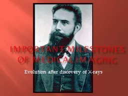
of medical imaging Evolution after discovery of Xrays Xrays   discovered by German physicist Wilhelm Conrad Roentgen He also produced the first xray picture of the body his wifes hand in 1895 ID: 378942

sri imaging radiography ray imaging sri ray radiography university developed radiographers system contrast vessels pet blood patient england image lanka spiral digital

Share:

Link:

Embed:

Download Presentation from below link

Download Presentation The PPT/PDF document "Important milestones" is the property of its rightful owner. Permission is granted to download and print the materials on this web site for personal, non-commercial use only, and to display it on your personal computer provided you do not modify the materials and that you retain all copyright notices contained in the materials. By downloading content from our website, you accept the terms of this agreement.


Presentation Transcript

Slide1

Important milestones

of medical imaging

Evolution after discovery of X-raysSlide2

X-rays


discovered

by German physicist Wilhelm Conrad Roentgen.

He also produced the first x-ray picture of the body (his wife's hand) in 1895.

1895Slide3

Chest x-raywidespread use of the chest x-ray made early detection of tuberculosis (which was the most common cause of death)

1900 Slide4

X-ray IV contrast medium First contrast filled image of the renal system (kidneys).

1906 Slide5

Barium sulfateIntroduction   as contrast agent for gastro-intestinal diagnosis.

1910Slide6

Theory of Radioactivity published by Marie Curie and investigation of  radiation for patient therapy 

(e.g. treatment of cancer). 1910/1912Slide7

Founded Society of Radiographers UK1920Slide8

Radiographic imaging of the gallbladder, bile duct and blood vessels for the first time. 1924Slide9

Sri Lanka

Start of Radiological services in Sri Lanka

Dr.

H.O.Goonawardane

– first Radiologist

1925Slide10

First trained Radiographers in Sri Lanka appointed

Mr. M.L.B.J.

Caspersz

& Mr. J.A.N.

Fernandopulle

1928Slide11

Cardiac catheterization first performed by Forssmann on himself.

1929Slide12

Publication “Positioning in radiography” by K.C.Clark - UK

1939Slide13

Coronary artery imaging Visualization of (blood vessels that feed the heart).

1945Slide14

In Sri Lanka

Founded the Government X-ray Technical Officers’ Association

Publication of the journal “Ceylon Radiographer”

1948Slide15

Publication “Atlas of Radiographic positions” by Vinita Merill - America

1949Slide16

Nuclear Medicine applied in imaging the kidneys, heart, and skeletal system.

1950Slide17

X-ray Image Intensifier-Television units To allow dynamic x-ray imaging of moving structures.

1955Slide18

OPGPanoramic x-ray images of the entire jaw and teeth.

1955Slide19

Inauguration of Sri Lanka School of Radiography

by Mr.

Jaundrell

Thompson B.Sc., F.S.R

1957Slide20

Ultrasound imaging using high frequency sound waves to look at the abdomen and kidneys, fetal baby, carotid blood vessels and heart.

1960Slide21

X-ray mammography finds widespread application in imaging the breasts.

1970Slide22

Computed Tomography (CT) scanning invented by British engineer Godfrey Hounsfield of EMI Laboratories, England, and South African born physicist Allan Cormack of Tufts University, Massachusetts.

1972Slide23

In Sri Lanka

Founded the Society of Radiographers – Sri Lanka

1975Slide24

DSA Digital subtraction Angiography

1977Slide25

Magnetic Resonance Imaging (MRI) of the brain was first done on a clinical patient. MRI was developed by Paul Lauterbur and scientists at Thorn-EMI Laboratories, England, and Nottingham University, England.

1980Slide26

Radiography training around the world changed from 2- year diplomas to 3-yearhigher diplomas and then to 3-4 year bachelors' degrees Slide27

CR first Computed radiography system introduced

1981Slide28

3-Dimensional image processing using digital computers and CT or MR data, three dimensional images of bones and organs were first made.

1984Slide29

Positron Emission Tomography (PET) Clinical scanning developed by scientists at the University of California

1985Slide30

PACSClinical Networks were first implemented to allow digital diagnostic images to be shared between physicians via computer network, allowing a doctor in Boston to review a CT examination from a patient in Beijing, China

1985Slide31

Spiral CT allows fast volume scanning of an entire organ during a single, short patient breath hold of 20 to 30 seconds. Spiral CT had caused a renaissance in CT and lead the way to significant

developments like CT Angiography.

1989Slide32

MR Angiography developed and clinically available to allow non-invasive imaging of the blood vessels without radiation or contrast injection.

1989Slide33

Echo Planar MR Imaging (EPI) Developed and clinically available to allow MR systems to provide early detection of acute stroke. EPI also makes possible functional imaging, for instance of brain activity allowing doctors to investigate the function of different centers of the mind

. 1993Slide34

Open MRI Developed to allow MR scanning of severely claustrophobic or obese patients who could not tolerate conventional MR imaging in a close bore system.

1993Slide35

PET/CT scanner

Invented by Dr. Ron Nutt and Dr. David Townsend

The first generation of PET/CT scanners included a single slice spiral CT integrated with a PET camera which utilized BGO detectors.

SPECT/CT also was

introduced

2000Slide36

Radiotherapy

Many advancements have taken place.(This will be dealt with in the lecture by a Medical Physicist today)Slide37

In Sri Lanka

Inauguration of First BSc. Radiography

programme

at University of

Peradeniya

2005Slide38

In sri Lankafirst ever BSc. graduate

radiographers pass out 2011Slide39

End ?What is awaiting TO BE DONE ?Slide40

The greatest tribute we can pay to roentgen is to provide a better service to the sick and the needy.UPGRADING THE TECHNOLOGY ALONE CANNOT ACHIEVE THAT UNLESS THE COMPETENCY OF THE USER IS OF HIGH STANDARDSlide41

In almost all

the countries radiography courses today are University degree courses.

This has become necessary to meet the advancement in technology and requirement in accurate diagnosis and therapy.Slide42

It is necessary to make avenues to advance our knowledge in keeping with international levels and standards.

An external degree programme is an appropriate and practical solution and is the need of the hour.Slide43

Thank you !