PDF-International Standards for the Classification of Spinal Cord Injury .
Author : stefany-barnette | Published Date : 2016-02-27
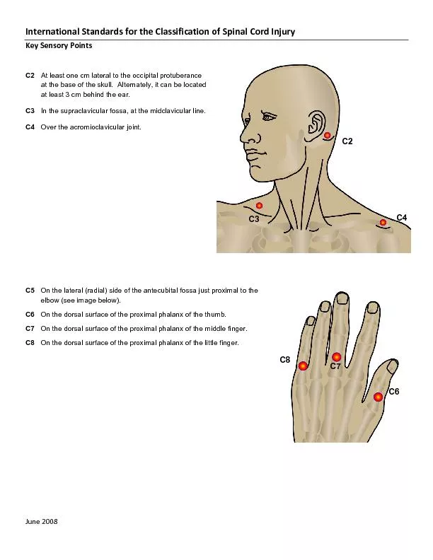
Key Sensory PointsJune 2008 At least one cm lateral to the occipital protuberance at the base of the skull Alternately it can be located at least 3 cm behind the
Presentation Embed Code
Download Presentation
Download Presentation The PPT/PDF document "International Standards for the Classifi..." is the property of its rightful owner. Permission is granted to download and print the materials on this website for personal, non-commercial use only, and to display it on your personal computer provided you do not modify the materials and that you retain all copyright notices contained in the materials. By downloading content from our website, you accept the terms of this agreement.
International Standards for the Classification of Spinal Cord Injury .: Transcript
Download Rules Of Document
"International Standards for the Classification of Spinal Cord Injury
."The content belongs to its owner. You may download and print it for personal use, without modification, and keep all copyright notices. By downloading, you agree to these terms.
Related Documents