/
JOURNAL CLUB DR DIMPI SINHA,MDRD  CYSTIC LESIONS OF PANCREAS-DIAGNOSIS JOURNAL CLUB DR DIMPI SINHA,MDRD  CYSTIC LESIONS OF PANCREAS-DIAGNOSIS

JOURNAL CLUB DR DIMPI SINHA,MDRD CYSTIC LESIONS OF PANCREAS-DIAGNOSIS - PowerPoint Presentation

stefany-barnette
stefany-barnette . @stefany-barnette
Follow
346 views
Uploaded On 2019-11-03

JOURNAL CLUB DR DIMPI SINHA,MDRD CYSTIC LESIONS OF PANCREAS-DIAGNOSIS - PPT Presentation

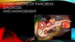
JOURNAL CLUB DR DIMPI SINHAMDRD CYSTIC LESIONS OF PANCREASDIAGNOSIS AND MANAGEMENT articles Pancreas Cystic LesionsDiagnosis and management Marc Engelbrecht Jennifer Bradshaw and Robin ID: 762768

cystic pancreatic duct cysts pancreatic cystic cysts duct lesions imaging solid neoplasm ipmn lesion cyst mucinous tumor pancreas neoplasms

Share:

Link:

Embed:

Download Presentation from below link

Download Presentation The PPT/PDF document "JOURNAL CLUB DR DIMPI SINHA,MDRD CYSTIC..." is the property of its rightful owner. Permission is granted to download and print the materials on this web site for personal, non-commercial use only, and to display it on your personal computer provided you do not modify the materials and that you retain all copyright notices contained in the materials. By downloading content from our website, you accept the terms of this agreement.


Presentation Transcript

JOURNAL CLUB DR DIMPI SINHA,MDRD

CYSTIC LESIONS OF PANCREAS-DIAGNOSIS AND MANAGEMENT

articles Pancreas - Cystic Lesions-Diagnosis and management Marc Engelbrecht , Jennifer Bradshaw and Robin Smithuis Radiology department of the Academical Medical Centre, Amsterdam and the Rijnland hospital in Leiderdorp , the Netherlands-published in 2012 in radiology assistant. Cystic Pancreatic Lesions: A Simple Imaging-based Classification System for Guiding Management published in R adiographics in 2005. Dushyant V. Sahani ,  Rajgopal Kadavigere ,  Anuradha Saokar ,  Carlos Fernandez-del Castillo ,  William R. Brugge ,  Peter F. Hahn

Morphologic classification of cysts based on imaging findings. Roadmap to approach pancreatic cystic lesion. Mangement of such lesions.

INTRODUCTION Cystic lesions of the pancreas encompass a variety of neoplastic and nonneoplastic processes that cause a range of clinical symptoms. With the increasing use of cross-sectional imaging, pancreatic cysts have become a common incidental finding. The accurate imaging-based characterization of pancreatic cystic lesions is necessary for appropriate patient triage and management, as some lesions require surgical intervention or follow-up imaging, whereas others require no further action.

Clinical presentation Pseudocysts typically occur with acute pancreatitis or trauma or may develop insidiously in the setting of chronic pancreatitis . A large number of pancreatic cysts are incidentally detected during imaging work-up for an unrelated medical problem. The majority of incidentally detected pancreatic cysts turn out to be cystic neoplasms. Symptomatic cysts are most likely to manifest with abdominal pain. Jaundice or recurrent pancreatitis indicating that the lesion is either in communication with the pancreatic ductal system or obstructing the pancreatic or biliary duct.

MRI VS CT CT will depict most pancreatic lesions, but is sometimes unable to depict the cystic component.  Currently , Abdominal MRI with MRCP is considered the modality of choice for demonstrating the 1.Cystic nature, 2. M orphologic features of the cyst (including septa and mural nodules), 3.Establishing the presence of communication between the cystic lesion and the pancreatic duct as is seen in IPMN, and 4.Evaluating the extent of pancreatic ductal dilatation. There are cases when CT can be helpful, since it better depicts a central calcification in SCN or peripheral calcification in a mucinous cystic neoplasm (MCN).

ROLE OF ENDOSCOPIC USG Guided aspiration. FNAC from cystic lesion.

DIFFERENTIALS OF PANCREATIC CYSTIC LESIONS

MORPHOLOGIC CLASSIFICATION Morphologic Classification of Cystic Pancreatic Lesions B ased on the imaging morphologic features of the cyst, Pancreatic cysts can be classified into four subtypes:  ( a)   U nilocular cysts,  (b) M icrocystic lesions,  ( c)   M acrocystic lesions, and  ( d)   Cysts with a solid component Reference: Cystic Pancreatic Lesions: A Simple Imaging-based Classification System for Guiding Management Dushyant V. Sahani ,  Rajgopal Kadavigere ,  Anuradha Saokar ,  Carlos Fernandez-del Castillo ,  William R. Brugge ,  Peter F. Hahn

UNILOCULAR CYST Pseudocyst Branch type IPMN Unilocular serous cystadenoma True epithelial cysts

PSEUDO CYST Collection of pancreatic enzymes, blood and necrotic tissue. Debris within a cystic lesion is a specific MR finding. History of pancreatitis or abdominal trauma. The imaging findings -pancreatic inflammation, atrophy or calcification of the pancreatic parenchyma, and dilatation of and calculi in a typically thin-walled pancreatic duct.   Cysts develop in 4-6 weeks - usually decrease in size over time - sometimes enlarge or become infected. Found in any part of the pancreas or anywhere within the abdomen and sometimes even in the chest .

MICROCYSTIC LESION

MICROCYSTIC LESIONS SEROUS CYSTADENOMA Typically in ’ GrandMother ' - median age: 60-70 years In 70% of cases-MICROCYSTIC PATTERN. E xternal lobulations , enahncing wall and sepattions . A fibrous central scar with or without a characteristic stellate pattern of calcification at CT or MR imaging is highly specific and is considered to be virtually pathognomonic for serous cystadenoma .Pancreatic ductal dilatation or communication with main pancraetic duct is not seen.Only cystic neoplasm with no malignant potential.

Branch duct type ipmn Branch-duct IPMN  - This tumor can look like a SCN, but has no scar or calcifications. Can be multifocal. Identification of a septated cyst that communicates with the main pancreatic duct is highly suggestive of a side-branch or mixed IPMN. Location: pancreatic head >> tail and corpus.

MACROCYSTIC LESION

MUCINOUS CYSTIC NEOPLASM Premalignant tumor - may transform into a mucinous cystadenocarcinoma Exclusively seen in women  - Typically in 'Mother' - median age: 40-50 years Macrocystic with thickened wall , septations and peripheral calcifications. The cysts may occasionally contain debris or hemorrhage. The complex internal architecture of the cyst, including septa and an internal wall, is best appreciated at MR imaging and endoscopic US, allowing differentiation from serous cystadenomas P eripheral calcifications seen in 25%. P eripheral eggshell calcification -specific finding for a mucinous cystic neoplasm and is highly predictive of malignancy.Location in the tail and body of the pancreas (95%).These tumors can be asymptomatic in up to 75% of cases .When present, symptoms are caused by the mass effect of these often large neoplasms. Because mucinous cystic neoplasms have a high potential for malignancy, surgical resection is usually advocated They do not communicate with the pancreatic duct, they can cause partial pancreatic ductal obstruction.

IPMN-MAIN DUCT TYPE key findings : Mucine producing tumor in main pancreatic duct. Abnormal dilatation of main pancraetic duct usually more than 8 mm, best seen on MRCP. Intraductal solid nodule is highly suggestive of IPMN. May present with pancreatitis. IPMN has premalignant potential. Signs of malignancy are: Pancreatic duct > 8 mm - as in this case. Solid node in duct. Mass around the pancreatic duct. Enlarged choledochal duct.

CYST WITH SOLID COMPONENTS Cysts with a solid component may be either unilocular or multilocular . True cystic tumors ( eg , mucinous cystic neoplasms, IPMNs) as well as solid pancreatic neoplasms associated with a cystic component or cystic degeneration are included in this category. Solid tumors associated with a cystic component include 1.Islet cell tumor 2.solid pseudopapillary tumor 3.adenocarcinoma of the pancreas, 4.Metastasis

SPEN Solid Pseudopapillary Neoplasm key findings : Very uncommon neoplasm seen in women 20-30 years ( Daughter ). Commonly located in pancreatic tail. Solid and cystic neoplasm with capsule and with early ' hemangioma -like' enhancement. Sometimes intratumoral hemorrhage.

Neuroendocrine tumor with cystic degeneration key findings : Non-functioning endocrine neoplasm Also called islet cell tumor Hypervascular with ring-enhancement. This is unlike serous cystic neoplasms that enhance from the center and more solid.

SYSTEMATIC APPROACH

AGE and gender Grandma  - Serous cystic adenoma Mother  - Mucinous cystic neoplasm Daughter  - SPEN IPMN -Male=female.

MANAGEMENT Histopathology of tumours,Age , life-expectancy and comorbidity should be considered in the possible surveillance or treatment.  Cysts smaller than 3 cm and no worrisome or high risk-features can be considered for follow-up with either MRI, CT or ultrasound.  Symptomatic cysts and asymptomatic cysts with obvious high risk stigmata should be considered for resection based on other comorbidities or risk factors. Pancreatic cysts are regarded  symptomatic  when there is hyperamylasemia , recent-onset diabetes, severe epigastric pain, weight loss, steatorrhea , or jaundice. 

THANK YOU