/
Weakness; Myopathy, Anterior horn cell disease,Neuropathies and Neurom Weakness; Myopathy, Anterior horn cell disease,Neuropathies and Neurom

Weakness; Myopathy, Anterior horn cell disease,Neuropathies and Neurom - PDF document

stefany-barnette
stefany-barnette . @stefany-barnette
Follow
441 views
Uploaded On 2015-12-05

Weakness; Myopathy, Anterior horn cell disease,Neuropathies and Neurom - PPT Presentation

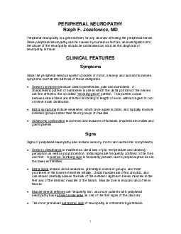
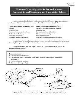
Weakness Lower motor neur Upper motor neur FlaccidSpasticity including a Babinski responseDecreased toneIncreased toneDecreased muscle stretch reflexesIncreased muscle stretch reflexesProfound mu ID: 215097

Weakness Lower motor neur Upper motor neur FlaccidSpasticity

Share:

Link:

Embed:

Download Presentation from below link

Download Pdf The PPT/PDF document "Weakness; Myopathy, Anterior horn cell d..." is the property of its rightful owner. Permission is granted to download and print the materials on this web site for personal, non-commercial use only, and to display it on your personal computer provided you do not modify the materials and that you retain all copyright notices contained in the materials. By downloading content from our website, you accept the terms of this agreement.


Presentation Transcript

Weakness Weakness; Myopathy, Anterior horn cell disease,Neuropathies and Neuromuscular transmission defectsupper motor neuronlower motor neuron weakness. The differences are tabulated below. Lower motor neur Upper motor neur FlaccidSpasticity, including a Babinski responseDecreased toneIncreased toneDecreased muscle stretch reflexesIncreased muscle stretch reflexesProfound muscle atrophyMinimal muscle atrophyFasciculations presentFasciculations absentMay have sensory disturbancesMay have associated sensory disturbancesIt is also customary, and very helpful, to classify LMN weakness on the basis of theanatomical station affected. These stations ar 1) The anterior (ventral) horn cell.2) The peripheral nerve, (ventral and dorsal nerve rootsneuropathy.)3) The neuromuscular junction.4) The muscle (i.e. myopathy). WeaknessII. Anatomy of the lower motor neuron The anterior horn cells are somatotopically organized in the spinal cord. That is, mediallyinnervate more distal muscles. The arrangement at cervical segments is shown in figure 2. Thisorganization means that diseases that destroy anterior horn cells can result in highly selectiveaffected. As a rule however the adjacent anterior horn cells will also be affected with weakness ofFigure 2. The somatotopic arrangement of anterior horn cells at cervical and the first thoraciclevels. Because the anterior horn cells that innervate different muscles in the upper and lowerextremities are present at different segments of the spinal cord, a whole extremity is not presented A reminder on the classification of dorsal and ventral root fibersdorsal rootsand their sizes. This has led to some confusion in the literature (and for medical students!!). The Thus, there are I, II, IIIand IV fiber types. You already have heard about the muscle spindles and are large and fast conducting. You also have heard that the associated with the Golgi tendon organs and are little smaller and slower conducting than the Weaknessslow conducting. Such fibers are associated with cooling and first pain. Finally, versus Roman numerals. The largest and fibers. (alpha) fibers are comparable to the Ias and Ibs. (alpha-beta) fibers are equivalent to II fibers in size and conduction velocities. A fibers, arelightly myelinated and are visceral afferents; they have no equivalent in the Roman numeral system.Finally, ventral root fibers. The processes of lower motor neurons that innervate fibers. Finally, there are axons in the ventral roots that innervate the extrafusal) fibers of the muscle spindles. These are called ARemember, A and B fibers are myelinated and Cs are not. In the Roman numeral system, justremember that only the IVs are not myelinated. This is important, since demyelinatingdiseases would affect the somatic and visceral afferents and efferent fibers in peripheralnerves, pain and temperature would not be affected. Weakness thousand muscle fibers. The muscle fibers innervated by a single anterior horn cell are collectively motor unit. The “territory” of such a motor unit spans 10-15 mm in a muscle however itis rare that directly adjacent muscle fibers are innervated by the same anterior horn cell/axon. Theindividual anterior horn cells. The clear muscle fibers below are innervated by a single anterior horncell and comprise a motor unit. The vertically oriented fibers are innervated by a different anteriorhorn cell constituting a second motor unit and the horizontally oriented represent yet another.We also need to distinguish between type 1 (slow contracting) muscle fibers and type 2 (fastcontracting) muscle fibers. The type of muscle fiber is dependent on the type of anterior horn cellthat innervates it. Thus if a muscle fiber is innervated by a type 1 anterior horn cell, it will contractslowly. Certain histochemical reactions, amongst others myosin ATPase, distinguish between type 1and type 2 fibers. Thus muscle reacted with myosin ATPase will normally exhibit a checkerboarddifferent fiber type (figure 4).Figure 4. The clear fibers in the figure above are myosin ATPase free and are all innervated by oneventral horn cell. The striped fibers are the ATPase rich and would look similar under a microscope.However, we want to illustrate that the ATPase rich fibers are innervated by two different ventralhorn cells (a and b; hence the different orientations of the stripes). Neur A muscle fiber is activated via a nerve impulse generated by an anterior horn cell. The impulse is Weaknessthe subsequent release of acetylcholine into the synaptic cleft. Acetylcholine binds to post-synapticacetylcholine receptors on the muscle membrane. This induces an end plate potential whichFigure 5. The neuromuscular junction.III. Diagnosis of the different lower motor neuron subgroupsdisease to one of the 4 anatomic stations. This can be accomplished by a combination of the In recording the history it is of particular importance to document the following. The timeof disease onset, the presence or absence of a family history of other similarly affectedof fatigability. The clinical examination serves to corroborate the clinical history, and to Weakness Histological examination of muscle or nerve biopsy specimensThese will be dealt with in more detail during the neuropathology section of your Pathologycourse. Muscle is not too smart and can only react in a limited number of ways to insult. Thus mostFigure 6. Typical, non-specific pathological findings in a primary myopathy. A necrotic fiber(asterix), and a hypercontracted muscle fiber (star), are shown. The entire muscle is shortened andthus, the hypercontracted fiber is thicker. The connective tissue between the muscle fibers is Weakness findings in the muscle! The poor muscle can only interpret these events as “I am denervated.” Thetype groupinggroup atrophyanterior horn cell or axon results in denervation of a number of muscle fibers. These muscle fibersadjacent muscle fibers. The end result is that now one axon innervates more muscle fibers thannormal, (a giant motor unit) and also the normal checkerboard pattern of innervation is lost. That is,a whole group of type 1 or 2 fibers can now be seen adjacent to one another (type grouping). With results in Wallerian degeneration, a bead-like disruptionThis is seen in diseases affecting the axons in the peripheral nerve, or in anterior horn cell disease. WeaknessFigure 8. Wallerian degeneration seen in axonal damage.healthy.Figure 9. A nerve fiber with shortened internodes that are hypomyelinated; typical findings in Since there are few pure motor nerves to study, motor nerve conduction (i.e. thenar muscle group). The appropriate nerveevoked responses measured. These evoked. The time it takes from stimulation to generation of the CMAP is the (e.g. the sural nerve is a pure sensory nerve). Stimulation of a sensory nerve leads to action. Furthermore, by stimulating the same nerve over different Weakness Weakness repetitive nerve stimulation studies. A routine motorneuromuscular transmission defects decreasesneuromuscular transmission defect (figure 12). This is called a decremental responsemechanism of the decremental response is complex and beyond the scope of this course!! Don’tFigure 12. Repetitive nerve stimulation study. Four CMAPs are shown in each tracing. Note that the Summar y of nerve conduction findings in dif fer ent disease gr Station 1- Anterior (Ventral) Horn CellThis results in low CMAP amplitudes in muscleinnervated by the dying anterior horn cells whose axons travel in the nerve being stimulated. Therebeing a smaller (than normal) CMAP. Since there is still a population of normal axons from otherthe median nerve) has normal axons that camouflage the dying ones. The sensory nerve conductionStation 2- Peripheral Nerve disease: sensory axons are affected. In most peripheral nerve diseases both become affected. If the changes appreciably as the remaining axons conduct at normal speed. There are however too fewnormal axons and thus, the evoked potentials in the muscle (CMAPs) are small. Weakness Station 3- Neuromuscular Junction disease: decremental CMAP Station 4- Muscle disease: Nerve conduction studies are normal, but the CMAP amplitudes will below, as there is loss of muscle fibers.b) The needle examinationactivation of the muscle. This test is accompanied by some discomfort, but if performedmotor unit potentials (MUPs; compare with CMAPs). A normal muscle and a normal MUP isFigure 13. Normal muscle. The above muscle fibers are innervated by three different lower (alpha)motor neurons. Think of the MUP as representing the action potentials of the muscle fibers potentials are of relatively normal amplitudes. REMEMBER: neuromuscular transmission defects result in decremental CMAP responseswith repetitive nerve stimulation.REMEMBER: primary muscle diseases result in low CMAP amplitudes, similar to the Weaknessresult from denervation (neurogenic). As described in the section on the anatomy of muscle fibers, muscle fibers are normally innervated by different anterior horn cells or motor axons. I know we haveWith damage to an anterior horn cellclose proximity to the EMG needle. This is seen histologically type groupinglarger MUPs (“neurogenic” MUPs), figure 14. You mightwonder why, if there is reinnervation (or “sprouting”) and larger MUPs, why are the CMAPs smaller?Well, that is because muscle fibers are also dying (remember group atrophy?)Figure 14. Neurogenic atrophy with type grouping. A large MUP is recorded. Think about thesingle active motor neuron in Figure 13 as innervating more muscle fibers. When it fires therewill be more muscle fiber action potentials and thus a larger MUP.Figure 15. Myopathic muscle. A small MUP is recorded. Weakness are noted clinically as a contraction of a small group of muscle fibers. Theyresult from the spontaneous discharge of an anterior horn cell or a motor axon with the subsequentFasciculations can also be recorded with the needle electrode. Clinically, fasciculations are seen after on the other hand are not visible through the skin. They are the smallmuscle fiber is denervated, several pathological changes take place. The acetylcholine receptorsend-plate. This spread may play a role in attracting new innervation to the denervated muscle fiberfrom adjacent nerve sprouts. The muscle fiber becomes much more sensitive to free acetylcholineIf the muscle fiber reinnervates sucessfully, these potentials disappear again.Figure 16. Fibrillation potentials. Each complex represents the spontaneous discharge of a singlemuscle fiber as recorded with a needle electrode in the muscle. Note that the discharge intervals are Neuromuscular transmission defects. acetylcholine receptor antibodiesWith muscle breakdown of any kind, creatine phosphokinase Genetic studiesThe genetic defects of many neuromuscular diseases are now known and can be detected inperipheral blood or in muscle. Weakness . Let us put this all together!1. Anterior poliomyelitis, motor neuron disease andspinal muscular atrophyspinal muscular atrophy will be discussed further. This is usuallyan autosomal recessively inherited disease with onset at any time from infancy to adulthood. Thethat they die usually from an associated lung infection. The reason for the progressive loss ofanterior horn cells is not clear, but the disease is associated with an abnormality on chromosome 4. Normal nerve conduction velocities, normal SNAP amplitudes, low CMAPamplitudes, large MUPs on needle examination, fasciculations. Type grouping and group atrophy. This encompasses a vast number of diseases and only a cursory overview will be attempted. Clinical featur Damage to the peripheral nervous system results in motor, sensory and autonomicdysfunction. A neuropathy is any disease of the nerves. There are a number of different classesDistal polyneuropathy: All the nerves are affected distally in the extremities. Clinically thein distal extremity muscles (e.g. ankle jerk). Longer nerves are affected more severely and thus thechanges predominate in the legs. Most distal polyneuropathies are purely sensory or affect thesensory and motor nerves together. Pure motor distal neuropathies are rare. Depending on theetiology, the neuropathies can be axonal (axis cylinder), demyelinating, or show features of both. a) Predominantly axonal disease:or absent CMAP and SNAP amplitudes. Needle examination shows large MUPs that result fromb) Predominantly demyelinating disease: Relatively normal CMAP and SNAP amplitudes with Type grouping and group atrophy only if there is axonal (axis cylinder) damage. Weakness Neur transmission defects will be discussed further. This disease is characterized byabnormal fatigue with exercise. Myasthenia gravis commonly affects young woman and has apredilection for ocular, facial, masticator and proximal upper extremity muscles. Typically thepatients recover to some degree after rest. Thus they feel much better in the morning, butbecome weaker as the day progresses. When the extraocular eye muscles are affected, diplopia(double vision) and ptosis (drooping of upper eyelid) are common and bothersome signs. This Neur transmission defects EMG findings:Normal nerve conduction velocities, CMAP and SNAP amplitudes.Decremental response on repetitive nerve stimulation. Biochemistry:Acetylcholine receptor antibodies are present in blood. Histology:Usually normal.4. Muscle dystrophiesonset at any time after birth. They are diagnosed on the pattern of muscle involvement. ForDuchenne muscle dystrophy is characterized by large calves, proximal muscle weaknessMyotonic dystrophy patients Normal motor and sensory nerve conduction studies. The CMAPs are low because of loss in muscle Biochemical findings:All progressive myopathies have increased CK blood levels indicating the breakdown of muscle. Non-specific myopathic features such as large fibers, necrotic fibers, and increased connective Weakness IN SUMMAR Y camouflage dead ones), large “neurogenic” MUPs (normal axons take over muscle fibers of dead ones),ge “neurogenic” MUPs (normal axons take over muscle fibers of dead ones),(become elevated if a muscle fiber breaks down, or if the muscle membrane becomes porous. Bothconditions allow CK to leak from the muscle fiber into the blood. In neurogenic atrophy (of anteriorhorn cell or peripheral [axonal] nerve origin) the muscle membranes remain intact and thus CK levelsremain normal). Fasciculations (grossly) and fibrillations (only upon needle exam) are present.Peripheral nerve diseases. Clinically characterized by the associated findings of sensory and autonomicabnormalities. EMG findings depend on whether it is primarily an axonal (axon cylinder) ordemyelinating neuropathy. Axonal EMG findings are those of low CMAP amplitudes (fewer axisnerve, relatively normal nerve conduction velocities (normal axons camouflage dead ones), large Weakness The EMG findings are those of relatively normal CMAP amplitudes (axis cylindersYou might wonder why normal myelinated fibers don’t camouflage the diseased fibers. In reality,demyelinating neuropathies affect multiple focal areas of every nerve and you have normal segmentsin between. Thus, you do not find “normal” and “abnormal fibers”, all the fibers are affected tosome degree. In axonal neuropathies some fibers are affected, others not. The normal fibersNeuromuscular transmission defects.“SPEED PLAY”Increased reflexes in a symptomatic limb suggest a central lesion, while reduced reflexesBilateral sensory and motor deficits throughtout the body below a roughly horizontal level in 106Radiculopathy RADICULOPATHIESprotrusion and bony spurs are common causes of radiculopathy. The motor, sensory and sundryother tracts, the spinal grey matter, and the spinal nerve roots should no longer be strangers to you.ventral or anteriordorsal or posterior sensory nerve root. The spinalDorsal and ventral rami contain three types of fibers; sensory, somatomotor and visceromotor. 107The distribution of sensory fibers in each spinal nerve is called a dermatome Root compression PAINparesthesia*dull pain is usually more proximal and difficult to localize The lumbar roots emerge from vertebrae. These roots are vulnerable just above their exitbelow). The intervertebral disc lying between vertebrae L4 andL5 is called the L4/5 disc. The disc between the L5 vertebraeand the sacrum is the L5/S1 disc. Since the L4 root emergesdamages the L5 root. Moreover, a lateral herniation of the L5/KNOW THIS!! compr essive radiculopathy.and does not require surgical therapy. One fifth of pain freestructural causes and surgical therapies only when there is an (L5 root; 50%) and at L5/S1 (S1 root; 46.3%) interspace. Consequently,compression5th lumbar nerve root Radiculopathy narrowerSyndromes associated with lumbosacral radiculopathies radiculopathy (nerve roots). When caused by SO, L5 = DORSUM BIG TOE, S1 = LATERAL FOOT. radiculopathy. The figure below illustrates the pain distribution in lumbar radiculopathies . You should notethat areas associated with each root are larger than illustrated in the dermatome charts. The reason for C5 root exits between the C4-C5 vertebrae and would be effected by a C4/5 disc herniation; 112Radiculopathy The term cauda equina (horse’s tail)the first lumbar vertebra. At that level, the floor). The actual act of voiding is under the control of higher cortical centers that develops asinterrupted or when there are physical problems with the pelvic floor and sphincter muscles. Whenneurogenic bladderbe investigated aggressively.the LCST. These bilateral projections terminate onbladder. These postganglionic parasympathetic cells in turn(all are not shown in the diagram). When the bladder fills,contraction of the bladder (voiding). This voiding reflex is AUTONOMIC DYSFUNCTION IN SPINAL CORD DISEASES You should be able to identify motor dysfunction of the bladder. Both resultin clinically divergent neurogenic bladders.Lesion 1 - Upper Motor Neuron Lesion:of the LCST. Spinal shock and flaccidity, right? Thisretention. The bag fills as there isno tone. There may be overflow incontinence when thebladder cannot physically hold any more urine. Withlesion of the LCST). This causes urinary frequencyand urgency (there are some sensory pathways intact);carried by afferents activate the parasympathetic motorvoiding. The bladder is things occur. First there is a flaccid bladder (acute),Lesion 2 - Lower Motor Neuron Lesion: (This is easier.)disrupted, then the lesion results in weakness, atrophy, and hyporeflexia. The bladder does notcontract and, if the sensory afferents are affected, no sensation of a full bladder will be perceived. Ifsensation is intact, but the motor efferents are affected, then there is an urge to void but goodroots in the cauda equina, the pelvic nerve, the pelvic plexus, or the second order, postganglionicparasympathetic neurons that innervate the detrussor. 115Autonomic dysfunction Remember, lesions of the spinal cord rostral to thesacral cord result first in a flaccid (atonic; acute) bladder,followed by a spastic ( chronic, automatic) bladder. LesionsONLYWe have already discussed deficits that result fromthese deficits differ from those following lesions of the conusand is in proximity to the nerve roots. Thus, injuries to this areadermatomes and myotomes of the affected segments. On theLet’s compare these lesions RESPIRATIONRespiratory section of your Physiology course. What is important in this Neuroscience course is thatsome spinal cord lesions have effects on respiration. Respiratory centers in the medulla and ponscontrol respiration via pathways to the spinal cord. These pathways influence the:Diaphragm-the primary muscle for breathing. When this dome-shaped muscle contracts, itflattens, descending into the abdominal cavity, causing the lungs to inflate.Intercostal muscles-They connect the ribs. When they contract, the chest wall is lifted up andAccessory muscles-Are located in the neck and shoulders. When they contract, the first twospace. These are muscles you use when you cough or sneeze.The way your breathing is effected following spinal cord injury will depend on the level ofyour injury, whether the injury is complete or incomplete and how much improvement or recovery thepatient may get. Everyone knows that there is voluntarily control of breathing. This voluntary controlpathways for control of breathing there are involuntary pathways from the medulla and pons. Themedullary and pontine pathways travel in the ventral funiculus and are also bilateral. Interestingly,Ondine’s curse is a condition where the involuntary descending pathways are damaged (or theircenters in the medulla and pons) while the voluntary pathway is OK. Thus, the patient can breathvoluntarily but not involuntarily.Keep in mind that the descending pathways from the medulla and pons are bilateral. Thus,isolated from their UMN control centers. This causes paralysis of all the muscles that you need tobreathe, including the diaphragm. This is rare but it may mean that you need a ventilator (respirator) totherefore you should be able to breathe on your own. However, your abdominal and intercostalmuscles will all be paralysed and you will not be able to breathe as well as you did before your injury.You will need help to cough to clear your sputum and are more likely to have problems with chestinfections from time to time. The higher the injury in your neck, the more difficulty you may find withyour breathing. What would happen if the lesion was in the ventral horn of C3, 4 and 5? If you have a high paraplegia (above T6), some of the/intercostal muscles and all of theyou may need assistance to be able to cough well. The lower the level of your paraplegia (T6-T12) theIf your injury is below T12 all of the respiratory muscles will be working and your breathing should beclose to as good as it was before your spinal cord injury.Ondine’s Curse (This is purely informational and is not meant to be sexist in any way)nymph’s immortality. If a nymph ever falls in love with a mortal and bears his child, she loses her giftWhen Lawrence saw her, he was, as they used to say, smitten by her beauty. He longed to know herto the knight’s visits. In time they met, they spoke, and they fell in love. As happens in most fairytales, these two attractive and special beings married. When they exchanged vows, Sir Lawrence said,against you.” Unfortunately, however, this tale is not one in which the couple lives happily ever after.A year after their marriage Ondine gave birth to Lawrence’s son. From that moment on shebegan to age. Her body became susceptible to the weathering effects of sun, wind, and time, and herby passion than by love. As Ondine’s physical attractiveness diminished, he began to develop awandering eye, with particular interest in some of the younger, prettier women living nearby.snoring of her husband. Amused by the fact that he had apparently fallen asleep in the middle of theday in this odd place, she decided to wake him up and take him home to finish his nap. When sheentered the stable, however, she saw Sir Lawrence lying on a pile of hay in the arms of some woman.Items of clothing strewn around the stable told the story. Ondine’s sacrifice of her immortality for thisman, who had now betrayed her, demanded retribution. Still retaining enough magic to achieve hervengeance, Ondine kicked her husband awake, pointed her finger at him, and uttered her curse: “Youswore faithfulness to me with every waking breath, and I accepted your oath. So be it. As long as youfrom you and you will die!” The tale ends with the favorite line of many old story tellers: “And so it Syndromes and Anatomic LocalizationThe basic principle of neurology is to define the anatomy of where the nervous system is affected andthe etiology for what is going wrong. The anatomy is defined by symptoms, patterns of neurologic loss,sites. Without some concept of where the lesion is, appropriate evaluation with modern imagingyou do not know where to look. The etiology for lesions of the nervous system relate more to the onsetconditions, e.g. B12 deficiency.foramen magnum. You should be familiar with the following anatomic patterns of neurologic loss.Affects specific muscles, usually proximal muscles giving weakness.dystrophies. An Neuromuscular JunctionWeakness that is variable and affects some muscles preferentially especially extraocular muscles andoropharyngeal muscles. No sensory loss. Abnormal decremental response to repetitive stimulation.You have already heard about distal polyneuropathies. Remember, a neuropathydistal polyneuropathybe affected. In the foot for example this could involve the common peroneal nerve (both superficialand deep branches) and the tibial nerve (medial and lateral plantar and calcaneal branches). The losscan be motor or sensory or both motor and sensory. Anatomic location Anatomic location 120There are also diseases of specific nerves either from compression or vascular disease (usuallyvasculitis or small infarctions associated with diabetes). Common nerves to be compressed are the median at the wrist (carpal tunnel), ulnar at the elbow, peroneal at the fibular head, lateral cutaneousnerve lesions. When multiple nerves are affected the term mononeuropathy multiplex is used. Anatomic location Pain, sensory, and motor loss. Referable to a dermatome and weakness in muscles innervated by the Anatomic location 122 Transverse lesion: bilateral sensory and Case historiesCASE PRESENTATIONThis 6 year old boy was brought to your office by his parents who were complaining that the boy had hadprogressive difficulty walking, climbing stairs and appeared “clumsy”. The child’s teacher also felt thatthe boy was always behind his peers in any physical activity. Academically he did well at school. Thechild was a product of normal pregnancy and normal delivery. His early developmental milestones wereThe family history was noncontributory.marked enlargement of both calves. There was evidence of contractures in his Achilles tendons. He hadproximal weakness, especially in his legs but also to some extent in his arms. The boy had markeddifficulty rising from the floor and did it by climbing up his thighs (positive Gowers’ sign). Sensation wasnormal. Tendon reflexes were decreased. Plantar reflexes were flexor. His gait was waddling.QUESTIONS CASE HISTORY I1)Where is the lesion/defect that might explain the findings on your clinical exam. Is the weakness CASE HISTORY I 2)What could be the possible etiology?3)What diagnostic tests would you order and why? A 25 year old woman came to your office complaining of intermittent double vision for the last threeweeks. She also has complained of fatigue. She has felt best during the early morning hours, but later,during the course of the day, she gradually develops double vision and diffuse weakness. Her boy-friendhas observed that her right eyelid has been drooping frequently. She used to play competitive basketballwhile in college, but now she has been short of breath after climbing only 2 flights of stairs. Also, on aexamination showed decreased movement in all directions in the right eye. There was also slightlyright sided ptosis got much worse. Motor examination showed normal muscle bulk and tone. Muscletesting revealed that she was initially strong, but rapidly became “tired” or weak with repeated effort. Shewas unable to hold her arms abducted at 90 degrees for more than 30 seconds. All sensory modalities,reflexes, coordination and gait examination were normal. Plantar reflexes were flexor.QUESTIONS CASE HISTORY II1) Where is the lesion and why? Is it in the corticospinal tract, anterior horn cells, peripheral nerves, CASE HISTORY II 2) What is the possible etiology?3) What diagnostic tests would you order and why? What results would you expect? This 20 year old college student came to the emergency room complaining of tingling in her feet andfingers. She appeared anxious. Neurological examination showed no abnormality and she was dischargedto home with a diagnosis of anxiety and hyperventilation. However, she returned to the emergency roomHer examination at this time showed mild diffuse weakness, decreased muscle tone and absent tendonreflexes. Plantar reflexes were flexor. Sensation to pain was slightly decreased in the feet. There was nosensory level. A sensory level is a region of the body below which a sensation(s) is lost and above whichsensation is normal (for a lesion of the spinothalamic tract at T1, the level is T3). Her respiration rate was26 (normal adult rate is 10-15). Mental status and cranial nerve examination was normal. She wasshe developed respiratory failure and had to be intubated and placed on a ventilator.QUESTIONS CASE HISTORY III1) Where is the lesion and why? Is it in the brain, spinal cord, nerve roots, peripheral nerves, neuromuscular CASE HISTORY III 2) What are the possible etiologies?3) What diagnostic tests would you order and why? What results would you expect? intermittent for the past 6 months. The pain is in the middle of the lower back and usually radiatesinto the left buttock. The pain is made worse by sneezing, coughing, or when he hits a pot holeinto the bottom lateral aspect of his foot. Over the past 6 weeks, he has noted that it is difficult forweakness of his left gastrocnemius. There is abnormal sensation over the lateral aspect of the leftfoot. He cannot stand on his toes of his left foot. When you have him lying down, you cannotQUESTIONS CASE HISTORY IV1) Where is the lesion?2) What would an EMG/NCV study show? CASE HISTORY IVCase histories 3) What diagnostic testing would you order? cramps. On a few occasions he choked on food. His wife noted diffuse twitching of muscles on his chestand upper back. Two months ago he developed a foot drop in his left leg. He has not complained of anysensory symptoms. There has been no cognitive decline. He has no difficulty with bowel or bladderfunction. His family history is noncontributory.Examination showed that the patient had normal mental status. Motor examination showed severe,bilateral diffuse muscle wasting in both upper and lower extremities. The most atrophied were the deltoid,triceps, biceps, hand muscles and quadriceps on either side and the left anterior tibialis. There wereprominent fasciculations in all muscle groups. The muscle tone was increased, (spastic) in both upperand lower extremities. There was diffuse weakness in all 4 extremities with complete left foot drop. NeckThe tendon reflexes were hyperactive in all four extremities. The plantar reflexes were extensor (BabinskiQUESTIONS CASE HISTORY V1) Where is the lesion and why? Does it involve the corticospinal tracts, anterior horn cells, nerve roots, CASE HISTORY VCase histories 2) What are the possible etiologies and why?3) What diagnostic tests would you order and why? What results would you expect? 133 CASE HISTORY VIA healthy 25 year old woman is brought to the emergency room after being stabbed in the neck.1) Where is the location of the lesion?Case histories 3) Why might atrophy of the upper arm develop over time?