/
Microbiology Laboratory testing OF Male urethritis Microbiology Laboratory testing OF Male urethritis

Microbiology Laboratory testing OF Male urethritis - PowerPoint Presentation

tatyana-admore
tatyana-admore . @tatyana-admore
Follow
433 views
Uploaded On 2017-07-02

Microbiology Laboratory testing OF Male urethritis - PPT Presentation

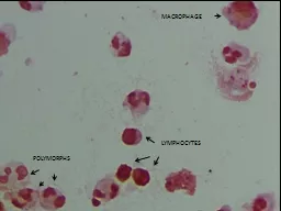
PATH 417A Case 2 Jen Yong Content overview Common pathogens that cause urethritis in men Required Specimens Lab tests performed amp Results 1 Causative pathogens Neisseria gonorrhea Intracellular ID: 565806

retrieved gram amp tests gram retrieved tests amp http stain detection jpg neisseria culture test giemsa trachomatis www urine

Share:

Link:

Embed:

Download Presentation from below link

Download Presentation The PPT/PDF document "Microbiology Laboratory testing OF Male ..." is the property of its rightful owner. Permission is granted to download and print the materials on this web site for personal, non-commercial use only, and to display it on your personal computer provided you do not modify the materials and that you retain all copyright notices contained in the materials. By downloading content from our website, you accept the terms of this agreement.


Presentation Transcript

Slide1

Microbiology Laboratory testing OF Male urethritis

PATH 417A Case #2

Jen YongSlide2

Content overview

Common pathogens that cause urethritis in men

Required Specimens

Lab tests performed & ResultsSlide3

1. Causative pathogensSlide4

Neisseria gonorrhea

Intracellular

Gram-negative

Cocci

Pili for adherence

Incubation period in males: 2-6 d

Common S/

Sx: urethral discharge, proctitis, dysuria, testicular pain

Chlamydia trachomatis

Common Pathogens

Obligate intracellular

18

serovars

(different outer membrane proteins), D-K implicated in urinary tract infection

Exist in two stages:

Elementary Bodies (EB) are non-replicating, infectious particles with a rigid cell wall that contain bacterial genome and plasmid

Reticulate Bodies (RB) are the non-infectious particles, that are formed as a result of binary fission within the endocytosed EB cytoplasmic vacuoleSlide5

Chlamydia developmental cycle

Retrieved from:

http://www.nature.com/nri/journal/v5/n2/fig_tab/nri1551_F2.html

https://www.google.ca/url?sa=i&rct=j&q=&esrc=s&source=images&cd=&cad=rja&uact=8&ved=0ahUKEwiO2OH135bSAhUK2GMKHehCDMYQjRwIBw&url=http%3A%2F%2Fmynotes4usmle.tumblr.com%2Fpost%2F47670799230%2Fusmlepathslides-elementary-body-of-chlamydia&psig=AFQjCNEeoAGUNoHFAVxpU9jjfSv3aAEYkA&ust=1487407767260374Slide6

Mycoplasma

genitalium

Gram-negative

Flask-shaped

15-25% of symptomatic non-gonococcal urethritis in males

Ureaplasma

urealyticum

Less common pathogens

Gram-negative

Spherical / ovoid

Cocci

Retrieved from: http://www.contraboli.ro/stuff/p/Ureaplasma.jpg

Retrieved from:

http://diseasespictures.com/wp-content/uploads/2015/11/Mycoplasma-Genitalium-Infection.jpgSlide7

2. Specimen requiredSlide8

Urethral swab

Pts should not pass urine for at least 2 hours prior to collection

2-4cm inside the urethra

Store frozen at -70’C, up to 60 days

urine specimen

Types of specimen

Pts should not pass urine for at least 1-2 hours prior to collection

10 mL of first catch urine

Store refrigerated at 2-8’C, up to 30 days

Retrieved from:

http://www.jadco.gov.jm/images/ace-img/Sample.jpg

Retrieved from:

http://images.slideplayer.com/25/8043625/slides/slide_16.jpgSlide9

Timely detection, diagnosis and treatment are essential to clearing urethritis and preventing severe complications (e.g. epididymitis associated infertility, Reiter syndrome)! (However, laboratory assessment of GU vs. NGU is not considered clinically essential, as results do not alter therapy.)Slide10

3. Laboratory tests & resultsSlide11

Culture tests

Gram stain

Differential media

Biochemical tests

Oxidase

Gonochek

II

Acid production detection

Non-culture tests

Types of Lab tests

Giemsa stain

Antigen detection by immunoassaysDirect Fluorescent AntibodyMolecular tests

Nucleic Acid Amplification tests (NAAT)Slide12

Gram stain

Add Crystal violet (basic dye)

Add iodine (mordant), forms an insoluble complex with crystal violet

Wash with alcohol (decolourizer)

Thick peptidoglycan layer becomes dehydrated and retains the violet complex

 Purple = Gram-Positive

Outer membrane becomes degraded and thin peptidoglycan layer cannot retain the violet complex

 decolourised = Gram-negative

Counterstain with safranin

 Gram-negative = pink

Retrieved from:

http://laboratoryinfo.com/wp-content/uploads/2016/01/gram-stain-procedure.pngSlide13

Gram stain results

Neisseria gonorrhea

Chlamydia trachomatis

pink

Not easily visualised (obligate intracellular)

 detection by Giemsa

N/A

Retrieved from:

http://hit-micrscopewb.hc.msu.edu/Microbiology/Images/5IIB2_penile_100X_3_tn.jpgSlide14

Thayer Martin Agar

2.0g beef extract

17.5g casein hydrolysate

1.5g starch

17.0g agar dissolved in 1 liter of distilled water, pH adjusted to neutral at 25 °C

5% chocolate sheep blood

+VCN inhibitor

Vancomycin kills most Gram-positive

Colistin kills non-Neisseria Gram-negativeNystatin kills most fungi

Differential media

Mycoplasma agar

Bacteriological peptone 10.0 g/L

Lab-

Lemco

powder 10.0 g/L

Sodium chloride 5.0 g/L

Mineral supplement 0.5 g/L

Agar 10.0 g/L

 selects

Mycoplasma

genitalium

McCoy cell monolayer

Selects for obligately intracellular

Chlamydia trachomatis

Expensive, slow, labour-intensive

Less preferred than non-culture detection methodsSlide15

Thayer-Martin agar

Neisseria gonorrhea

Chlamydia trachomatis

Growth

No growthSlide16

Mycoplasma agar

Neisseria gonorrhea

Chlamydia trachomatis

Mycoplasma

genitalium

No growth

No growth

Growth

N/A

N/A

Retrieved from: http://www.cell.com/cms/attachment/2007951303/2030487012/gr1.jpgSlide17

McCoy Cell monolayer

Neisseria gonorrhea

Chlamydia trachomatis

No growth

Growth

N/A

Retrieved from: http://bestpractice.bmj.com/best-practice/images/bp/en-gb/510-4_default.jpgSlide18

Oxidase

Positive for aerobic bacteria

Cytochrome C oxidase oxidises the test reagent (

tetramethyl

-p-phenylenediamine) to a purple coloured redox product (indophenol).

Positive = purple

Negative = no colour change

Biochemical tests

Gonochek

II

Differentiates

Neisseria

species

Single test tube contains:

B-

galactoside

(X-gal)

G-glutamyl-p-nitroanilide (GPNA)

Prolyl-4-methoxynaphthylamide (PMNA)

Blue =

N.

lactamica

Yellow =

N. meningitides

Pink =

N. gonorrhoeae

No colour =

M.

catarrhalis

Acid production

Positive for

Neisseria

pH indicator (phenol red) changes colour if organism produces sufficient amounts of acid from carbohydrate

Yellow =

Neisseria

Red = otherSlide19

Culture tests

Gram stain

Differential media

Biochemical tests

Oxidase

Gonochek

II

Acid production detection

Non-culture tests

Types of Lab tests

Giemsa stain

Antigen detection by immunoassaysDirect Fluorescent AntibodyMolecular tests

Nucleic Acid Amplification tests (NAAT)Slide20

Giemsa stain

Giemsa stain is useful for

Chlamydia trachomatis

since the Gram-negative bacteria is not easily visualized by Gram stain, and culture of the strains are expensive and labour-intensive.

Giemsa staining method enables direct visualization of the organism in urethral smear, and eliminates requirement for culture.Slide21

Giemsa stain results

Neisseria gonorrhea

Chlamydia trachomatis

No staining

Purple-blue

N/A

Retrieved from:

http://ecfrancis-microbiology.blogspot.ca/p/pathogenic-species.htmlSlide22

Antigen detection by immunoassay (IA)

Bacterial antigen (e.g. LPS of

C. trachomatis

) can be detected using anti-LPS capture antibodies, monoclonal anti-LPS primary antibodies, and enzyme-linked polyclonal secondary antibodies. Substrate is added, and the product is measured. The signal is translated into the level of antigen by using a calibration curve.

Sample specimen may be urine or urethral smear.

This type of assay is susceptible to false-positives, since antigens such as LPS may originate from bacteria other than

C. trachomatis

.

Retrieved from:

http://www.rajaha.com/wp-content/uploads/2011/12/Sand-wich-elisa-test.jpgSlide23

Direct fluorescent Antibody (DFA)

Similar to IA, DFA method detects bacterial antigens by the use of antibodies. However, in this case, the signal generated is fluorescence from the dye attached to the antibacterial antibody.

Sample specimen may be urine or urethral smear.

This type of assay is susceptible to false-positives, since antigens such as LPS may originate from bacteria other than

C. trachomatis

.

Retrieved from:

https://image.slidesharecdn.com/identificationofbacterialpathogens-111126232752-phpapp01/95/identification-of-bacterial-pathogens-32-728.jpg?cb=1322351153Slide24

Nucleic Acid Amplification test (NAAT)

Amplification and detection of the RNA/DNA of the bacteria

Used to identify the bacteria

Currently most sensitive and specific

Easier, quicker, cheaper than culture

Urine sample is sufficient Slide25

Retrieved from:

https://aws.labome.com/figure/te-127-4.pngSlide26

Summary of results

Lab Test

If

Neisseria gonorrhoeae only

If

Chlamydia trachomatis only

Gram stain

Pink

N/A

Thayer-Martin medium

Growth

No growthMcCoy cell monolayerNo growthGrowthOxidase testPositive (purple)Negative (No change)Gonochek II testN/APinkAcid production testRedYellowGiemsa stainN/APurpleAg detection by immunoassay++++DFA++++

NAAT

+++

+++