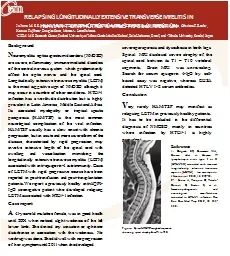
PPT-MRI Examination Transverse images –
Author : tatyana-admore | Published Date : 2018-12-15
Proton Density and STIR MRI interpretation There is hyperintensity on STIR sequences associated with the second metacarpal bone along its palmar abaxial aspect
Presentation Embed Code
Download Presentation
Download Presentation The PPT/PDF document "MRI Examination Transverse images –" is the property of its rightful owner. Permission is granted to download and print the materials on this website for personal, non-commercial use only, and to display it on your personal computer provided you do not modify the materials and that you retain all copyright notices contained in the materials. By downloading content from our website, you accept the terms of this agreement.
MRI Examination Transverse images –: Transcript
Proton Density and STIR MRI interpretation There is hyperintensity on STIR sequences associated with the second metacarpal bone along its palmar abaxial aspect as well as the palmar aspects of C2 and C3 medially . ) . and hypokineses.. Cardiac MRI demonstrating LV ejection fraction 49%(57-77), RV ejection fraction 31% (47-67%. ) . and . hypokineses. .. Cardiac MRI demonstrating LV ejection fraction 49%(57-77), RV ejection fraction 31% (47-67%. Background. N. euromyelitis. . optica. spectrum disorders (NMOSD) are severe, inflammatory, immune-mediated disorders of the central nervous system which predominantly affect the optic nerves and the spinal cord. Longitudinally extensive transverse . CHRISTINA SIEBENEICHER. ANIMAL HISTORY-SIGNALMENT. Waffle is a 8 year old spayed female dachshund. Over the past few months waffle has been trouble getting around and yelps in pain when picked up. Waffle can no longer jump onto the couch and doesn’t want to go on her nightly walks anymore. : . Improving. Patient Outcomes. Neil K. Sanghvi, M.D., F.A.C.C.,. F.H.R.S.. Cardiac Electrophysiology. Jacksonville, FL. Office: 904-423-0010. Disclosures. Received honoraria and travel expenses from . QUIZ. 3. : . Shoulder. Imaging. What is the . mean goal of . T1WS ?. :. A : to analyze the . trophicity. and . /or the fatty . degeneration of the . muscles . of rotator cuff. Daniela Batista de Almeida . Freitas. Hospital . Alian. ça. Salvador, Bahia, Brazil. Purposes. To describe the spectrum of brain magnetic resonance imaging (MRI) findings in neonates in a tertiary hospital. . Daniel Wannemacher MD. . and Humberto Morales MD. Department of Radiology. University of Cincinnati Medical Center. Control # 1594. Presentation # eP-66. Disclosure. The authors have no disclosures. Safety Background. The MRI scanner is a very large and powerful magnet. Most clinical scanners are 1.5 - 3 Tesla scanners. 3 Tesla = 30,000 gauss. Earths magnetic field ~ 0.5 gauss. MRI SAFETY. Magnetic Resonance Imaging (MRI) is one of the most powerful medical diagnostic technologies, combining strong magnetic fields with powerful computer image processing.. hh. 10/8/2015. FCC-hh RF and Transverse Feedback System / W. Hofle 08 October 2015. 1. As presented at the FCC Week. With some additional remarks. Wolfgang Hofle. BE-RF-FB. Start with where we stand . R. Apsimon. Lancaster University / Cockcroft Institute. Motivation. Currently bunch length and energy spread measurements done with separate diagnostics. Can’t reconstruct the longitudinal phase space. Nassir F. Marrouche, MD. , Oussama . Wazni. , MD, Tom Greene, PhD, J Michael Dean, MD, Eugene . Kholmovski. , PhD, Moussa Mansour, MD, Hugh Calkins, MD, Francis . Marchlinski. , MD, David Wilber, MD, Gerhard . Introduction You have been advised by your doctor to have a MRI arthrogram. This leaflet will explain what the examination involves. It may not answer all your questions, so if you have any que ries Magnetic Resonance Imaging (MRI) is a painless and harmless examination. This type of scan uses a powerful magnetic field and radio frequencies. It does not use Xrays. It isa technique that allows do A108 of Femoral Head Banuprakash S 1 1 Associate Professor, Department of Radio-Diagnosis, Sri Siddhartha Medical College, Agalakote, B H Road, Tumkur- 572107, India Corresponding author: Dr Banupra
Download Document
Here is the link to download the presentation.
"MRI Examination Transverse images –"The content belongs to its owner. You may download and print it for personal use, without modification, and keep all copyright notices. By downloading, you agree to these terms.
Related Documents