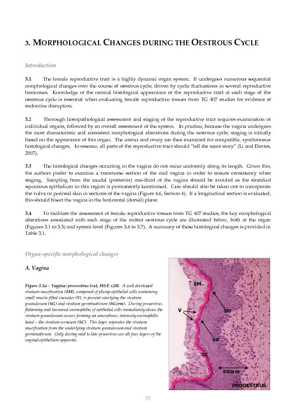
/
ORPHOLOGICAL ORPHOLOGICAL

ORPHOLOGICAL - PDF document

tatyana-admore
tatyana-admore . @tatyana-admore
Follow
381 views
Uploaded On 2016-07-02

ORPHOLOGICAL - PPT Presentation

75 SGerm SG SG SM V PROOESTRUS SC 3 M C HANGES DURING THE O ESTROUS C YCLE Introduction 31 The female reproductive tract is a highly dynamic organ system It undergoes numerous sequential morpho ID: 387896

75 SGerm SG SG SM V PROOESTRUS SC 3. M C HANGES DURING THE O ESTROUS C YCLE Introduction 3.1 The

Share:

Link:

Embed:

Download Presentation from below link

Download Pdf The PPT/PDF document "ORPHOLOGICAL" is the property of its rightful owner. Permission is granted to download and print the materials on this web site for personal, non-commercial use only, and to display it on your personal computer provided you do not modify the materials and that you retain all copyright notices contained in the materials. By downloading content from our website, you accept the terms of this agreement.


Presentation Transcript

75 SGerm SG SG SM V PROOESTRUS SC 3. M ORPHOLOGICAL C HANGES DURING THE O ESTROUS C YCLE Introduction 3.1 The female reproductive tract is a highly dynamic organ system. It undergoes numerous sequential morphological changes over the course of oestrous cycle, driven by cyclic fluctuations in several reproductive hormones. Knowledge of the normal histological appearance of the reproductive tract at each stage of the oestrous cycle is essential when evaluating female reproductive tissues from TG 40 7 studies for evidence of endocrine disrupti on. 3. 2 Thorough histopathological assessment and staging of the reproductive tract requires examination of individual organs, followed by an overall assessment of the system. In practice, because the vagina undergoes the most characteristic and consis tent morphological altera tions during the oestr o us cycle, staging is initially based on the appearance of this organ. The uterus and ovary are then examined for compatible, synchronous histological changes. In essence, all parts of the reproductive trac t should “ tell the same story ” (Li and Davies, 2007) . 3.3 The histological changes occurring in the vagina do not occur uniformly alon g its length. Given this, the authors prefer to examine a transv erse section of the mid vagina in order to ensure consis tency when staging. Sampling from the caudal (posterior) one - third of the vagina should be avoided as the stratified squamous epithelium in this r egion is permanently keratinised. Care should also be taken not to incorporate the vulva or perineal skin in sections of the vagina (Figure 4.6, Section 4) . If a longitudinal section is evaluated , this should bisect the vagina in the horizontal (dorsal) plane. 3.4 To facilitate the assessment of female reproductive tissues from TG 40 7 studies, the key morpholo gical alterations associated with each stage of the rodent oestrous cycle are illustrated below, both at the organ (Figures 3.1 to 3.3) and system level (Figures 3.4 to 3.7). A summary of these histological changes is provided in Table 3.1. Organ - specif ic morphological changes A. Vagina Figure 3.1a – Vagina: prooestrus (rat, H&E x20) . A well developed stratum mucification ( SM ), composed of plump epithelial cells containing small mucin - filled vacuoles ( V ), is present overlying the stratum granulosu m ( SG ) and stratum germinativum ( SGerm ) . During prooestrus, flattening and increased eosinophilia of epit helial cells immediately above the stratum granulosum occurs forming an amorphous, intensely eosinophilic band – the stratum corneum ( SC ). This layer separates the stratum mucification from the underlying stratum granulosum and stratum germinativum . Only during mid to late prooestrus are all four layer s of the vaginal epithelium apparent. SC 76 Figure 3.1b – Vagina: oestrus (rat, H&E x20) . The stra tum mucification has sloughed away to reveal the fully formed stratum corneum ( SC ). Large , anuclear cornified epithelial cells ( CE ) are present within the vaginal lumen. As oestrus progresses, dehiscence of the stratum corneum occurs; this exposes the un derlying stratum granulosum ( arrowhead ) and releases large numbers of cornified epithelial cells into the vaginal lumen. Figure 3.1c – Vagina: metoestrus (rat, H&E x20 and x10 ) . Complete dehiscence of the stratum corneum has occurred and only a small number of cornified epithelial cells ( CE ) remain in the vaginal lumen. Th e stratum granulosum is absent – only the stratum germinativum remains. A prominent polymorphonuclear cell infiltration ( N ) is present in the superficial epithelial cell layers; var iable numbers of polymorphonuclear cells are also present within the vaginal lumen. Note the attenuation of the stratum germinativum ( A ) and reduced number s of polymorphonuclear c ells towards the end of metoestrus . END OF METOESTRUS N A 77 Figure 3.1d – Vagina: dioestrus (rat, H&E x20). The epithelial cells ( P ) in the superficial layers of the stratum germanitivum ( SGerm ) become polygonal and plump, reflecting early mucification ( M ) . During dioestrus, the attenuated epithelium of metoestrus increases in thickness from approxim ately 4 - 6 to 8 - 10 cell layers. B. Uterus Figure 3.2a – Uterus: prooestrus (rat, H&E x20). The endometrial epithelium is composed of medium - sized, tall columnar cells. Low numbers of mitotic figures ( M ) may be observed in t he luminal uterine epith elium. P olymorphonuclear cells ( N ) infiltrate the lamina propria, increasing in number during late prooestrus. Obliquely sectioned areas of normal endometrial epithelium ( Ob ) are frequently seen in histological sections of the uterus. Luminal dilatation is also commonly noted during this stage of the oestrous cycle. 78 Figure 3.2b – Uterus: oestrus (rat, H&E x20 & x40). The endometrial epithelium is composed of very large, tall columnar cell s. Vacuolar degeneration ( D ) and apoptosis ( A ) are frequen tly observed within the lu minal and glandular epithelium, but mitotic figures are rare. Large numbers of polymorphonuclear cells ( N ) infiltrate the lamina propria and endometrial glands . Figure 3.2c – Uterus: metoestrus (rat, H&E x2 0 & x40 ). The end ometrial epithelium is reduced in height c ompared with oestrus. The stromal polymorphonuclear cell infiltration ( N ) and epithel ial cell degeneration/apoptosis observed during oe strus persist during this stage . Epithelial regeneration, characterised by in creased mitotic activity ( M ), begins during metoestrus. Figure 3.2d – Uterus: dioestrus (rat, H&E x40 & x20). The luminal and glandular epithelium are reduced in height; both are composed of small, low columnar cells. Occasional apoptotic/degenerate e pithelial cells may be present and mitotic figures ( M ) are numerous. Low numbers of polymorphonuclear cells ( N ) remain within the lamina propria during this stage. Note the plump, round to oval stromal fibroblasts ( S ), activated by high levels of circu lating progesterone. 79 C. Ovary Figure 3. 3a – Ovary: prooestrus (rat, H&E x10 ). Preovulatory (Graafian) follicles ( GF ) are observed during this stage; the absence of free - floating primary oocytes within the follicular lumina is a sectional artefact. The corpora lutea ( CL ) of preceding cycles are typically degenerate, characterised by luteal cell vacuolation ( V, inset ) and varying degrees of fibrous tissue proliferation. Figure 3. 3b – Ovary: oestrus (rat, H&E x10 & x20 ). Newly formed corpo ra lutea ( CL ) are present during oestrus . These early postovulatory follicles are small in size and composed of small basophilic cells that are morphologically similar to the follicular granulosa cells f rom which they originate. Figure 3. 3c – Ovary: met oestrus and dioestrus (rat, H&E x40 ) . In metoestrus , t he luteal cells of the recently formed corpora lutea are large and plump, and contain moderate amounts of foamy eosinophilic cytoplasm. Note the absence of cytoplasmic vacuolation an d fibrous tissue proliferation. During dioestrus , small cytoplasmic vacuoles ( white arrows ) may form within the luteal cells. Early fibrous tissue proliferation is often evident during this stage. x40 80 Key histological features – prooestrus  S tratum mucification and stratum granulosum present throughout prooestrus (vagina)  S tratum corneum present during mid to late prooestrus (vagina)  Medium - sized tall columnar endometrial epithel ial cells lining dilated lumen (uterus)  No epithelial degeneration/apoptosis present but mitotic figures observed (uterus)  Preovulatory (Graafian) follicles (ovary) SM stratum mucifica tion SC stratum corneum SG stratum granulosum SGerm stratum germinativum V mucin - filled cytoplasmic vacuole M mitotic figure Ob oblique section through luminal endometrial epithelium GF preovulatory (Graafian) follicle SGerm Stage - specific reproductive tract morphological changes Prooestrus Figure 3.4 – clockwise from left: vagina, uterus and ovary during prooestrus (rat, H&E). x10 x10 x20 PROOESTRUS SGerm SC SG SM V x20 81 Key histological features – oestrus  Stratum mucification absent (vagina)  W ell developed stratum corneum (vagi na)  C ornified epithelial cells typically present in lumen (vagina)  Very large, tall columnar endome trial epithelial cells (uterus)  Marked epithelial degeneration/apoptosis (uterus)  Immature, basophilic corpora lutea (ovary) SC stratum corneum CE cornif ied epithelial cell D vacuolar degeneration CL immature corpus luteum Oestrus Figure 3.5 – clockwise from left: vagina, uterus and ovary during oestrus (rat, H&E). x20 x10 x20 82 Key histological features – metoestrus  Stratum mucification and stratum corneum absent (vagina)  Epithelial polymorphonuclear cell infiltration (vagina)  Progressive a ttenuation of stratum germinativum (vagina)  Large, tall columnar endome trial epithelial cells (uterus)  Epithelial degeneration/apoptos is and mitotic figures present (uterus)  No fibrous tissue proliferation within most recently formed corpora lutea (ovary)  Plump luteal cells with foamy eosinophilic cytoplasm ( ovary) A attenuated stratum germinativum CE cornified epithelial cell N polymorphonuclear cell M mitotic figure Metoestrus Figure 3.6 – clockwise from left: vagina, uterus and ovary during metoestrus (rat, H&E). x40 x20 x2 0 x40 x20 83 Dioestrus Figure 3.7 – clockwise from left: vagina, uterus and ovary during dioestrus (rat, H&E). x20 x20 x40 Key histological features – dioestrus  Stratum mucif ication, stratum corneum and stratum granulosum absent (vagina)  Early mucification of stratum germinativum (vagina)  Small, low columnar endometrial epithelial cells with nu merous mitotic figures (uterus)  Early fibrous tissue proliferation within most recently formed corpora lutea (ovary) SGerm stratum germinativum P plump superficial epithelial cell (early mucification) N neutrophil M mitotic figure early lip id vacuolation 84 Table 3.1 – Typical histological changes observed in the female rat reproductive tract during the oestrous cycle. ORGAN PROOESTRUS OESTRUS METOESTRUS DIOESTRUS VAGINA Stratum mucification and stratum granulosum present throughout S tratum c orn eum present mid to late prooestrus Occasional polymorphonuclear cell s may be present in epithelium Stratum mucification absent Well developed stratum cor neum with cornified ep ithelial cells in lumen Few polymorphonuclear cells present in epithelium a t start of oestrus but numbers progressively increase Stratum mucification, stratum granulosum and stratum corneum all absent; c ornified epithelial cells may be present in lumen Attenuation of stratum germinativum to 4 - 6 cell laye rs thick marks end of me toest r us Marked epithelial polymorphonuclear cell infiltrati on at start of metoestrus but progressively decreases Early mucification of stratum germinativum (plump and polygonal superficial cells) Attenuated stratum germinativ um increases in thickness f rom 4 - 6 to 8 - 10 cell layers Few polymorphonuclear cells may remain scatte red in epithelium UTERUS Medium - sized, tall columnar endometrial epithelial cells lining dilated lumen Vacuolar d egeneration/apoptosis absent Frequent epithelial mitotic figures but numbers progressively decrease Minimal polymorphonuclear cell infiltration of lamina propria but progressively increases Very large, tall columnar endometrial epithelial cells Marked v acuolar degeneration/apoptosis of endometrial epithelium Rar e epithelial mitotic figures Marked p olymorphonuclear cell infiltration of lamina propria & endometrial glands Large, tall columnar endometrial epithelial cells Epithelial v acuolar d egeneration/apoptosis persists Frequent epithelial mitotic figures Moderate p olymorphonuclear cell infiltration of lamina propria & endometrial glands but progressively decreases Small, low columnar endometrial epithelial cells Scant vacuolar degeneration/apoptosis of endometrial epithelium High numbers of epithelial mitotic figures Minimal polymorphonuclear cell infiltration of lamina propria OVARY Preovulatory (Graafian) follicles Degenerate corpora lutea of preceding cycles, characterised by luteal cell vacuolation and varying degrees of fibrous tissue prolife ration, are present Immature, basophilic corpora lutea Central cavity/haemorrhage may be present Recently formed corpora lutea: Plump luteal cells with foamy eosinophilic cytoplasm No fibrous tissue proliferation Recently formed corpora lutea: Small lipid vacuoles within plump luteal cells may be present Earl y fibrous tissue proliferation

Related Contents


Next Show more