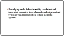
Presented by Dr Reza Hatam Dental pulp as a connective tissue How respond to irritants What makes it prone to degeneration Unique environment of dental pulp Unyielding walls of dentin ID: 535469
Download Presentation The PPT/PDF document "Pulpal Irritants and Dentin-Pulp Reactio..." is the property of its rightful owner. Permission is granted to download and print the materials on this web site for personal, non-commercial use only, and to display it on your personal computer provided you do not modify the materials and that you retain all copyright notices contained in the materials. By downloading content from our website, you accept the terms of this agreement.
Slide1
Pulpal Irritants and Dentin-Pulp Reactions
Presented by: Dr. Reza
HatamSlide2
Dental pulp as a connective tissue:
How respond to irritants?
What makes it prone to degeneration?Slide3
Unique environment of dental pulp
Unyielding walls of dentin
Resistant and resilient fiber-reinforced ground substance
Does not have a consistent effective collateral circulationSlide4
Pulpal Reactions
Caries
Local Anesthetics
Restorative Procedures
Dental Materials
Vital BleachingPeriodontal ProceduresOrthodontic Movement Slide5Slide6
Permeability of DentinSlide7Slide8
The role of odontoblastsSlide9
Hydrodynamic Theory Slide10Slide11
Pulpal Reaction to Caries
Decrease in dentin permeability
Tertiary dentin formation
Inflammatory and immune reactionsSlide12
Formation of Sclerotic Dentin Slide13Slide14
Bioactive Molecules Sequestered During
Dentinogenesis
Heparin-binding growth factor
Transforming growth factor (TGF)-
β
1, β3Insulin-like growth factorPlatelet-derived growth factor
Bone morphogenetic protein (BMP)
The TGF-
β
super family in particular seems to be importantSlide15
Formation of Tertiary Dentin Slide16
Pulpal Immune Response
The early inflammatory response is accumulation of chronic inflammatory cellsSlide17
Dental caries stimulates the accumulation of pulpal dendritic cells in and around
odontoblastic
layerSlide18Slide19
Calcium hydroxide produces an inflammatory response that stimulates dentinal bridge formationSlide20
The high PH can liberate bioactive moleculesSlide21Slide22Slide23
Vital Pulp Therapy Slide24
Definition
“
Treatment initiated to preserve and maintain pulpal tissue in a healthy state, tissue that has been compromised by caries, trauma or restorative procedures’’Slide25
Goals of pulp therapy
Primary objective is to maintain
the integrity and health of
the oral tissues
Stimulate the formation of reparative dentin to retain the tooth as a functional unit
Apexogenesis
of the immature permanent toothSlide26
Correct diagnosis - important in planning for treatment
Reversible pulp
pathosis
Irreversible pulp
pathosis
Aim – preservation of pulp vitalitySlide27
Potentially Reversible
Probably Irreversible
Pain
Momentary-dissipates readily after stimulus is removed (e.g. cold)(A-
fiber stimulation
Continous-persists for minutes to hours after stimulus is removed;presence of internal(secondary) irritant (C fibers stimulation)
Throbbing-may be present;due to arterial pulsation in area of increased pulpal pressure(C fibers stimulation)
Stimulus
Requires external stimulus
Spontaneous does not require external stimulus; dead or injured pulp tissue present in chamber or canal
Intermittent-spontaneous pain of short duration
History
Patient may have undergone recent dental procedures
Patient may have had extensive restoration, pulp capping, deep caries or traumaSlide28
Potentially Reversible
Probably Irreversible
Electric pulp test
May be premature response
May be premature, delayed or mixed response
Percussion
Negative response
May respond in advanced stages of pulpitis when concomitant acute apical periodontitis is present
Lying down
Negative because of minimally affected pulp tissue
Common finding because increase in cephalic blood pressure increases already excessive intrapulpal pressure
Color
Negative
May be present as a result of tissue lysis and intra pulpal hemorrhage
Radiograph
Probable cause (e.g. restoration or caries)
Peri-apex negative
Probable cause (e.g. restoration or caries)
Peri-apex- may be slight widening of apical periodontal spaceSlide29
Treatment Modalities
Indirect pulp capping therapy
Direct pulp capping therapy
Pulpotomy
Pulpectomy
Apexogenesis
ApexificationSlide30
Definition
Indirect pulp capping therapy
The procedure involving a tooth with a deep carious lesion where carious dentin removal is left incomplete, and the decay process is treated with a biocompatible material for sometime in order to avoid pulp tissue exposureSlide31
Indirect Pulp Capping Therapy
Rationale Slide32
Outer layer
Irreversibly denatured
Non remineralizable
Infected
Should be removed
Inner layer
Reversibly denatured
Remineralizable
Not infected
Should be preservedSlide33
Necrotic, soft brown, teeming with bacteria and not painful to remove
Firm but still softened discolored dentin with fewer bacteria, painful to remove
Sound dentin, discolored area, minimal amount of bacteria, painful to instrumentation
Three dentinal layers encountered in active caries Slide34
Indirect Pulp Capping Therapy
Objectives
Arrest the carious process
Remineralization
of carious or pre-carious dentin
Reduction in anaerobic bacteria
Formation of reparative dentin
Vital pulp maintenance
Continued normal root closureSlide35
Indirect Pulp Capping Therapy
Indications
Pain history
No extremes
May be associated with eating specially carbohydrates
Sometimes dull
Clinical examination
Large carious lesion
Normal mobility
No gingival pathologic condition
Normal color of tooth
Radiographic examination
Probable carious exposure
Normal peri-apical tissuesSlide36
Indirect Pulp Capping Therapy
Contraindications
Pain history
Sharp, penetrating pulpalgia indicating acute pulp inflammation and/or necrosis
Prolonged night pain
Clinical examination
Mobility of tooth
Discoloration of tooth
Negative reaction to electric pulp testing
Radiographic examination
large carious lesion producing definite pulp exposure
Interrupted lamina dura
Widened periodontal ligament space radiolucencySlide37
Procedure
First appointment
Isolation with rubber dam
Cavity outline with high speed water spray hand piece
Remove superficial debris and soft necrotic dentin with slow speed hand piece and large round burs, but do not expose the pulp
Potentially exposed site is covered with a commercial hard set calcium hydroxide or other capping material.
Seal the cavity for 1-3 monthsSlide38
Procedure
Second appointment
Between appointment history should be negative and restoration intact
Take bitewing radiograph-sclerotic dentin
Isolation
Carefully
remove temporary dressing
Remaining carious dentin “flaky” and easily removed
Calcium hydroxide dressing
Reinforced zinc oxide
eugenol
or glass
ionomer
cementSlide39
Evaluation of therapy
Minimally 1-3 months – to produce adequate
remineralization
of the cavity floor
Good long lasting seal of restoration
Tooth asymptomatic
Carious decalcified dentin
Rhythmic layers of irregular reparative dentin
Regular tubular dentin
Normal pulp with slight increase in fibrous elementsSlide40
One step approach
1. local anesthetic
2. Isolation
3. Removal of caries at enamel-dentine junction.
4. Judicious removal of soft, deep carious dentin
5. placement of lining material6. final restoration Slide41
The major difficulties
Determine at what point excavation halt
Voids under restorative material
Restoration failure and reactivation of lesionSlide42
Definition
Direct pulp capping therapy
“
treatment of an exposed vital pulp by sealing the pulpal wound with a dental material placed directly on a mechanical or traumatized exposure to facilitate formation of reparative dentin and maintenance of vital pulp.”
During cavity preparation
Traumatic injury
Due to cariesSlide43
Direct Pulp Capping Therapy
Contraindications
Severe toothaches at night
Spontaneous pain
Tooth mobility
Thickening of periodontal ligament
Radiographic evidence of pulp or peri-radicular degeneration
Excess of hemorrhage at the time of exposure
Purulent or serous exudate from the exposureSlide44
Clinical success
Maintenance of pulp vitality
Absence of sensitivity or pain
Minimal pulp inflammatory responses
Absence of radiographic signs of dystrophic changesSlide45
Direct Pulp Capping Therapy
Treatment considerations
Debridement
Hemorrhage and clotting
Exposure enlargement
Bacterial decontamination
Medications and materialsSlide46Slide47Slide48
Direct Pulp Capping Therapy
Medicaments
Zinc oxide
eugenol
cement (Glass & Zander 1949)
Corticosteroids and antibiotics
Polycarboxylate
cements
Inert materials
Isobutyl cyanoacrylate (
Berkman
1971)
Tricalcium
phosphate ceramic (Heller 1975)
Collagen fibers
Formocresol
Adhesive dentin bonding agentsSlide49
Direct Pulp Capping Therapy –
calcium hydroxide
Herman 1930 , seltzer & Bender 1958Slide50
An ideal pulp capping material
Stimulate reparative dentin formation
Maintain pulpal vitality
Release fluoride to prevent secondary caries
Bactericidal or bacteriostatic
Adhere to dentinAdhere to restorative materialResist forces during restoration placementSterile
Radiopaque
Provide bacterial sealSlide51
MTA
Sustain alkaline PH after curing
Small particle size
Slow release of calcium ions
Induce pulpal cell proliferation and hard tissue formation
A gap free interface precludes microleakageResist compression when final restoration Slide52Slide53Slide54
Partial pulpotomy
“Surgical removal of small portion of the coronal pulp as a means of preserving the remaining coronal and radicular pulp”Slide55
Prognosis is extremely good (94-96%)Slide56
Definitions
Pulpotomy
“
The complete removal of the coronal portion of the dental pulp, followed by placement of a suitable dressing or a medicament that will promote healing and preserve vitality of the tooth”
this technique is advocated for deciduous teeth.Slide57
Caries Removal
Dental dam isolation and Under magnification and using dye are critical.
Propylene glycol solution of Acid Red52.
The retained caries affected of dentin allows for remineralization by calcium phosphate from pulp.Slide58
Hemostasis
The one significant measurable variable to predict the outcome.
If hemostasis is not within 5-10 min, diagnosis of irriversible pulpitis.
Ferric sulfate, epinephrine, H2O2, NaOCL
NaOCL clearance of dentin chips, biofilm removal, chemical removal of blood cloth, disinfection of the cavity Slide59
Another emerging potential hemostatic agent is MTAD
An irrigant and antimicrobial agent for removal of smear layer
Doxycycline, acid citric and Tween 80Slide60
Postoperative Follow-up
When MTA is used as pulp cap next visit can be at 5-10 days. If treatment appears successful next follow-up 6 weeks, then 6 and 12 months.
In immature permanent teeth (apexogenesis) the tooth compared with contralateral tooth. Slide61
Regenerative Endodontics
The interplay among
stem cells
growth factors
scaffolds (biologic materials)Slide62Slide63Slide64Slide65Slide66
Future researches…