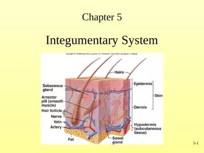
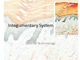
Epidermis Dermis Hypodermis Epidermis The Outermost layer of skin Avascular Complete regeneration approximately 35 days Dermis Also called corium or true skin Contains blood vessels nerves involuntary muscle sweat and oil glands and hair follicles ID: 740317
Download Presentation The PPT/PDF document "Integumentary System Three main layers o..." is the property of its rightful owner. Permission is granted to download and print the materials on this web site for personal, non-commercial use only, and to display it on your personal computer provided you do not modify the materials and that you retain all copyright notices contained in the materials. By downloading content from our website, you accept the terms of this agreement.
Slide1
Integumentary SystemSlide2
Three main layers of tissue make up the skin
Epidermis
Dermis
HypodermisSlide3
Epidermis
The Outermost layer of skin
Avascular
Complete regeneration approximately 35 daysSlide4
Dermis
Also called corium or “true skin”.
Contains blood vessels, nerves, involuntary muscle, sweat and oil glands and hair follicles.Slide5
Hypodermis
(Subcutaneous Fascia)
Innermost layer of
skin
Made
of elastic and fibrous connective tissue and adipose
tissue
Connects skin to underlying
musclesSlide6
Two main types of glands
Sudoriferous Glands
Sebaceous GlandsSlide7
Sudoriferous Glands
(Sweat Glands)
Coiled tubes.
Extend through dermis and open on surface of skin at pores.
Sweat eliminated by these glands.Slide8
Sebaceous Glands
Oil glands.
Usually open to hair follicles.
Produces sebum which is an oil that keeps skin and hair from becoming dry and brittle.Slide9
Hair & Nails
Hair
Each hair consists of root which grows in hollow tube (follicle) and a hair shaft.
Helps protect body.Slide10
Hair & Nails
Nails
Consist of dead epithelial cells packed closely together to form thick, dense surface.
Formed in
nailbed
.Slide11
Function of Integumentary System
Protection
Serves as barrier to sun’s ultraviolet rays and invasion of pathogens (germs).
Hold moisture in .
Prevents deeper tissues from drying out.Slide12
Sensory Perception
Nerves in skin help body to respond to pain, pressure, temperature, and touch sensations.Slide13
Body Temperature Regulation
Blood vessels dilate- excess heat from blood escapes through skin.
Blood vessels constrict – heat is retained in body.Slide14
Waste Disposal
Excretion of oil, water, sodium and carbon dioxide.
Slide15
Vitamin D production.Slide16
Pigmentation
Melanin
Only pigment made in skin.
Yellow to reddish-brown to black
Absorbs ultraviolet light to tan skin.
G
ives color to hair, skin, and the irisSlide17
K
arotene
Yellowish-red pigment
Can help
determine skin color.Slide18
Abnormal Skin Colors
Jaundice
Yellow discoloration of skin.
Can indicate bile in blood due to liver or gallbladder diseaseSlide19
Erythema
Reddish color of skin.
Due to burns or congestion of blood vesselsSlide20
Cyanosis
Bluish discoloration of skin.
Caused by insufficient oxygen.
Associated with heart, lung, and circulatory diseases or disorders.Slide21
Acne – overactive secretion of sebaceous glands.
Pimples and blackheads.
Teens to early twenties.
Rx: Thorough washing, steroid creams, UV light, avoidance of certain foods, chemical face peel.
Skin Diseases and Disorders Slide22
Eczema
Vesicles
or reddened skin which burst and weep a crust ( dried pus and blood)
Most
common inflammatory disorder of the skin
Rx: Tranquilizers, antihistamines, wet dressings, starch baths.Slide23
Psoriasis
Psoriasis – Red thick areas covered with white or silver scales
Chronic, noncontagious, inherited skin disease
Rx: No Cure
Cortisone Ointments
Ultraviolet Light
Removal of ScalesSlide24
Contact Dermatitis
Redness, itching, blisters, edema
Caused by poison ivy, poison oak, cleansing agents, cosmetics, etc.
Rx: Wash with soap and water then apply alcohol and antipruritic (relieves itching) lotions.Slide25
Impetigo
Erythema, vesicles with sticky yellow crusts
Very contagious.
Infection with staph or strep
Rx: Remove crusts and apply
antibiotic
ointmentSlide26
Warts
Caused by virus.
Painless except for plantar warts.
Rx: Nitric or sulfuric acid deep into root of wart or freezing with liquid nitrogen.Slide27
Scleroderma
systemic autoimmune disease of skin, muscles, bones, heart, lungs
. Skin
becomes hard and tight.
Progressive disease. Mainly affects women in child bearing years. Considered an auto immune disease
Rx: Ointment, heat, massage, steroids.Slide28
Skin Cancer
Definition: neoplasms or abnormal growth of cells that originate in the epidermis.
More than 800,000 new cases each year in the USA
One in five people in the US will develop skin cancer in his/her lifetime. This number jumps to one in three in the Sunbelt states
.
Three Major Types of Skin Cancer
Basal Cell, Squamous Cell, Malignant MelanomaSlide29
Skin Cancer
Basal
Cell Carcinoma
Most common type of skin cancer
Malignancy begins in cells at the base of the epidermis and most often appears on the nose and face
Incidence increases after age 40
Basal cell tumors rarely metastasize but may cause wide-spread destruction of normal tissue if left untreatedSlide30
Skin Cancer
Squamous Cell Carcinoma
Slow-growing
Arises from the epidermis
Most frequently occurs in middle-aged and elderly individuals
Typically found on sun-exposed areas of skin
May metastasize but is not likely to spread to other body areas.Slide31
Skin Cancer
Malignant Melanoma
Most deadly of all skin cancers
Steady increase in incidence of 4% per year over last 20 years
Median age of diagnosis is 53 years
Sometimes develops from a pigmented Nevus (mole) to become a dark spreading lesion
Most likely to metastasizeSlide32
Skin Cancer
Malignant MelanomaSlide33
Skin Cancer
http://www.skincancer.org/
“ABCD” Rule of Self-Examination of Moles
Asymmetry: Lesion halves are not mirror images of each other giving a lopsided appearance
Border: Irregular or indistinct borders
Color: Unevenly colored, exhibiting a mixture of shades or colors
Diameter: By the time lesions exhibit characteristics A, B, and C, it is probably larger than 6mm or ¼ inchSlide34
BURNS
Burn is an injury that can be caused by fire, heat, chemical agents, radiation and/or electricity
Classification of burns: Severity of burn is determined by depth of lesion and percent of body surface burned.Slide35
Burns
First-degree or superficial
Least severe type of burn
Involves only top layer of skin, the epidermis
Usually heals in 5 to 6 days without permanent scarring
Skin is reddened or discolored
May have some mild swelling
Victim feels pain
Three common causes
Overexposure to sun or mild sunburn
Brief contact with hot objects or steam
Exposure of skin to weak acid or alkaliSlide36
Burns
Second-degree or partial-thickness
Usually causes injury to top layers of skin and involves both epidermis and dermis
Blister or vesicle forms
Skin is red or has mottled appearance
Swelling occurs along with severe pain
Surface of skin appears to be wet
Painful burn that may take 3 to 4 weeks to heal
Three common causes
Excessive exposure to sunlamp or artificial radiation or severe sunburn
Contact with hot or boiling liquids
Burns from firesSlide37
Burns
Third-degree or full-thickness
Most severe type of burn
Involves injury to all layers of skin in addition to underlying tissue
Area has a white or charred appearance
Can be extremely painful or relatively painless if nerve endings are destroyed
Can be life-threatening because of fluid loss, infection, and
shock
Common causes
Exposure to fire or flames
Prolonged contact with hot objects
Contact with electricity
Immersion in hot or boiling liquids
Slide38
Burns
Methods to Determine Percent of Body Surface Burned
“
Rule of Palms”:
Based on the assumption that palm size of burn victim is about 1% of body surface. Estimating the number of “palms” burned will approximate the percentage of body surface involved
.Slide39
Burns
Methods to Determine Percent of Body Surface Burned
“Rule of Nines”:
9% of total skin area covers head and each upper extremity, including front and back
surfaces
18%of total skin area covers each of the following:
front of trunk
back of trunk
each lower extremity including front and back surfacesSlide40
Burns
Methods to Determine Percent of Body Surface Burned
Lund-Browder Charts
:
Permits more accurate estimates of burned surface area in children
Makes allowances for large percent of surface are in certain body regions in children such as the headSlide41
Burns
Medical help for burns
Usually not required for first-degree or superficial burns
Should be obtained if:
More than 15% of adult’s body is burned
More than 10% of child’s body is burned
Rule of nines is used to calculate the percentage of body surface burned
Burns affect face or respiratory tract
Victim is having difficulty breathing
Burns cover more than one body part
Victim has a partial-thickness burn and is under 5 or over 60 years of age
Burns result from chemicals, explosions, or electricitySlide42
Burns
All third-degree or full-thickness burns should receive medical care
First aid for superficial and mild partial-thickness burns with closed blisters
Cool area by flushing with large amounts of cool water
Do not use ice or ice water because it causes body to lose heat
Use dry, sterile gauze to blot area dry
Apply dry, sterile dressing (
nonadhesive
or nonstick is best) to prevent infection
Elevate affected part if possible to reduce swelling
Do not apply oils, grease, butter or other substances unless instructed to do so by physician
Do not break or open any blisters as this creates an open wound prone to infectionSlide43
Burns
First aid for severe second-degree and full-thickness or third-degree burns
Call for medical help immediately
Cover burned area with thick, sterile dressings
Elevate Hands or feet if they are burned
Do not allow victim to walk if feet or legs are burned
Do not attempt to remove particles of clothing from burn
Watch for respiratory distress or signs of
shockSlide44
Burns