/
pulmonary embolism Dr.  Zaki pulmonary embolism Dr.  Zaki

pulmonary embolism Dr. Zaki - PowerPoint Presentation

taylor
taylor . @taylor
Follow
0 views
Uploaded On 2024-03-13

pulmonary embolism Dr. Zaki - PPT Presentation

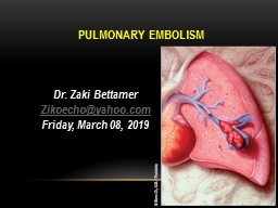
Bettamer Zikoechoyahoocom Friday March 08 2019 67 years Old male presented with acute dyspnea Pleuritic Right side chest pain and hemoptysis with history of recent pelvic fracture post RTA one week back ID: 1047779

embolism pulmonary lung diagnosis pulmonary embolism diagnosis lung patient dimer dvt amp massive test clot emboli increased clots death

Share:

Link:

Embed:

Download Presentation from below link

Download Presentation The PPT/PDF document "pulmonary embolism Dr. Zaki" is the property of its rightful owner. Permission is granted to download and print the materials on this web site for personal, non-commercial use only, and to display it on your personal computer provided you do not modify the materials and that you retain all copyright notices contained in the materials. By downloading content from our website, you accept the terms of this agreement.


Presentation Transcript

1. pulmonary embolismDr. Zaki BettamerZikoecho@yahoo.comFriday, March 08, 2019

2. 67 years Old male presented with acute dyspnea, Pleuritic Right side chest pain and hemoptysis , with history of recent pelvic fracture post RTA one week back.On examination : Left leg swelling and tender, BP: 90/60, pulse : 140 beat / minute, regular.Chest examination: normalThe commonest arrhythmia expected in this patient

3.

4. Definition:It is an obstructive lesion in the pulmonary vascular tree. It is usually caused by a insoluble thrombus that has travelled from a distant site. A blood clot that forms and remain in a vein is called thrombus, a clot that travels to another part is called embolus.

5. Incidence: The majority (80%) of pulmonary emboli arise from the propagation of lower limb DVT The annual incidence is about 60-70 per 100 000 population.Rare causes include septic emboli (from endocarditis affecting the tricuspid or pulmonary valves), tumour (especially choriocarcinoma), fat, air, amniotic fluid and placenta.It is a common mode of death in patients with cancer, stroke and pregnancy.

6. Correct diagnosis is made only in 10-30 %. 60 % of autopsies showed some evidence of post PE. 15 % of postoperative deaths are related to PE or its sequels. PE is a commonest cause of maternal death in the UK.

7. Predisposing factorsRecent surgery especially pelvic, large joint and extensive abdominal . Following pelvic and leg fractures. Pregnancy and child birth. Old age: > 70 years. Smoking . Inherited clotting disorders: a. Hypercoagulable state. b. Platelets abnormalities. c. Protein S & C deficiency.

8. Long periods of immobility: a. Prolonged hospitalization. b. Long plane or car trips. c. Prolonged bed rest. Severe debilitating illness: a. Severe injures. b. Cardiac diseases. c. Malignancies.

9. Increased estrogen level: a. Contraceptive pills. b. Hormonal therapy. Increased levels of clotting factors: a. pancreatic, lung and ovarian malignancies (increased procoagulant). b. Female hormones.

10. Family history. Pace makers or venous catheterization either central or peripheral. Being overweight: There is a link between the formation of clots formation to leptin ( hormone produced by fat cells). Previous proved history of venous thromboembolism.

11. Cardiac problems: a. Congenital. b. Congestive. c. Hypertensive. Others: a. Nephrotic syndrome. b. Thrombotic and myeloproliferative disorders. c. Inflammatory bowel diseases.

12. Sources and origin of PEThe source can be indentified in only 50 – 70 % of cases. 70 – 90 % of the identified cases, clots come from IVC. 10 – 20 % of the identified cases, clotscome from SVC.

13. Pathological leading causesDeep vein thrombosis. Air embolism.Amniotic fluid embolism.Fat embolism.Rare : Tumor emboli & Bacterial clumps.

14. Progression of pulmonary embolism.The course of events depend on the size of the clot causing PE.A. Small to medium sized PE.Cut off the circulation to a part of the lung.It heals with fibrous scar.Can travel more distally infracting the lung.

15. B. Massive pulmonary embolism.Embolize the main pulmonary artery Blocking off almost blood flow to lungs.The emboli may happen suddenly causing Massive PE or gradual causing recurrent PE state.Lodge at the bifurcation of the main pulmonary artery causing hemodynamic collapse or death.

16. Probable outcome of PE.10 % of clinically presented PE are fatal. 30 % of patients with PE will suffer a second attack.70 % will had PE complications.

17. Differential diagnosis of PEMyocardial infarction. Pneumonia.Congestive heart failure. Asthma. COPD. Intrathoracic neoplasm. Rib fractures. Pneumothorax. Musculoskeletal pains.

18. AcuteLarge sized. Massive PE.It involves > 50 % of the main PASudden death.ChronicSmall sized.Pulmonary infarction with episodes of pleuritic chest pain And haemoptysis.Presentations of PE. According to the size of venous emboli:

19. Postoperative presentations.Classical time: 2nd postoperative week !Sudden death Cyanosis DysponeaTackyponea Cough HemoptysisChest pains Low EtCO2

20. Completely normal. Chest pain Pleuritic pain Dysponea Apprehension Cough Hemoptysis DiaphoresisSymptoms More clear in patient with pre-existing lung diseases.Possible presentations:

21. Syncope TachyponeaRalesTachycardiaFever > 37.8PhlebitisCyanosisLeg swellingLeg painWheezing Angina pain. AFComa Pul.infection Sweating

22. Chronic effects of PE 1. Increased PVR. 2. Decrease gasexchange. 3. Alveolar hyperventilation. 4. Increased AWP. 5. Decreased lung compliance. 6. Right side ventricular dysfunction & failure.

23. Screening and Diagnosis. PE is difficult to diagnose especially with underlying lung or cardiac diseases. More than one test should be done to increase the efficacy of correct diagnosis.

24. 1st line of investigations. ECG. Nonspecific changes are common. Sinus tachycardia is the commonest among changes. Possible changes: AF, RBBB, T wave inversion & P pulmonale. S1 , Q3, T3

25. inverted T in III S wave in lead 1 Q wave in III (S1, Q3, T3) Signs of right ventricular strainECG

26. normal. Atelectasis or density.Pleural effusion.Pleural opacity. Elevated diaphragm. Prominent PA.Cardiomegally.Pulmonary edema. Chest X ray.Possible findings

27. Wedge shaped density. Pleural effusion

28. ABGsMay be normal ! Hypoxia. Respiratory hypocarbia. Increased A-a gradient. * Gradient > 15-20 is considered abnormal.

29. D – dimer test.D – dimer are released as a result of fibrinolysis & indicate the presence of intravascular thrombus. It reflects the breakdown of plasmin. Useful, to exclude the PE. Has > 99% sensitivity in acute PE or DVT..

30. Negative D – dimer Test. Exclude PE in Patient with low pre-test Probability. Helpful in patient with a high clinical Probability. Positive D – dimer test. Needs further investigations to confirm the diagnosis Less useful in patients with longer hospitalization. False positive can result from the clot formation at venepuncture sites.

31. False positive d – dimer testMalignant Patient.Recent Surgery.Chronic Inflammatory ProcessSepsisVenepuncture Due to clot formation

32. Pulmonary angiographyShows pulmonary vascular obstructionGold standard of diagnosis of PE, Thrombolytic therapy can be given through the catheter afterwards.

33. Computed tomographic pulmonary angiogramHas sensitivity > 90 % Dilatation of pulmonary vessels proximal to the embolus with collapse of distal vessels.

34.

35. Isotope lung scan. ( ventilation/perfusion scan)Use of radioactive tracers to study air flow( ventilation ) and blood flow (perfusion).

36. 2nd line of investigations. Leg ultrasound scan doppler for detection of DVT. Around 70 % of patient proved PE will have a proximal DVT.. Echocardiogram.Diagnostic only in the massive PE.Of prognostic information's.The findings may be: * RV overload & dysfunction. * Dilatation of IVC. * Dilated pulmonary arties. * Disturbed flow velocity. * Direct visualization of clots in the prox. PA, RV, or RA.

37. Spiral “ Helical “ computerized tomography.Three dimensional series of very thin x ray beams using contrast dye.

38. Lung ultrasound. 1. It is noninvasive sonar. 2. Uses high frequency sound waves to check for blood clots. 3. It is cheaper than V/Q scan Venography. Invasive procedure to reveal clot blockage at any point of the vein course using contrast.

39.

40. Magnetic resonance ( MRI ). Expensive. Reserved for pregnant woman and renal patient. TEE.Intraoperative diagnosis of PE.

41. c. Intraoperative prophylaxisDecrease venousstasis: 1. Raising legs. 2. Avoid leg trauma. 3. Use of pneumatic leg compression. 4. Use of electrical calf muscle stimulation.

42. When pulmonary embolism is life threatening ?Massive PE. Worsening the cardiopulmonary underlying diseases. Treatment plan are not effective.

43. Treatment overview of PE1. Preventive strategies plan: A. Preventive steps in the hospital: 1. Correction of the risk factors. 2. Adequate hydration. 3. Graduated compression stockings. 4. Use of pneumatic compression. 5. Early physical activity.

44. 6. Anticoagulant therapy. a. Heparin: 5000 iu, 8-12 hˉ1 , 2h before operation. b. Ultra low dose heparin: 1 unit.kg.hˉ1 infusion. c. Adjusted heparin dose: start 3500 iu subcut. Every 8 hˉ1 to keep ATT 1.5 X normal. d. Low molecular heparin: one dose, 12hˉ1 subcut.untill patient ambulate.

45. B. Preventive steps while travelling.Take a walk. Exercise while you set. Wear support stockings. Drink plenty of fluids. Ask for medical advise: * If you had DVT before. * If the travelling time is more than 6 hours. * If you at any risk.

46. II. Active strategies plans.A. Treatment of DVT: 1. Heparin. * 5000 IU bolus, followed by 40000 IU over 24 h for 7 days. * adjust the dose: APTT 1.5-2 2. Oral anticoagulants: * Warfarin for 3 – 6 month. * Keep PT 1.2-1.5 ( INR : 2 – 3 )

47. B. Thrombolytic therapy.indicated for acute massive PE causing collapse or arrest. 1. Streptokinase: 250000 U IV over 30 min, then 100000 U hˉ1 for 24 hours. 2. Tissue type plasmogen activator 100 mg IV over 2 hours. 3. Urokinase: 4400 IU Kgˉ1 over 10 min. followed by 4400 IU Kgˉ1.hˉ1 for 12 -24 hours.

48. Actilyse ” Alteplase” * Action: It is an enzyme that increase plasmin that break down clots.Recommendations a. Should be given as soon as possible. b. Given as infusion over 2-3 hours. c. 10 mg bolus over one or two minutes followed by 90 mg.

49. C. Surgical pulmonary embolectomy. * Under CP bypass. * Postoperative & operative mortality is 20 – 50 % * Long term survival is 71 % after 5 years. * Indicated in acute massive PE with lifeD. I.V.C umbrella filter: * Placed percutaneously under local analgesia. * Aim: to avoid recurrence of more emboli.

50. D. I.V.C umbrella filter: * Indications: a. Contraindication to anticoagulant. b. Failure of anticoagulant therapy. c. Chronic recurrent PE d. Emergency treatment following massive PE.

51. E. Cather throbectomy. * Indications: A. Contraindication for frinolysis. B. Surgical embolectomy is not available.

52. Patients at risk of DVT should be encouraged to keep mobile. Please don’t forget

53. Regarding the D – dimer test in diagnosis of Pulmonary Embolism :Presence of D – Dimer is not indicate the presence of intravascular thrombus.Has <2 % sensitivity in acute Pulmonary Embolism or DVT.Useful to exclude the Pulmonary Embolism.Negative D – dimer Needs further investigations to confirm the diagnosis.Positive D – dimer test Exclude Pulmonary Embolism in Patient with high pretest Probability.

54. One of the following is Chronic effects of Pulmonary Embolism :Decrease Pulmonary Vascular Resistant .Increase gas exchange.Decrease A Wedge Pressure .Increase lung compliance.Right side ventricular dysfunction & failure.

55. THANK YOU