/
Islamic University of Gaza Islamic University of Gaza

Islamic University of Gaza - PowerPoint Presentation

test
test . @test
Follow
386 views
Uploaded On 2016-05-19

Islamic University of Gaza - PPT Presentation

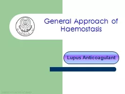
Lupus Anticoagulant General Approach of Haemostasis Anti Phospholipids antibodies Anti phospholipid antibodies are a heterogeneous family of autoantibodies that react with epitopes ID: 325831

lupus test time phospholipid test lupus phospholipid time presence plasma dimer anticoagulants aptt antibody factor results antibodies coagulation platelet anticoagulant anti phospholipids

Share:

Link:

Embed:

Download Presentation from below link

Download Presentation The PPT/PDF document "Islamic University of Gaza" is the property of its rightful owner. Permission is granted to download and print the materials on this web site for personal, non-commercial use only, and to display it on your personal computer provided you do not modify the materials and that you retain all copyright notices contained in the materials. By downloading content from our website, you accept the terms of this agreement.


Presentation Transcript

Slide1

Islamic University of Gaza

Lupus Anticoagulant

General Approach of HaemostasisSlide2

Anti-Phospholipids

antibodies.

Anti-phospholipid antibodies are a

heterogeneous

family of

autoantibodies

that react with

epitopes

on proteins that are themselves

complexed

with negatively charged phospholipids.

The blood vessel problems can then lead to complications such as stroke, heart attack, and miscarriage.

The two most commonly measured kinds of Antibodies are:

Lupus anticoagulant

Anticardiolipin

antibody Slide3

Lupus Anticoagulants

(lupus antibody, LA, or lupus inhibitors)

Lupus anticoagulants (LA) are heterogeneous IgG or IgM autoantibodies

which

inhibit

phospholipid

-dependent assays

of blood coagulation by binding to plasma

phospholipid

-binding proteins such as beta 2-glycoprotein I

(β2-

GPI) or

prothrombin

Lupus Anticoagulant was found in patient with SLE.

Most patients with a lupus anticoagulant do not actually have lupus

Erythematosus

, and only a small proportion will proceed to develop this

disease.

It is a

prothrombotic

agent

, that is, presence of Lupus anticoagulant antibodies precipitates the formation of thrombi in vivo.

In vivo, it is thought to interact with platelet membrane phospholipids, increasing adhesion and aggregation of platelets; thus its in vivo

prothrombotic

characteristics.Slide4

The term "anticoagulant" accurately describes its function in vitro, but in vivo, it is now known that it functions as a coagulant

Lupus anticoagulants impair the in vitro phospholipid dependent activation of factor X, factor IX, and Prothrombin. Presence of these antibodies causes an increase in aPTT.

The initial workup of a prolonged PTT is a mixing test.

If the mixing test indicates an inhibitor, diagnosis of a lupus anticoagulant is then confirmed with phospholipid-sensitive functional clotting testing, such as the

dilute Russell's viper venom time

, or the Kaolin clotting time

The presence of an LA is usually not associated with a bleeding problem unless accompanied by

Thrombocytopenia,

Factor II deficiency,

Platelet dysfunction

Or drug administration (e.g., aspirin). Slide5

The

ISTH(International Society on Thrombosis and Haemostasis ) recommends that the laboratory diagnosis of lupus anticoagulants (LA) should be carried out on double-centrifuged plasma following a four-step procedure adhering to these principles:

Prolongation of a phospholipid-dependent coagulation test.

Extrinsic

(

dPT

),

Intrinsic

(aPTT, dilute aPTT, KCT, colloidal silica clotting time),

Final common pathway

(

dRVVT

,

Taipan

venom time,

Textarin

and

Ecarin

time)

Evidence of inhibitory activity on mixing tests.

Evidence of phospholipid dependence.

Correction of the prolonged coagulation time after addition of excess phospholipid or platelets

Lack of specificity for any one coagulation factor

Lack of

specifi

c inhibition of one coagulation factor (such as FVIII:C,FIX:C, or FXI).Slide6
Slide7

There

is many classes of anticardiolipin antibody, abbreviated as

IgG,

IgM

, and

IgA

.

The

anticardiolipin

antibody is measured in an ELISA test.

The

IgG type of anticardiolipin antibody is the type most often associated with complications.Some lupus patients with very high IgM anticardiolipin antibody have a problem called hemolytic anemia, in which their immune system attacks their red blood cells.

Anti-

Cardiolipin

Antibodies.Slide8
Slide9

Anti-Phospholipid Antibody Syndrome

APS is present if at least one

of the clinical criteria and one of the

laboratory criteria

that follow are met:

Clinical criteria:

vascular thrombosis or pregnancy morbidity

Laboratory criteria:

Lupus anticoagulant present in plasma on two or more occasions at least 12 weeks apart.

Anticardiolipin antibody,

IgG

or IgM positive in medium or high titer on two or more occasions at least 12 weeks apart Anti-β2GPl antibody, IgG or IgM present on two or more occasions at least 12 weeks apart. The positive laboratory criteria and the clinical criteria should occur within 5 years of each other.Slide10

Laboratory Investigations

Immunological AssaysIndividuals with Lupus anticoagulants may also show the presence of other anti phospholipid antibodies. The most frequent finding is the presence of Anti- cardiolipin antibodies. The commonly employed method is the ELISA technique where the solid phase is coated with cardiolipin and β-2 GPI (as a cofactor). The ELISA method detects IgM, IgG and IgA class of anti-cardiolipin antibodies.

An important point to note is that Lupus anticoagulants and anti-cardiolipin antibodies differ in their phospholipid-binding characteristic hence detection of anti cardiolipin antibodies is not specific for the presence of Lupus anticoagulants though they may be present in the same patient.Slide11

Clot based assays

APTT ( Activated Partial Thromboplastin Time)

TTI (Tissue Thromboplastin Inhibition

test)

KCT

(Kaolin Clotting

Time)

PNP

(Platelet Neutralization

procedure)

Hexagonal

Phase Phospholipids (Staclot-LA)Slide12

APTT ( Activated Partial Thromboplastin Time)

The specificity of the APTT to LA is reduced:

Different reagents

have varying

sensitivity

for the presence of

Lupus anticoagulants

on the basis of their phospholipid

composition ( variability of the test)

The APTT test is affected by the presence of

inhibitors to Factor

VIII, IX and XI. The APTT test is also the test of choice for monitoring heparin therapy. Slide13

TTI (Tissue Thromboplastin Inhibition test)

The Tissue Thromboplastin Inhibition test utilizes

a diluted PT reagent. The results are expressed as ratio of patient values to normal control values.

The TTI test is affected by numerous variables:

Species and tissue origin of thromboplastin

can affect the test results as different sources of thromboplastin have varying sensitivity and responsiveness.

Choice of “Normal” reference plasma

is the most critical variable, because depending on the laboratory the choice of reference plasma could be lyophilized plasma, a frozen plasma pool or fresh plasma. The ratio of patient to normal can therefore change according to the choice of “normal” plasma.

Some

IgM

Lupus anticoagulants

do not prolong the TTI testSlide14

KCT (Kaolin Clotting Time)

KCT is similar to APTT, the difference being that KCT reagent is devoid

of phospholipids and incorporates Kaolin as contact activator. The test is

performed on

a range of mixture of normal and patient’s plasma

. Different patterns of

response are

obtained indicating the presence of Lupus anticoagulants or the deficiency

of one

or more coagulation factors.

The KCT test though sensitive is

not specific for LA, additionally:It cannot be automizedThe test shows prolonged results with factor VIII, IX, XI & XII deficiency or corresponding inhibitorsThe test is also highly sensitive for the presence of heparin.Slide15

PNP (Platelet Neutralization procedure)

The platelet neutralization procedure (PNP) is based on the ability of

platelets to significantly correct in vitro coagulation abnormalities.

The disrupted platelet membranes present in the freeze-thawed platelet suspension neutralize

phospholipid

antibodies present in the plasma of patients with LA.

After the patient plasma is mixed with the freeze-thawed platelet suspension, the APTT will be shortened when compared with the original baseline APTT. But if an inhibitor is directed against specific coagulation factor, the clotting time is not shortened.

A correction of the baseline APTT of a defined amount of time (i.e., 3 to 5 seconds or more) by the platelet suspension as compared with the control is indicative of the presence of an LA. Slide16

Due to limited stability the platelet preparations loose their activities on storage hence do not show reproducible results

.Slide17

Hexagonal Phospholipid Neutralization Assay

The

hexagonal phospholipid neutralization assay uses the same principle as the

PNP assay

, normalization of the aPTT in the presence of added phospholipid, but this assay

specifically

uses a

phospholipid in a hexagonal conformation,

Neutralization

by this hexagonal form in an assay with a very lupus-anticoagulant sensitive aPTT reagent, is a more

sensitive confirmation test than the PNP. Comment Specimen collection, centrifugation, and processing are critical when testing for the presence of an LA. Slide18

Confirmatory Tests

for Lupus Anticoagulants Confirmatory tests to identify an LA include those that utilize a low concentration of phospholipid in the test system, thereby increasing the LA effect such as

The tissue thromboplastin inhibition test (TTIT), Dilute Russell's viper venom time (dRVVT)

,

And the kaolin clotting time (KCT), or

Those that increase the phospholipid, thereby neutralizing the LA effect, such as the platelet neutralization Slide19

D

ilute Russell’s

V

iper

V

enom

T

ime

(DRVVT)dRVVT : The test of choice for screening and confirmation of LAIndicating the phospholipid dependence of LAAchieving maximum sensitivty for the precence of LA’s.In general dRVVT based tests comprise of:

SCREENING

REAGENT, containing limited amount of phospholipids with

RVV (Russell’s

Viper Venom)

CONFIRMATION

REAGENT, containing additional phospholipids with

same amount

of Russell’s Viper Venom, to confirm the presence of

phospholipid dependent

Lupus anticoagulants.Slide20

Principle of

dRVVT for LA detectionRussell’s Viper Venom directly activates Factor V and X in presence of phospholipid and calcium ions, bypassing Factor VII of the extrinsic pathway and the contact and antihaemophilic factors of the intrinsic pathway.

In normal plasma, in the absence of lupus anticoagulants, Factor V and X is directly activated by Russell’s Viper Venom, which in presence of phospholipid and calcium ion leads to clot formation.Slide21

In patients with LA, autoantibodies bind the epitopes

of reagent phospholipids thereby preventing the activation of prothrombinase complex. This results in a prolongation of clotting time with SCREENING reagent. The CONFIRMATION reagent incorporates additional phospholipids to neutralize the LA. Once LA are neutralized clot formation proceeds relatively uninterrupted achieving a lower clotting time, to prove the phospholipids dependence of the

autoantibodies.Slide22

Interpretation of results with dRVVT

testIf SCREEN TIME is prolonged, to confirm the presence of

lupus anticoagulants the plasma sample is tested with CONFIRMATION REAGENT.

If CONFIRM TIME results shows a lower clotting time as compared to

SCREEN TIME

, it indicates the presence of phospholipid dependant Lupus Anticoagulants.

Also the results can be expressed as ratio

,

The

results expressed, as ratio is further useful in classifying the patient as

normal, moderate

, high and very high LA.If results of the ratio are borderline, mixing studies may be done further with the sample specimen. The mixing studies should be carried out with a 50:50 mixture of test plasma and normal plasma.Slide23

General Approach of Haemostasis

D-

DimerSlide24

D-

dimer(Fragment D-dimer; Fibrin

degradation (

fragment

D-dimer is a fibrin degradation product (or FDP), a small protein fragment present in the blood after a blood clot is degraded by fibrinolysis. It is so named because it contains two crosslinked D fragments of the fibrinogen protein.

D-dimer test used to diagnose

thrombosis,

disseminated intravascular coagulation.

The circulating enzyme plasminncleaves the fibrin gel in a number of places. The resultant fragments, "high molecular weight polymers", are digested several times more by plasmin to lead to intermediate and then to small polymers (fibrin degradation products or FDPs). The cross-link between two D fragments remains intact, however, and these are exposed on the surface when the fibrin fragments are sufficiently digested. The typical D-dimer containing fragment contains two D domains and one E domain of the original fibrinogen moleculeSlide25

the D-dimer assay depends on the binding of a monoclonal antibody to a particular epitope on the D-dimer fragment. Several detection kits are commercially available; all of them rely on a different monoclonal antibody against D-dimer. Of some of these it is known to which area on the D-dimer the antibody binds. The binding of the antibody is then measured quantitatively by one of various laboratory methods.

D-dimer testing is of clinical use when: There is a suspicion of deep Venous Thrombosis (DVT)Pulmonary Embolism (PE) Disseminated Intravascular Coagulation (DIC).It can also rise Postoperatively.

It is under investigation in the diagnosis of Aortic DissectionSlide26

Interpretations of Results

A very high score, or pretest probability, a D-dimer will make little difference and anticoagulant therapy will be initiated regardless of test results, and additional testing for DVT or pulmonary embolism may be performed.For a moderate or low score, or pretest probabilityA negative D-dimer test will virtually rule out thromboembolism: the degree to which the D-dimer reduces the probability of thrombotic disease is dependent on the test properties of the specific test used in the clinical setting: most available D-dimer tests with a negative result will reduce the probability of thromboembolic disease to less than 1% if the pretest probability is less than 15-20%

If the D-dimer reads high, then further testing (ultrasound of the leg veins or lung scintigraphy or CT scanning) is required to confirm the presence of thrombus. Anticoagulant therapy may be started at this point or withheld until further tests confirm the diagnosis, depending on the clinical situation.Slide27

False positive readings can be due to various causes: liver disease, high rheumatoid factor,

inflammation, malignancy, trauma, pregnancy, recent surgery as well as advanced age[citation needed]False negative readings can occur if the sample is taken either too early after thrombus formation or if testing is delayed for several days. Additionally, the presence of anti-coagulation can render the test negative because it prevents thrombus extension.

Likelihood ratios are derived from sensitivity and specificity to adjust pretest probability.Slide28

Thank

you