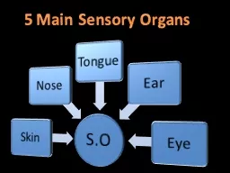
Distortion due to variation in the size and density of sensory neuron receptive fields The Stimuli of Somatosensation SKIN body surface Mechanical pressure this is touch Vibration Hz this is generally texture ID: 626989
Download Presentation The PPT/PDF document "The Skin: Largest Organ in the Body" is the property of its rightful owner. Permission is granted to download and print the materials on this web site for personal, non-commercial use only, and to display it on your personal computer provided you do not modify the materials and that you retain all copyright notices contained in the materials. By downloading content from our website, you accept the terms of this agreement.
Slide1
The Skin: Largest Organ in the Body
Distortion due to variation in the size and density of sensory neuron
‘
receptive fields
’Slide2
The Stimuli of
SomatosensationSKIN (body surface)Mechanical pressure: this is ‘touch’
Vibration (Hz): this is generally ‘texture’Damage/Temp (pain/hot/warm/cold)Chemical (example is menthol)
MUSCLES &TENDONS (body position)Stretch, Tension
Kinesthesis, ProprioceptionBODY SURFACE + BODY POSITION = “HAPTIC PERCEPTION”Slide3
Receptors can be characterized in terms of:
Stimulation Type
(pressure, vibration, temp, damage)
Size of Receptive Field
(amount of branching)
Rate of Adaptation
(slow, medium, fast)
Basket Cell
Pacinian
corpuscle
Free Nerve
EndingsSlide4Slide5
Pacinian Corpuscles
Detect Vibration (texture)
Pressure
Stimulus
Pacinian
ActivitySlide6
Muscle Spindle
– Stretch Receptor
Golgi Tendon Organ
– Tension Receptor
Receptors in Muscles and Tendons
Encode Body Position
Muscle fiber
Red is motor
Blue is sensory
Sensory fiber
attached to tendon
Bone
MuscleSlide7
This is a Muscle spindle,
but other
Mechanoreceptors
(i.e., Basket Cells and
Pacinian
Corpuscles) also work this way
The
cytoskeletal strands
are like the ‘tip links’ of inner hair
cells. Ion channels ‘pulled open’ by mechanical force.Slide8
Skin Senses: 2 Pathways to Cortex
Lemniscal Pathway (mechanorecepetors)Tactile, pressure, Basket Cell
(detect)Tactile, vibration/texture, Pacinian Corpuscle (ID)
Spinothalamic Pathway (free nerve endings) Tissue Damage, pain,
Nociceptor (detect)Temperature, hot/cold, Thermal Receptor (ID)This pathway is ‘gated’ in the spinal cordSlide9
D o r s a l – S e n s o r y
V e n t r a l – M o t o r
Hypothalamus
Thalamus
Spinal Cord
Thalamus
Cortex
parietal
Hindbrain
Dorsal
Ventral
Dorsal
Ventral
Thinking in 3D
Dorsal/Ventral Organization
Left/Right Crossing
Cortex
frontal
D
O
R
S
A
L
SENSORY NEURONS
MUSCLES
MUSCLESSlide10
Somatotopic
OrganizationSlide11
2D Receptor
Array
Thalamus
Primary Cortex
However…Slide12
Dense 2D
Receptor Array
Thalamus
Cortex Is
‘Modular’
Means the size and density of cortical columns is fixedSlide13
Lemniscal System
(
mechanoreceptors
)
Spinal Cord
Thalamus
Cortex
parietal
Hindbrain
Cortex
frontal
PACINIAN
CORPUSCLE
FuzzySlide14
Receptive fields
periphery vs. cortex
Stimulation anywhere within this large receptive field goes to one
cortical
column
Stimulation within this tiny receptive field goes to one
cortical
columnSlide15
Two-Point Discrimination
Converging neurons =
Less discrimination,
Lower threshold
Less convergence=
More discrimination,Higher threshold
DetectIdentifySlide16
Sensory
(
parietal
)
Motor
(
frontal
)
The brain has no ‘sense’ of itselfSlide17
Two-Point Thresholds
Where best for Braille?Slide18
Pressure
(Detect)
Two-Point
(ID)
Thresholds for Detection and IdentificationSlide19
Receptive fields
periphery vs. cortexSlide20Slide21
Monkey
CortexSlide22
Experience Changes Cortical Maps
what happens if you lose a finger?cortical maps will readjustexperience alone can readjust - the example of violin training - young vs. oldSlide23
What is the result of all this ‘experience’?
Smart? Dumb? Dumber?Slide24
Variation in Cortical Maps
Overall brain weight differs by ~30%
Size of primary cortical areas
differs by as much as 100%Slide25
Touch
Vision
Audition
Blind
Normal
Cortex is allocated based on use
The beauty of modular architecture“Columns is Columns”Slide26
Nociceptors
respond to AND release chemical
stimuli
(
the basis of inflammation
)
Pain
Is A
Perception:
The Stimulus is Tissue Damage
Pain
Pain
Pain
PainSlide27
Convergent
Excitation: lower thresholds (better detection) come at a cost of lousy ID.
Heart Attack?
Lung Pain?
Arm Pain?Slide28
Spinal Gate Theory:
Two Ways to Inhibit
L-fibers are mechanoreceptors
S-fibers are free nerve endings
1.
2.Slide29
Spinothalamic System
(
free nerve endings
)
1
.
Nociceptor
(excitatory)
3
. Modulatory Brainstem neurons (excitatory)
Spinal Cord
Thalamus
Cortex
parietal
Hindbrain
Cortex
frontal
Opiate Neuron (inhibitory)
2
. Mechanoreceptors (excitatory)
2
.
3
.
1
.
OUCH!Slide30
Spinothalamic System
(
free nerve endings
)
1
.
Nociceptor
(excitatory)
3
. Modulatory Brainstem neurons (excitatory)
Spinal Cord
Thalamus
Cortex
parietal
Hindbrain
Cortex
frontal
Opiate Neuron (inhibitory)
2
.
Mechanoreceptors
(excitatory)
2
.
3
.
1
.Slide31
Spinothalamic System
(
free nerve endings
)
1
.
Nociceptor
(excitatory)
3
.
Modulatory Brainstem neurons
(excitatory)
Spinal Cord
Thalamus
Cortex
parietal
Hindbrain
Cortex
frontal
Opiate Neuron (inhibitory)
2
. Mechanoreceptors (excitatory)
2
.
3
.
1
.Slide32
Hindbrain
(5-HT, NE)
Midbrain
(DA)
Dopamine (DA)
Serotonin (5-HT)
Norepinephrine (NE)
“one-to-many”
Architecture of
‘Modulatory’ SystemsSlide33
Endogenous Opiates
Common practice to name neurotransmitters after the plant-derived chemical that mimics their action in the brainMorphine-like neurotransmittersEndorphins, Enkephalins
Role in suppressing pain (i.e., inhibiting input from nociceptors)Opiate neurons found throughout brain and spinal cord – not all are involved in painSlide34
EPSP: glutamate
IPSP:
gaba
or endogenous opiate
Synaptic potentials are brief (a few
milliseconds
) positive or negative changes in voltage.
Occur at dendrites – positive and negative summate.
Caused by neurotransmitter released from a presynaptic neuron.
EPSPs increase likelihood of action potentials, IPSPs decrease that likelihood.
+
-
YES!
NO!
Drug effect: excitatory agonist
Drug effect: inhibitory agonist
Agonist drugs can also produce positive or negative changes in voltage.
They do so by mimicking the chemical structure of naturally-occurring neurotransmitters.
However, the DURATION of their effects can be
minutes or
hours.
Neurons respond to this stimulation by making themselves more or less excitable.
Leads to dose tolerance and with persistent use, chemical dependence.
YEEEEEEEEEEEEEEEEEEEEEEEEEEEEEESSSSSSSSSSSSSSSSSSSSSSSSSS!
NOOOOOOOOOOOOOOOOOOOOOOOOOOOOOOOOOOOOOOO!Slide35
Short term effect of morphine is increase in K
+
current
Long term effect of morphine is increase in Na
+
current
Why drugs produce chemical dependence. . .
. . . even ‘good’ onesSlide36
The ‘Spinal Gate’ in action
Cell bodies in
brainstem
dorsal
ventralSlide37
Activation of a NociceptorSlide38
Activation of a Nociceptor: Inhibition by Enkephalin / MorphineSlide39
Cone Snail Venom
Venom consists of a
‘cocktail’ of proteins
One of these venom
proteins blocks ‘N’
type calcium channels
Nociceptors
have ‘N’
type calcium channels
on their axon terminalsSlide40
Neurotransmitters
Ca
++
K
+
Na
+
Where a venom (or drug) could work. . .
Receptor Agonists / Antagonists
Reuptake Inhibitors