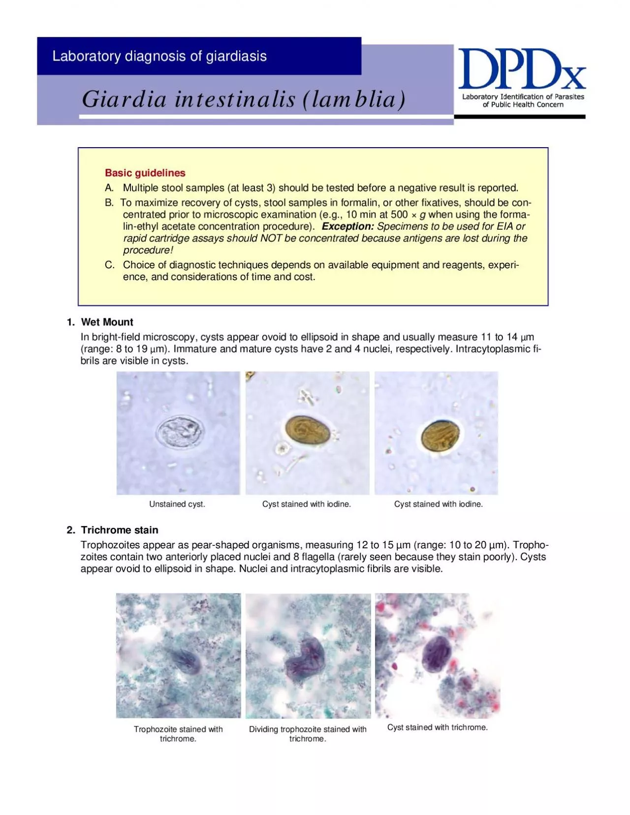
Unstained cyst Cyst stained with iodine Cyst stained with iodine 2 Trichrome stain Trophozoites appear as pearshaped organisms measuring 12 to 15 µm range 10 to 20 µm Trophozoites conta ID: 958683
Download Pdf The PPT/PDF document "Basic guidelines A Multiple stool sample..." is the property of its rightful owner. Permission is granted to download and print the materials on this web site for personal, non-commercial use only, and to display it on your personal computer provided you do not modify the materials and that you retain all copyright notices contained in the materials. By downloading content from our website, you accept the terms of this agreement.
Basic guidelines A. Multiple stool samples (at least 3) should be tested before a negative result is reported. B. To maximize recovery of cysts, stool samples in formalin, or other fixatives, should be concentrated prior to microscopic examination (e.g., 10 min at 500 × when using the formalin-ethyl acetate concentration procedure). Exception: Specimens to be used for EIA or rapid cartridge assays should NOT be concentrated because antigens are lost during the procedure! C. Choice of diagnostic techniques depends on available equipment and reagents, experience, and considerations of time and cost. 1. Wet Mount In bright-field microscopy, cysts appear ovoid to ellipsoid in shape and usually measure 11 to 14 (range: 8 to 19 m). Immature and mature cysts have 2 and 4 nuclei, respectively. Intracytoplasmic fibrils are visible in cysts. Unstained cyst. Cyst stained with iodine. Cyst stained with iodine. 2. Trichrome stain Trophozoites appear as pear-shaped organisms, measuring 12 to 15 µm (range: 10 to 20 µm). Trophozoites contain two anteriorly placed nuclei and 8 flagella (rarely seen because they stain poorly). Cysts appear ovoid to ellipsoid in shape. Nuclei and intracytoplasmic fibrils are visible. Trophozoite stained with Dividing trophozoite stained with Cyst stained with trichrome. 4. Enzyme immunoassay (EIA) The EIA does not rely on microscopy and is useful for screening large numbers of specimens. Borderline po
sitives and questionable negatives obtained with this technique should be further confirmed by DFA. Antigens of are detected in the feces using this method; therefore, specimens should not be concentrated prior to testing. However, special equipment (microplate reader) and commercially available test kits are required. Rapid immunochromatographic cartridge assays The rapid cartridge assays may be used with preserved specimens and are quick and easy to perform. Antigens of are detected in the feces using this method; therefore, specimens should not be concentrated prior to testing. Borderline positives and questionable negatives obtained with this technique should be further confirmed by DFA. No special equipment is needed. 3. Direct fluorescent antibody (DFA) assay This technique offers the highest combination of sensitivity and specificity and is considered the gold standard by many laboratories. For commercial DFA kits, it is recommended that a concentrated stool specimen be used to intion of low numbers of cysts. However, special equipment (fluorescence microscope) and commercially available test kits are required and it does not provide a permanently stained slide that can be archived. labeled with fluorescent antibodies. The quality of reagents in commercially available kits may be variable or deteriorate under storage condi-tions; for that reason external controls are necessary to determine whether the kit is properly performing.