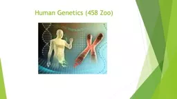
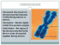
In humans each cell normally contains 23 pairs of chromosomes for a total of 46 Twentytwo 22 of these pairs called autosomes look the same in both males and females The 23rd ID: 912083
Download Presentation The PPT/PDF document "Dr. Rana S.Jawad Human chromosome Nom..." is the property of its rightful owner. Permission is granted to download and print the materials on this web site for personal, non-commercial use only, and to display it on your personal computer provided you do not modify the materials and that you retain all copyright notices contained in the materials. By downloading content from our website, you accept the terms of this agreement.
Slide1
Dr. Rana S.Jawad
Human chromosome Nomenclature
Slide2In humans, each cell normally contains 23 pairs of chromosomes, for a total of 46. Twenty-two (22) of these pairs, called autosomes, look the same in both males and females. The 23rd pair, the
sex chromosomes
, differ between males and females
.
Females have two copies of the X chromosome, while males have one X and one Y chromosome.
Slide3A chromosome may be: metacentric, with its centromere in the middle; submetacentric, with the
centromere
closer
to one end of the
chromosome
acrocentric
, in which
the
centromere
is near one end of the chromosome and the short
arm is essentially comprised of repetitive DNA that constitutes the satellites and
nucleolar
organizing regions
Slide4Chromosomes 1 and 3 are examples of metacentric chromosomes,chromosomes 4 and 5 are large submetacentric chromosomes,and chromosomes
13–15
are considered medium sized
acrocentric
chromosomes
Slide5Slide6Slide7Slide8Each chromosome has a constriction point called the centromere, which divides the chromosome into two sections, or “arms.” The short arm of the chromosome is labeled the “p arm.”
The
long
arm of the chromosome is labeled the “
q arm
.”
Slide9Slide10The location of the centromere on each chromosome gives the chromosome its characteristic shape, and can be used to help describe the location of specific genes.
Slide11Each chromosome arm is divided into regions, or cytogenetic bands, that can be seen using a microscope and special stains. The cytogenetic bands are labeled p1, p2, p3, q1, q2, q3, etc., counting from the centromere out toward the telomeres. At higher resolutions, sub-bands can be seen within the bands. The sub-bands are also numbered from the centromere out toward the telomere.
Slide12Slide13For example, the cytogenetic map location of the CFTR gene is 7q31.2, which indicates it is on chromosome 7, q arm, band 3, sub-band 1, and sub-sub-band 2.The ends of the chromosomes are labeled ptel and qtel. For example, the notation 7qtel refers to the end of the long arm of chromosome 7.
Slide14The commonly used G-, Q-, and R-bandingtechniques show bands distributed along the entire chromosome, whereas the C-, T-, or NOR-banding techniques areused to identify speci fi c chromosome structures that areheritable features
Slide15A band isdefined as a part of the chromosome that is clearly distinguishable from its adjacent segments based on its stainingproperties.As a general rule, a chromosome band contains ~5–10
mega bases
(Mb) of DNA
Slide16Giemsa or G-banding is the most common banding method employed in North American cytogenetics laboratories.Facilitate the identification of structural abnormalities
G-dark
(positive) bands are AT rich, gene poor, and late replicating.
The
early replicating
G-light
(negative) bands are
GC rich, gene rich, and late replicating
Slide17C-banding is particularly useful whenidentifying the morphologically variable heterochromatinregions of the Y chromosome and chromosomes 1, 9, and 16.Reverse
or R-banding
are useful for analyzing deletion or translocation that involve the telomeres of chromosome