/
Anatomy of the Digestive System Organs of the Alimenta Anatomy of the Digestive System Organs of the Alimenta

Anatomy of the Digestive System Organs of the Alimenta - PDF document

trish-goza
trish-goza . @trish-goza
Follow
427 views
Uploaded On 2015-04-25

Anatomy of the Digestive System Organs of the Alimenta - PPT Presentation

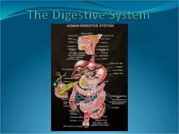
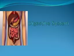
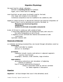
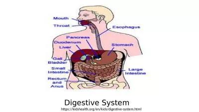
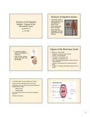
412423 Divisions of Digestive System 1 Alimentary Canal or Gastrointestinal Tract GIdigests and absorbs food coiled hollow tube with 2 openings mouth pharynx esophagus stomach small intestine large intestine 2 Accessory Organs assist in breakdown ID: 55248

412423 Divisions Digestive

Share:

Link:

Embed:

Download Presentation from below link

Download Pdf The PPT/PDF document "Anatomy of the Digestive System Organs o..." is the property of its rightful owner. Permission is granted to download and print the materials on this web site for personal, non-commercial use only, and to display it on your personal computer provided you do not modify the materials and that you retain all copyright notices contained in the materials. By downloading content from our website, you accept the terms of this agreement.


Presentation Transcript

1 Anatomy of the Digestive System: Organs of the Alimentary CanalCHAPTER 14p. 412-423 Divisions of Digestive System1. Alimentary Canal or Gastrointestinal Tract (GI)-digests and absorbs food•coiled hollow tube with 2 openings•mouth, pharynx, esophagus, stomach, small intestine, large intestine 2. Accessory Organs -assist in breakdown of molecules•teeth, tongue, salivary glands, pancreas, liver, gall bladder Organs of the Alimentary Canal1. Mouth or oral cavity–lips (labia) -protect oral cavity opening–cheeks -forms lateral walls–hard palate -forms anterior roof–soft palate -forms posterior roof–uvula -fleshy fingerlike projection; closes nasal cavity when swallowing–vestibule -area between lips & cheeks and teeth & gums–Frenulum–membrane that attaches tongue to floor of mouth -oral cavity proper: the area contained by the teeth-tongue: muscle occupying the floor of the mouthThe posterior end of the oral cavity are paired masses of lymphatic tissue (tonsils)palatine tonsilslinguinaltonsilsFood is mixed with saliva in the mouth and breakdown starts hereChewing = mastication 2 Subdivisions:–oropharynx-posterior to oral cavity–nasopharynx-part of respiratory passageway–laryngopharynx-continues to esophagus; larynx -voice boxWalls are made up of two alternating muscular layers allowing for peristalsis (propulsion of food) 2. Pharynx: common throat 3. Esophagus: gulletRuns from pharynx through the diaphragm to the stomach•25 cm long•smooth muscle•propels food to stomach through peristalsis http://www.westga.edu/~lkral/peristalsis/ Tissue Layers of the Alimentary CanalFrom esophagus to large intestine the walls contain the same 4 layers (tunics):1.Mucosa (innermost): epithelium, CT, thin muscle layer2.Submucosa: soft CT, has BV, nerves and lymph3.Muscularisexterna: muscle (circular/longitudinal)4.Serosa: layer of serous producing cells (visceral peritoneum). Held to parietal peritoneum by the mesentary Innervationof the Alimentary Wall•Submucosalnerve plexus•Myentericnerve plexus•Subserousnerve plexus–These help regulate the mobility and secretoryactivity of GI organs 4. StomachA. Structure–Cardiac –surrounds junction between esophagus & stomach Fundus–lateral to cardiac region–Body –midportion; lined with folded walls called rugaethat disappear when stomach is full–Pylorus –terminal portion 3 B. Valves or sphincters–Cardioesophageal(cardiac) –leads food into stomach–Pyloric –leads partial digested food (chyme) from body into small intestine–Curvatures:•Greater –outer (lateral) curve•Lesser –inner (medial) curve C. Mechanical Digestion3 muscle layers•segmentation of food–1st–longitudinal–2nd–circular–3rd–oblique D. Chemical digestionWalls lined with gastric pits that lead to the gastric gland which contains 3 types of cells1. Mucus neck cells –secrete alkaline sticky mucus to protect stomach walls2. Parietal cells –produces HClwhich activates enzyme production3. Chief cells –produce protein digestive enzymes called pepsinogen(inactive form of pepsin) Sequence of events in chemical digestion:1. food enters stomach2. Parietal cells secrete HClwhich lowers pH 3. low pH triggers chief cells to produce pepsinogen4. pepsinogenconverted to its active form of pepsin 4 5. Small intestineA. Structure•Extends from the pyloric sphincter to the ileocecalvalve•2 m long•Mesentary–web like membrane that coils small intestine & holds it intact 3 subdivisions:–Duodenum –curves around the pancreas; receives chymefrom stomach, enzymes from pancreas & bile from liver–Jejunum –middle portion; bulk of digestion & absorption–Ileum –terminal portion B. Chemical Digestion in sm. intestine1. pyloric sphincter controls amount of food entering from stomach2. pancreas produces enzymes that are secreted to small intestines through pancreatic duct3. Bile formed in liver is secreted through bile duct4. Pancreatic & bile ducts join to form hepatopancreaticampulla5. together enzymes,bileand bicarbonate (neutralize acids) enter duodenum through duodenal papilla C. Absorption in the sm. intestine•Nearly all absorption occurs here•Peyer’sPatches –collection of lymphatic tissue that increases toward end of small intestine that prevents absorption of bacteria •Surface area increased by 3 structures:–Circular folds (plicaecircularis) –deep folds of inner walls 5 –Villi–fingerlike projections that contain bv& lymphatic duct called the lacteal–Microvilli–“brush border”; projections of the cell membrane that give a fuzzy appearance 6. Large Intestine•Extends from ileocecalvalve to anus•Absorption of water & elimination of waste•No villi•Lined with goblet cells that produce mucus for lubrication•Outer walls puckered with haustrawhich aids in contraction •Cecum–1stportion ; ileocecalvalve that allows indigestible material from sm. Intestine•Appendix –sac extending from cecum •Ascending colon –travels up right side of abdominal cavity ; turns at the hepatic flexure•Transverse colon –travels across the abdomen; turns at the splenicflexture •Descending colon –travels down left side of abdomen•Sigmoid colon –S shaped•Rectum –storage chamber•Anal sphincter –external & internal; voluntary & involuntary muscles; opens during defecation