/
Department of Surgery, Buddhist Tzu Chi General Hospital, Hualien. Sch Department of Surgery, Buddhist Tzu Chi General Hospital, Hualien. Sch

Department of Surgery, Buddhist Tzu Chi General Hospital, Hualien. Sch - PDF document

trish-goza
trish-goza . @trish-goza
Follow
451 views
Uploaded On 2016-05-02

Department of Surgery, Buddhist Tzu Chi General Hospital, Hualien. Sch - PPT Presentation

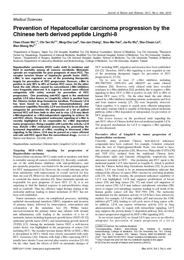
Institute of Medical Department of Laboratory Medicine and Biotechnology College of Medicine Tzu Chi University Hualien Taiwan Hepatocellular carcinoma HCC ranks sixth in incidence and third ID: 302207

& Institute Medical Department

Share:

Link:

Embed:

Download Presentation from below link

Download Pdf The PPT/PDF document "Department of Surgery, Buddhist Tzu Chi ..." is the property of its rightful owner. Permission is granted to download and print the materials on this web site for personal, non-commercial use only, and to display it on your personal computer provided you do not modify the materials and that you retain all copyright notices contained in the materials. By downloading content from our website, you accept the terms of this agreement.


Presentation Transcript

Department of Surgery, Buddhist Tzu Chi General Hospital, Hualien. School of Medicine, Tzu Chi University. & Institute of Medical Department of Laboratory Medicine and Biotechnology, College of Medicine, Tzu Chi University, Hualien, Taiwan Hepatocellular carcinoma (HCC) ranks sixth in incidence and third in mortality among all cancers worldwide. Metastatic spreads are responsible for poor prognosis of most HCC. The receptor tyrosine kinase of hepatocyte growth factor (HGF), c-Met, is now regarded as one of the promising therapeutic targets for prevention of HCC progression. However, c-Met is positive in only 20% to 48% of human HCC cases. On the other hand, the side effects caused by conventional c-Met inhibitors were frequently observed. It is urgent to search more efficient P atients with high c-Met expressing HCCs usually have shorter 5-year survival after curative resection [15-19]. On the other hand, the effects of HGF on metastatic changes of HCC including EMT, migration and invasion have been established [20-22]. Therefore,c-Met signaling is now regarded as one of the promising therapeutic targets for prevention of HCC progression [2, 23-25]. Up to now, at least 17 c-Met inhibitors including GEN-203, and ARQ197 are under clinical evaluation [26]. However, early clinical trials have revealed resistance to c-Met inhibition [24], probably due to negative c-Met signaling in these HCC (c-Met is positive in only 20% to 48% of In our recent report [44], we found LZ-8 may serve as an effective antagonist for prevention of tumor progression of a lot of _________ Conflict of interest: No conflicts declared. 1 Corresponding Author: Wen-Sheng Wu. Institute of medical biotechnology, College of Medicine, Tzu Chi University No. 701, Chung Yang Rd, Sec 3, Hualien 970, Taiwan. Phone: 8867-03-8565301 ext 2327Fax: 8867-03-8571917. Email: wuwstcu1234@yahoo.com.tw. % Chao-Chuan Wu and Chi-Tan Hu contribute equally to the paper © 2015 by the Journal of Nature and Science (JNSCI). Journal of Nature and Science, Vol.1, No.3, e50, 2015 patient derived HCC cell lines. These cell lines have been characterized for the status of c-Met signaling. As demonstrated in the report, cell migration of both c-Met positive and c-Met negative HCC, HCC372 and HCC329, respectively, can be suppressed by LZ-8. In the animal experiment, LZ-8 suppressed intrahepatic metastasis of HCC329 [44] and another c-Met positive HCC, HCC340 (unpublished result), in SCID mice. Fig.1 LZ-8 suppress tumor progression of HCC via blockade of c-Met dependent or in dependent signaling.LZ-8 may block c-Met-dependent or EGFR-dependent signaling for suppressing the progression of c-Met- positive and negative HCCs. The anti-HCC effects of LZ-8 on HCC progression were found to be closely associated with the blockade of critical signal transduction [44]. For the c-Met positive HCC372, LZ-8 suppressed both the expression of c-Met and phosphorylation of c-Met (at Tyr1234), which accompanied the decrease of the downstream ERK signaling and the inhibition of cell migration. On the other hand, to identify the signaling pathway mediating cell migration of the c-Met-negative HCC329, receptor array was used to screen the activities of RTKs that are deregulated in this cell. Among the 49 phosphorylated RTKs (p-RTKs) examined, p-EGFR was found to be the most highly upregulated in HCC329. Importantly, treatment of HCC329 with LZ-8 for 16-24 h specifically decreased p-EGFR by 70%. Consistently, the EGFR inhibitor AG1748 suppress cell migration of HCC329 as effective as LZ-8. In addition, LZ-8 may significantly decreased cell survival of both HCC372 and HCC 329. Taken together, LZ-8 may block c-Met-dependent or c-Met-independent signaling for anti-HCC progression (summarized in Fig.1). Proposed mechanisms for LZ-8 to block c-Met signaling: implication of the c-Met endocytosis The underlying mechanisms for LZ-8 to block c-Met signaling are being investigated. Since LZ-8 may decrease both expression and phosphorylation of c-Met, it is probable that LZ-8 can affect the gene transcription of c-Met. However, this was excluded by our recent experiments demonstrating that LZ-8 didn’t decrease the c-Met mRNA level in HCC372 (unpublished result). Therefore, LZ-8 may influence the stability of c-Met on the posttranslational level. Recently, the HGF-triggered c-Met endosomal signaling [for review 45] is highlighted in tumor progression [46]. Increased endocytosis /recycling of c-Met coupled with decreased lysosomal degradation of c-Met were responsible for enhanced activation of the GTPase Rac1, leading to increased cell migration, invasion and metastasis of breast cancer cells [46]. Therefore, modifications in the balance between degradation and recycling of c-Met may be a promising strategy for blocking c-Met signaling. We have found LZ-8 and Con A , a mannose specific Lectin, may prevent HGF-induced tumor progression of the HepG2 via blockade of c-Met endosomal signaling (unpublished result). Interestingly, the major biological activities of LZ-8 resemble the mitogenic Lectin which is capable of inducing cellular aggregate formation [47, 48]. Therefore, it is tempting to speculate that LZ-8 may disturb c-Met endosomal processing in HCC372 by promoting the lysosomal degradation of c-Met and decreased c-Met signaling. Conclusion and perspective LZ-8 acquires broad capabilities of suppressing the critical signal pathways, either c-Met- dependent or independent, mediating the progression of diverse HCCs. Being an active component of the Chinese herbal Lingzhi, the safety concern of LZ-8 has been approved. Whether LZ-8 can be a more efficient anti-HCC agent than the conventional drugs is worthy of further clinical investigation. 1. Ferlay J, Shin HR, Bray F, Forman D, Mathers C, et al. (2008) Estimates of worldwide burden of cancer in 2008: GLOBOCAN 2008. Int J Cancer 127: 2893-2917. 2. Llovet JM, Ricci S, Mazzaferro V, Hilgard P, Gane E, et al. (2008) Sorafenib in advanced hepatocellular carcinoma. N Engl J Med 359: 378-390. 3. Cheng AL, Kang YK, Chen Z, Tsao CJ, Qin S, et al. (2009) Efficacy and safety of sorafenib in patients in the Asia-Pacific region with advanced hepatocellular carcinoma: a phase III randomised, double-blind, placebo-controlled trial. Lancet Oncol 10: 25-34. 4. Llovet JM, Ducreux M, Lencioni R, Di Bisceglie AM, Galle PR, et al. (2012) EASL-EORTC clinical practice guidelines: management of hepatocellular carcinoma. J Hepatol 56: 908-943. 5. Zhu AX (2012) Molecularly targeted therapy for advanced hepatocellular carcinoma in 2012: current status and future perspectives. Semin Oncol 39: 493-502. 6. Zhai B, Sun XY (2013) Mechanisms of resistance to sorafenib and the corresponding strategies in hepatocellular carcinoma. World J Hepatol 5: 345-352. 7. Bosch FX, Ribes J, Diaz M, Cleries R (2004) Primary liver cancer: worldwide incidence and trends. Gastroenterology 127: S5-S16. 8. Tang ZY, Ye SL, Liu YK, Qin LX, Sun HC, et al. (2004) A decade's studies on metastasis of hepatocellular carcinoma. J Cancer Res Clin Oncol 130: 187-196. 9. Gupta GP, Massague J (2006) Cancer metastasis: building a framework. Cell 127: 679-695. 10. Liotta LA, Kohn EC (2001) The microenvironment of the tumour-host interface. Nature 411: 375-379. 11. Christofori G (2006) New signals from the invasive front. Nature 441: 444-450. 12. Gao CF, Vande Woude GF (2005) HGF/SF-Met signaling in tumor progression. Cell Res 15: 49-51. 13. Buijs JT, Stayrook KR, Guise TA (2011) TGF-beta in the Bone Microenvironment: Role in Breast Cancer Metastases. Cancer Microenviron 4: 261-281. 14. Gherardi E, Birchmeier W, Birchmeier C, Vande Woude G (2012) Targeting MET in cancer: rationale and progress. Nat Rev Cancer 12: 89-103. 15. Ueki T, Fujimoto J, Suzuki T, Yamamoto H, Okamoto E (1997) Expression of hepatocyte growth factor and its receptor c-met proto-oncogene in hepatocellular carcinoma. Hepatology 25: 862-866. 16. Osada S, Kanematsu M, Imai H, Goshima S (2008) Clinical significance of serum HGF and c-Met expression in tumor tissue for evaluation of properties and treatment of hepatocellular carcinoma. Hepatogastroenterology 55: 544-549. 17. Ke AW, Shi GM, Zhou J, Wu FZ, Ding ZB, et al. (2009) Role of overexpression of CD151 and/or c-Met in predicting prognosis of hepatocellular carcinoma. Hepatology 49: 491-503. 18. Wang ZL, Liang P, Dong BW, Yu XL, Yu de J (2008) Prognostic factors and recurrence of small hepatocellular carcinoma after hepatic resection or microwave ablation: a retrospective study. J Gastrointest Surg 12: 327-337. 19. Kaposi-Novak P (2009) [Comparative genomic classification of human hepatocellular carcinoma]. Magy Onkol 53: 61-67. 20. Ogunwobi OO, Liu C (2011) Hepatocyte growth factor upregulation promotes carcinogenesis and epithelial-mesenchymal transition in hepatocellular carcinoma via Akt and COX-2 pathways. Clin Exp Metastasis 28: 721-731. 21. Jiang Y, Xu W, Lu J, He F, Yang X (2001) Invasiveness of hepatocellular carcinoma cell lines: contribution of hepatocyte growth factor, c-met, and transcription factor Ets-1. Biochem Biophys Res Commun 286: 1123-1130. 22. Ogunwobi OO, Wang T, Zhang L, Liu C (2012) Cyclooxygenase-2 and Akt mediate multiple growth-factor-induced epithelial-mesenchymal ISSN 2377-2700 | www.jnsci.org/content/50 2 Mar 2015, Vol.1, No.3, e50