/
Pastillation Technology Based Design & Development of Oral Modified Release Multiparticulate Pastillation Technology Based Design & Development of Oral Modified Release Multiparticulate

Pastillation Technology Based Design & Development of Oral Modified Release Multiparticulate - PowerPoint Presentation

trish-goza
trish-goza . @trish-goza
Follow
352 views
Uploaded On 2018-11-12

Pastillation Technology Based Design & Development of Oral Modified Release Multiparticulate - PPT Presentation

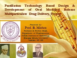
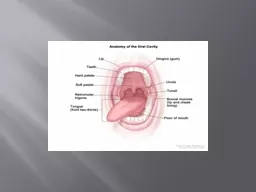
Presented by Prof B Mishra Professor amp Former Head Department of Pharmaceutics Indian Institute of Technology Banaras Hindu University Varanasi 221 005 Components of Research Background ID: 728541

drug pastilles peg release pastilles drug release peg study enteric contact coated gamma angle pharmacokinetic based amp doxofylline formulation

Share:

Link:

Embed:

Download Presentation from below link

Download Presentation The PPT/PDF document "Pastillation Technology Based Design &am..." is the property of its rightful owner. Permission is granted to download and print the materials on this web site for personal, non-commercial use only, and to display it on your personal computer provided you do not modify the materials and that you retain all copyright notices contained in the materials. By downloading content from our website, you accept the terms of this agreement.


Presentation Transcript

Slide1

Pastillation Technology Based Design & Development of Oral Modified Release Multiparticulate Drug Delivery System

Presented by:Prof. B. MishraProfessor & Former Head,Department of Pharmaceutics,Indian Institute of Technology(Banaras Hindu University)Varanasi- 221 005Slide2

Components of Research

Background Lipid based multiparticulate drug delivery systemDoxofylline as potential anti-asthmatic agentExperimental workDevelopment of Immediate and controlled release pastillesDevelopment of pulsatile release pastillesIn vivo study2Slide3

Lipid based multiparticulate system

Nanoparticles

Granules

Microparticles

Pellets/Beads

3Slide4

Pastillation

Pastillation is a widely used technique in chemical, petrochemical and agrochemical industriesIt is used for the solidification of dusty hazardous powders of chemicals into pastilles (hemispherical solidified units of uniform size) which eases their handling. In this process, the drops of chemical substances in molten state are deposited on a cooled stainless steel surface for rapid solidification to generate pastilles of uniform dimensions.Depending on the size of the drops and the physical properties of the melt, the drops flatten to a certain extent. The solidified droplet, therefore, has the typical pastille-like shape. The production process can be easily carried out at large scale with the help of specially designed equipments called ‘Rotoformer’.4Slide5

Pastillation

equipment

5Slide6

Suitable for hygroscopic drug as the processing of the ingredients is absolutely free from use of water.

Environment friendly as involves no use of organic solvents.Pastilles are stable and highly uniform in shape.Pastilles can be produced in a wide range of sizes with diameters ranging from 1 to 30mm.Pastilles have higher bulk density and better packing properties than powders and therefore, ideal for handling, filling and packaging.The conversion of a bulk molten liquid directly into individual solidified units provides dust free working environment. Single step process involving one equipment (melting of lipid, mixing of drug and excipients followed by solidification).Reduced energy costs due to absence of number of processes of formulation.Ease of packaging as the pastilles of smaller dimension can be capsule filled while the larger ones can either be strip packed or filled directly in sachets/bottles.Why pastillation in DDS?

6Slide7

Components of Research

Background Lipid based multiparticulate drug delivery systemDoxofylline as potential anti-asthmatic agent

Experimental work

Development of immediate and controlled release pastilles

Development of pulsatile release pastilles

In vivo study

7Slide8

Chemical Name:

2-(7-theophyllinemethyl)-1,3 dioxolaneMolecular Formula: C11H14N4O4Formula weight: 266Melting Point: 144-145.5°CSolubility: Freely Soluble in chloroform and Dichloromethane soluble in acetone, sparingly soluble in water and ethyl acetateStorage: Store in cool, dark and dry place.

Doxofylline

Ist

Marketed product in India

Product:

DOXOBID (

Doxofylline

tablets 400mg)

Company:

Dr. Reddy’s Labs.

Label Claim:

Each uncoated tablet contains:

Doxofylline

400mg

Indication :

used as Bronchodilator in Asthma

& chronic obstructive pulmonary

disease(COPD)

Dose :

400mg IR is given 2-3 times daily

8Slide9

Absorption

Absorption Bioavailability : 62.6%Therapeutic Drug ConcentrationChronic Bronchitis : 8-20 µg/mlTime to peak concentration (tmax) : 1.19 hours

The steady state is reached within 6hrs : 9.43 µg/ml

Area under the curve, AUC : 69.5 hr×µg

/mlDistribution

Protein Binding :

48%

Distribution Half Life :

0.19 hr

Volume of distribution :

1 L/kg

Metabolism

Metabolism sites & kinetics: Liver >90%

Metabolites:

Hydroxyethyltheophylline

(inactive)

Excretion

Kidney: Less than 4% of an administered dose of

doxofylline

is excreted unchanged in the urine

Total Body Clearance: 444-806µg/ml

Elimination Half life: Parent compound – 7 to 10hrs

Pharmacokinetic profile

9Slide10

Objectives of the research work

PART ITo design a immediate and controlled release formulation of doxofylline using pastillation technology

PART II

To design a pulsatile release formulation of doxofylline using pastillation technology

PART III

To evaluate

pharmacokinetic behavior of

the

developed formulations in animal model

To explore pastillation to design a platform technology for the development of novel and unique modified release drug delivery system

10Slide11

In-house laboratory scale device for pastillation

TransformerGlass syringeNeedlePastillesCold plate

Ice tray

Shaft

Ceramic insulation

Heating coil

11Slide12

Operating parameters

Sl No.FactorsLow (-)High (+)

1.

Needle dimensions (X1)

16G

20G

2.

Dropping height (X

2

)

1 cm

3 cm

3.

Temperature of plate (X

3

)

4 °C

25 °C

Factorial design using MINITAB

®

12Slide13

Evaluating parameter

Contact angle measurementMethod of analysis (Photographic method)The photographs of the pastilles were taken from the horizontal side at their contact with the plate and the snaps were then proportionally magnified and processed using Adobe Photoshop® software. The angle of contact was determined manually and confirmed mathematically using the following equation:θ = 2tan−12h/dWhere h is the height of the drop from the plate and d is the diameter of the drop. Both of these dimensions can be measured from the photograph for calculating the contact angle.

13Slide14

Contact angle of pastilles

14Slide15

Formulation batches (for optimization)

Sl. No.BatchesX1X2

X3Avg. Contact angle (Y

1)

1.

A1

16G

1 cm

4 °C

121°

2.

A2

16G

1 cm

25 °C

110°

3.

A3

16G

3 cm

4 °C

100°

4.

A4

16G

3 cm

25 °C

80°

5.

A5

20G

1 cm

4 °C

120°

6.

A6

20G

1 cm

25 °C

115°

7.

A7

20G

3 cm

4 °C

95°

8.

A8

20G

3 cm

25 °C

85°

15Slide16

Effect of needle size and dropping height on contact angle

(A) Response Surface 3D plot (B) Contour plot16Slide17

Effect of temperature of plate and dropping height on contact angle

(A) Response Surface 3D plot (B) Contour plot17Slide18

Effect of needle size and temperature of plate on contact angle

(A) Response Surface 3D plot (B) Contour plot18Slide19

Flow property of pastilles based on their contact angle

Flow propertyContact AnglePoor60-85°Fair

85-105°Good

105-125°

19Slide20

Optimized parameters for highest achievable (desirable) contact

angle Sl No.Optimized parameters1.

Needle dimensions (X1

)

20 G

2.

Dropping height (X

2

)

1

cm

3.

Temperature of plate (X

3

)

4 °C

20Slide21

Formulation chart

Compositiong/batch

B-1

B-2

B-3

B-4

B-5

B-6

B-7

B-8

B-9

B-10

DOX

0.5

0.5

0.5

0.5

0.5

0.3

0.4

0.5

0.5

0.5

Stearic acid

2.0

2.0

2.0

2.0

2.0

2.0

1.7

1.7

1.7

Benefat

-

-

-

-

-

-

0.3

1.5

0.75

PEG 4000

2.0

-

0.3

-

-

0.3

0.3

-

-

-

PEG 6000

-

-

0.3

-

-

-

-

-

-

PEG 400

-

-

-

0.3

-

-

-

-

-

Colloidal

silicon

dioxide

75

Drug content Uniformity (%)

100.56 ± 0.93

98.89 ± 1.23

21Slide22

Evaluation

Drug content uniformity (20 ml water added to pastilles eq. to 10 mg drug and heated at 75°, sonicated, cooled and volume made upto 25 ml. 5 ml filtered and measured spectrophotometrically)Drug release study (USP Appt. II, 500 ml of 0.1 N HCl, 50 rpm, 37±0.5°C for 2 h followed by pH 6.8 phosphate buffer for next 22 h)Scanning electron microscopy (The morphological structure of the prepared pastilles was observed using scanning electron microscope (FEI Quantum 200E Instrument)Stability studies (pastilles packed in 30 ml HDPE bottles kept in 40°C/75%RH for 3 months storage conditions in stability chamber

(Narang Scientific Works Pvt. Ltd., New Delhi, India))

22Slide23

Analytical method

UV–VIS spectrophotometry (Hitachi U-1800)Standard curves of doxofylline were prepared in water, 0.1 N HCl (pH 1.2) and phosphate buffer solutions (pH 6.8) in the concentration range of 5–35 μg/ml.A UV visible spectrum of doxofylline showed a characteristic peak at 273 nm in all the solutions.The standard curve was plotted as drug concentration (μg/ ml) vs. absorbance plot. Curve fitting was done by linear regression analysis using Microsoft Excel program 23Slide24

Standard Curves of Doxofylline

24Slide25

Effect of pore former & type of buffer media on drug release

25Slide26

Effect of drug load on drug release

26Slide27

Effect of benefat (lipid pore former) concentration on drug release behavior

R2=0.98327Slide28

Scanning electron microscopy

Batch B1 (B) Batch B1 at higher magnification (C) Batch B10 (D) Batch B10 at higher magnification28Slide29

Drug release profiles of initial and 3 months stored samples

Drug content uniformity1M-98.66%2M- 97.12%3M-96.46%Stability study at 40°C/75%RH indicate stable formulation with no change in physical appearance, drug release and drug content29Slide30

Components

of ResearchBackground Drug delivery systemTherapeutic applicationExperimental workDevelopment of immediate, controlled and pulsatile release pastillesIn vivo animal study30Slide31

Formulation chart

Ingredients P-IP-IIP-IIIP-IV

P-VP-VI

DOX (mg)

500

500

500

500

500

500

PEG 4000 (mg)

2000

2000

2000

2000

2000

2000

Colloidal silicon dioxide (mg)

-

75

150

75

75

75

Enteric coat

1

(5 g

Eudragit

L100 55 and 0.25 g

triethyl

citrate (plasticizer)

& 2% talc in 100ml methanol

-

-

-

10 ± 5 %*

-

-

Enteric coat

2

(5 g

Eudragit

L100 55 and 0.5 g

triethyl

citrate (plasticizer)

& 2% talc in 100ml methanol

10 ± 5 %*

10 ± 5%*

Floating

layer

(1g HPMC K15M, 0.1 g

triethyl

citrate (plasticizer) in 100 ml IPA DCM

mixture (60:40 v/v). NaHCO

3

crushed and passed through #100 mesh & 2% talc was dispersed in the above solution

-

-

-

-

-

20 ± 5%#

* Amount of enteric coat applied was calculated in terms of percentage weight gain with respect to the weight of uncoated pastilles

# Amount of floating coat applied was calculated in terms of percentage weight gain with respect to the weight of enteric coated pastilles

31Slide32

Evaluation

AssayDrug content uniformityDrug release study (USP Appt. II, 500 ml of 0.1 N HCl, 50 rpm, 37±0.5°C for 2 h followed by 2 h followed by pH 6.8 phosphate buffer for next 2 h)Scanning electron microscopyStability studies (40°C/75%RH for 3 months)32Slide33

Contact angle

Pastilles with A) desired contact angle (above 85°), B) with contact angle ≤ 45°, C) with contact angle above 70°33Slide34

Explanation for formation offlat pastilles

Friability B-I :0.596%B-II :0.104%Contact angle improvement of PEG pastilles at large scale34Slide35

Assay and drug content uniformity

 Assay (%)Drug content uniformityP-I100.12 ± 1.11

100.19 ± 2.13

P-II100.09 ± 1.91

99.98 ± 2.21

P-III

99.61 ± 2.17

98.89 ± 1.81

P-IV

98.12 ± 1.21

99.01 ± 0.91

P-V

98.08 ± 2.19

98.21 ± 1.27

P-VI

99.06 ± 1.98

99.12 ± 2.12

35Slide36

Drug release profile

36Slide37

Scanning electron microscopy

Surface morphology of coated pastilles using SEM A) Batch P-IV, B) Batch P-V, C) Batch P-VI37Slide38

Coated PEG pastilles floating in dissolution medium

38Slide39

Dissolution of initial and three months stored samples

39Slide40

Conclusion

A novel technology ‘pastillation’ was successfully employed for the development of immediate release pastilles. This dosage form after coating with enteric and floating coat were also found to be effective to achieve the required delay in drug release for treatment of nocturnal asthma. The prepared formulations showed desired drug release profile with an initial lag phase in the in vitro drug release study. The in vivo pharmacokinetic study would be helpful in further evaluating the potential of this formulation in the chronotherapeutic treatment of nocturnal asthma.40Slide41

Components of Research

Background Drug delivery systemTherapeutic applicationExperimental workDevelopment of controlled release pastillesDevelopment of pulsatile release pastillesIn vivo animal study41Slide42

In-vivo animal study

Animal study protocol were approved by the Animal Ethical Committee of Banaras Hindu University.(No. 2010-11/153) Male albino rats of 250 ± 20 g12 h fasting prior to dosing5.70 mg drug/kg body weight administered orallyPastilles were administered with 5.0 ml of 1.0% aqueous polyvinyl alcohol solutionBlood (0.5 mL) was collected via retro-orbital vein0, 0.25, 0.50, 1, 1.5, 2, 3, 4, 6, 8, 12 and 24 h

Blood samples were allowed to clotThey were centrifuged for 10 min at 3000 rpm The serum obtained was transferred to clean tube for storage

at −20 °C until analysis

42Slide43

Study design of pharmacokinetic studies

PEG Pastilles (IR)Stearic acid pastilles (CR)For controlled release pastilles

For pulsatile release pastilles

PEG Pastilles (uncoated)

PEG pastilles (enteric coated with floating layer)

PEG Pastilles (enteric coated)

43Slide44

Serum drug estimation

Drug separation from serum by liquid-liquid extraction method Serum (containing drug) (500 μl) + methanol (400 μl) Supernatant (400 μl)

Residue + mobile phase (200 μl

)

Reconstituted sample (20 μl

) injected for HPLC analysis

Vortexed (8 min) &

centrifuged at 3500 rpm for 10 min

Evaporated in vacuum oven at 40°C

Reconstituted

44Slide45

Serum drug estimation

Serum drug estimation by Reverse phase HPLC methodChromatographic conditions:Column: C18 reverse-phase 250×4.6 mm 5 μm ODS2 column (Waters, Ireland)Mobile phase: 18:82 acetonitrile–12.5 mM potassium dihydrogen orthophosphate buffer (pH adjusted to 3.0 with

orthophosphoric acid) Flow rate: 1 ml/min

Injection volume: 20 μl

λ

max: 275 nm

Retention Time: 9.75 min

45Slide46

Pharmacokinetic estimation

Cmaxpeak serum concentration Tmaxtime to reach peak concentrationAUC0-tarea under the curve from time zero to last measured concentration

HVD t50% Cmax

time span during which the serum concentrations were at least 50% of the C

max

R∆

ratio between the HVD

t50% Cmax

values of the test formulation and the drug suspension

Kinetica

®

software

GraphPad Prism

®

software

46Slide47

HPLC chromatograms and Calibration curve of doxophylline extracted from serum

47Slide48

Pharmacokinetic profile of PEG and lipid based pastilles

48Slide49

Pharmacokinetic data of PEG and lipid based pastilles

Pharmacokinetic parametersPEG based pastillesLipid based pastillesCmax (

ng/ml)

31.83 ± 1.28

16.32 ± 3.69

T

max

(h)

0.75 ± 0.06

6.0 ± 1.58

AUC

last

(

ng

/ml*h)

182.56 ± 19.98

210.39 ± 59.6

HVD (h)

3.18 ± 0.21

11.43 ± 1.52

$

R

-

3.59

$

R∆ =of 1.5, 2 and >3 indicates, low, intermediate and strong sustained release effect, respectively

Data are shown as mean

+

SEM

49Slide50

Pharmacokinetic profile of uncoated and coated PEG pastilles

50Slide51

Pharmacokinetic data of uncoated and coated PEG pastilles

Pharmacokinetic parametersP-II (Uncoated pastilles)P-V (Enteric coated pastilles)

P-VI

(Enteric & floating coated pastilles)C

max (

ng/ml)

31.83 ± 1.28

30.92 ± 2.12

25.12± 2.41

T

max

(h)

0.75 ± 0.06

3.0 ± 0.27

6.0 ± 0.82

AUC

last

(

ng

/ml*h)

182.56 ± 19.98

201.47 ± 29.7

241.68 ± 42.7

HVD

t50% Cmax

(h)

3.18 ± 0.21

4.23 ± 0.15

6.70 ± 0.13

R∆

-

1.33

2.11

Data are shown as mean

+

SEM

51Slide52

Gamma scintigraphic method

Radiolabeling of PastillesTechnetium (99mTc) was chosen for radio-labeling of the pastilles because of its short half-life of 6 hrs and very less amount of electron emission TLC (Silica Gel) After 2 min After 99% reduction

100 ml 99m

TC5 mg stannous chloride

dihydrate (1 mg/ml in 10% acetic acid) of pH 7.5 (adjusted with 0.5M NaHCO3

1g pastilles + 5 ml water

5 mg stannous chloride

dihydrate

(1 mg/ml in 10% acetic acid) of pH 7.5 (adjusted with 0.5M NaHCO

3

radiolabeling

efficiency was evaluated by TLC-SG strips as stationary phase & acetone as mobile phase

52Slide53

Gamma scintigraphic method

Stability of radiolabeled pastilles pH 1.2 0.1N HCl 1g pastilles pH 6.8 buffer pH 7.2 buffer

0.2 ml filtered solution checked for radioactivity by auto gamma counter

Kept under stirring in a water bath maintained at 37° C for 6 hr

53Slide54

Study design of gamma scintigraphy

For pulsatile release pastillesMale albino rats of 250 ± 20 g, 12 h fasting prior to dosing5.70 mg drug per kg body weight administered orallyAfter light anaesthetizationserial scintigraphic examination was done at 0.5, 1, 1.5, 2, 3, 4 and 5, 6 hrs depending on type of formulation using a large field view gamma camera Images were recorded for a preset time of 1 min/view to include the 140 keV photopeak of 99m

Tc

PEG Pastilles (uncoated)

PEG pastilles (enteric coated with floating layer)

PEG Pastilles (enteric coated)

54Slide55

Gamma scintigraphic set up

A

B

C

D

55Slide56

Stability data of radiolabeled pastilles

pHP-II(Uncoated pastilles)P-V(Enteric coated pastilles)

P-VI

(Enteric & floating coated pastilles)1.2

0.13

0.21

0.16

6.8

0.25

0.32

0.27

7.2

0.36

0.34

0.31

56Slide57

Gamma scintigraphy study of uncoated PEG pastilles

Gamma Scintigraphy study of uncoated PEG pastilles on rats at time point (A) 0.5 hr and (B) 1 hr57ABSlide58

Gamma scintigraphy study of uncoated PEG pastilles

OBSERVATION: Attenuation of radioactivity within 0.5 hrs. INFERENCE: In the presence of gastric fluid, PEG matrix dissolved completely and behaved as an immediate release dosage form. 58Slide59

Gamma scintigraphy study of enteric coated PEG pastilles

Gamma Scintigraphy study of enteric coated PEG pastilles on rats at time point (A) 0.5 hr and (B) 1 hr, (C) 1.5 hr, (D) 2 hr and (E) 3 hr 59AB

C

D

ESlide60

Gamma scintigraphy study of enteric coated PEG pastilles

OBSERVATION: The pastille maintained its matrix integrity till 1.5 hrs in the gastric region. At 2 hrs the pastille was located in the jejunum area where it started to dissociate.INFERENCE: No influence of gastric fluid on the enteric coating applied on the pastilles. 60Slide61

Gamma scintigraphy study of pastilles with floating coat

AB

CD

E

F

Time point (A)1 hr, (B) 2 hr and (C) 3 hr, (D) 4 hr, (E) 5 hr and (F) 6 hr

61Slide62

Gamma scintigraphy study of pastilles with floating coat

OBSERVATION: The pastille was retained in the stomach for 2 h. In the next hour, the intact pastille migrated into the jejunum. Further, in the 4th hour the dosage form was found to reach the ileum region where it started to disperse. The image of 6th hour shows complete disintegration of the dosage form. INFERENCE: This indicates that the floating coat is not only valuable to retain the dosage form in the stomach for more than two hours but also to protect the enteric coat from alkali environment for an hour. 62Slide63

Conclusion

The pharmacokinetic and gamma scintigraphic imaging confirms the ability of the formulations to release the drug only after a desired period of time as specifically required for the treatment of noctural asthma. The results are also in agreement with the in vitro drug release study which indicates the efficiency of the coating system. The present study also confirms the ability of PEG based pastilles to act as an immediate release dosage forms. Further coating of the PEG pastilles with appropriate polymers can significantly impart functional properties to modify the release of the drug in a pre-determined fashion.

63Slide64

Major findings

Use of pastillation technology for the first time in pharmaceutical fieldNovel drug delivery system “PASTILLES” were successfully formulated as: immediate release dosage form pulsatile release dosage form sustained release dosage form64Slide65

Important references

Cheboyina, S. and Wyandt, C.M., Wax-based sustained release matrix pellets prepared by a novel freeze pelletization Technique . I. Formulation and process variables affecting pellet characteristics, Int

J Pharm

, 359, 158-166, 2008.Reitz, C. and

Kleinebudde, P., Spheronization of solid lipid

extrudates, Powder

Technol

, 189 238-244, 2009.

Van G.F.,

Rao

Y.M., Modified release composition comprising doxofylline, Patent application No.WO2009/112436A1, 2009.

Huang, H.F., Lu, Y., He, H.B., and Tang, X., Preparation and bioavailability of sustained-release doxofylline pellets in beagle dogs, Drug Dev

Ind

Pharm

, 34, 676-682, 2008.

Gils, P.S., Ray D.,

Sahoo

P.K., Controlled release of doxofylline from biopolymer based

hydrogels

, Am J Biomed

Sci

, 2, 373-383, 2010.

Gannu

, R.,

Bandari

S,.

Sudke

S.G.,

Rao

, M. and Shankar, B.P., Development and validation of a stability-indicating RP-HPLC method for analysis of doxofylline in human serum. Application of the method to a pharmacokinetic study,

Acta

Chromatographica

, 9,149-160, 2007

Sandvik

Materials Technology. Available

at:http

://

www.processsystems.sandvik.com/ Rxlist. Available at: www.rxlist.comCipla Therapeutic Index, Respiratory drugs, Zordox.

CiplaDoc

. Available at: http://www.cipladoc.com/therapeutic/admin.php?mode=prod&action=disp&id=650

Aulton

, M.E., Dyer, A.M. and Khan, K.A., The strength and compaction of

millispheres

, Drug Dev

Ind

Pharm

, 20, 3069-3104, 1994.

Breitenbach

, J., Melt extrusion: from process to drug delivery technology,

Eur

J

Pharm

Biopharm

, 54, 107-117, 2002.

65Slide66

Publications from research work

S. NoPublication Details

Impact Factor

1.

Shukla

D,

Chakraborty

S, Singh S,

Mishra

B

.

Doxofylline

: a promising

methylxanthine

derivative for the treatment of asthma and chronic obstructive pulmonary disease.

Expert

Opin

Pharmacother

.

10(14): 2343-2356,

2009.

3.205

2.

Shukla

D,

Chakraborty S, Singh S,

Mishra B

.

Lipid based oral multiparticulate formulations – Advantages, technological advances and industrial applications.

Expert

Opin

Drug

Deliv

. 8(2):207-224,

2011

.

4.896

3.

Shukla

D,

Chakraborty

S, Singh S,

Mishra

B

.

Pastillation: A novel technology for development of oral lipid based multiparticulate controlled release formulation.

Powder Technol.

209 (1-3): 65-72,

2011.

2.080

4.

Shukla D, Chakraborty S and

Mishra B.

In vitro and in vivo evaluation of multilayered pastilles for

chronotherapeutic

management of nocturnal asthma.

Expert

Opin

Drug

Deliv

9(1):9-18,

2012

4.896

5.

Shukla

D,

Chakraborty

S and

Mishra

B.

Evaluation of in vivo behavior of controlled and pulsatile release pastilles using pharmacokinetic and γ-

scintigraphic

techniques.

Expert

Opin

Drug

Deliv

9(11):

1333-1345,

2012.

4.896

66Slide67

Dr. (Mrs) Dali Shukla

This Presentation is taken from the Ph.D. Thesis of Mrs Dali Shukla who has worked under my direct supervision. Slide68
Slide69

Varanasi GhatsSlide70

Lord Vishwanath (Varanasi)Slide71

BHU GateSlide72

Vishwanath Temple (in BHU)Slide73

IIT(BHU), VaranasiSlide74

Department of PharmaceuticsSlide75

THANK YOU