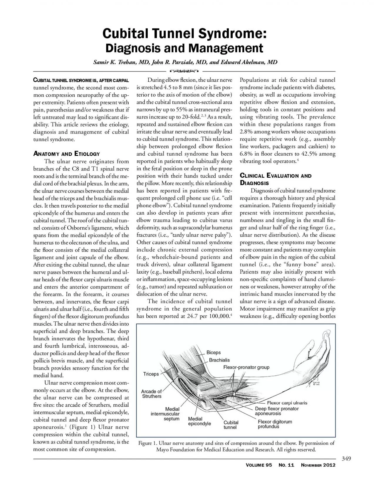
/
OLUME 11     OVEMBER 2012 OLUME 11     OVEMBER 2012

OLUME 11 OVEMBER 2012 - PDF document

udeline
udeline . @udeline
Follow
342 views
Uploaded On 2022-08-26

OLUME 11 OVEMBER 2012 - PPT Presentation

UBTALTUNNELSYDROMEAFTERARPALtunnel syndrome the second most common compression neuropathy of the upper extremity Patients often present with pain paresthesias andor weakness that if left untreated ID: 941892

nerve ulnar tunnel elbow ulnar nerve elbow tunnel cubital syndrome patients muscle disease 150 neuropathy pain hand compression atrophy

Share:

Link:

Embed:

Download Presentation from below link

Download Pdf The PPT/PDF document "OLUME 11 OVEMBER 2012" is the property of its rightful owner. Permission is granted to download and print the materials on this web site for personal, non-commercial use only, and to display it on your personal computer provided you do not modify the materials and that you retain all copyright notices contained in the materials. By downloading content from our website, you accept the terms of this agreement.


Presentation Transcript

OLUME 11 OVEMBER 2012 UBTALTUNNELSYDROMEAFTERARPALtunnel syndrome, the second most common compression neuropathy of the upper extremity. Patients often present with pain, paresthesias and/or weakness that if left untreated may lead to signicant disability. This article reviews the etiology, diagnosis and management of cubital tunnel syndrome. Figure 1. Ulnar nerve anatomy and sites of compression around the elbow. By permission of Mayo Foundation for Medical Education and Research. All rights reserved. EDICIEALT or jars), hand clumsiness (e.g., difculty typing) or difculty with precision pinch Since patients with mild disease may have no symptoms at the time of examination, various provocative exam techniques may aid in diagnosis of these patients. The elbow exion test is performed by placing the elbow in maximal flexion and full supination. The test is positive if paresthesias, numbness or tingling are reproduced in the ulnar nerve distribution. This test has been reported to be 75% sensitive after one minute. Tinel’s test, in which the cubital tunnel is tapped by the examiner’s nger, may also reproduce symptoms and has been reported to be 70% sensitive. Finally, compression of the nerve for one minute just proximal to the cubital tunnel with the elbow in 20° exion and full supination is 89% sensitive when performed alone and 98% sensitive when performed in combination with the elbow exion test.With advanced disease, objective ndings of weakness in the muscles innervated by the ulnar nerve may be noted on examination. Patients may have weak nger abduction secondary to interosseus muscle atrophy. In particular, the rst dorsal interosseous muscle can be examined by asking the patient to abduct the index nger against resistance. Small nger abduction following extension of the digits may also be noted (Wartenberg sign), which patients may notice by the small nger being caught when trying to place the hand inside of a pant pocket. Patients may also be unable to grasp with a key-pinch grip and instead compensate with a ngertip grip (Froment sign) secondary to adductor pollicis, rst dorsal interosseous and flexor pollicis brevis atrophy. (Figure 2) Finally, severe clawing of the ring and small ngers (i.e. exion of the interphalyngeal joints with extension of the metacarpophalyngeal joints) may be noted secondary to lumbrical and interosseous muscle atrophy.Physical examination must also include investigation of potential underlying causes for cubital tunnel syndrome. Thus, the elbow should be examined for range of motion, crepitus, ligament stability and deformity. In particular, patients whose chief complaint is medial elbow pain (as opposed to paresthesias, numbness, tingling or hand clumsiness) should be evaluated for medial epicondylitis and elbow instability.During the work-up of patients with suspected cubital tunnel syndrome, it is important to consider other potential sites of ulnar nerve compression, C8 radiculopathy, thoracic outlet syndrome, vascular disease, amyotrophic lateral sclerosis and peripheral neuropathy (which can be secondary to chronic alcoholism, diabetes, vitamin B deciency and hypothyroidism among other causes). Patients with C8 radiculopathy can have co-existent cubital tunnel syndrome—a phenomenon referred to as “double crush”—and therefore one diagnosis does not preclude the other.Although no disease-specific outcome measures have been validated for cub

ital tunnel syndrome, numerous severity scales have been reported based on ndings from history and physical McGowan rst classied cubital tunnel syndrome severity into three categories: mild, moderate and severe. Mild disease is dened as occasional paresthesias, positive Tinel’s sign and subjective weakness. Moderate disease is dened as occasional paresthesias, positive Tinel’s sign and objective weakness. Severe disease is dened as constant paresthesias ABORATORYADOGRAPHICLETRODAGOSTICSSESSMEDiagnostic testing may be helpful in patients with suspected cubital tunnel syndrome. Radiographs of the elbow may identify osteophytes, bone fragments or malalignment in patients with arthritis or a history of trauma. Electromyographic and nerve conduction studies may be helpful in conrming the diagnosis of ulnar neuropathy at the elbow, assisting in precise localization of the compressive lesion (e.g., proximal versus distal to the innervation of the exor carpi ulnaris), quantifying the degree of the neurologic decit and/or identifying alternate sites of nerve dysfunction simulating cubital tunnel syndrome such as cervical radiculopathy, brachial plexopathy and/or ulnar nerve compression at the wrist at Guyon’s canal. Ulnar nerve compression can be diagnosed if motor nerve conduction velocity (NCV) across the elbow is less than 50 m/s. Performing NCV studies with the elbow in moderate exion (i.e., 70 to 90 degrees from the horizontal) maximizes test sensitivity by providing the greatest correlation between the skin surface measurement and true nerve length.1, 9Needle EMG examination should always include the rst dorsal interosseous muscle, which is the most frequent muscle to rst demonstrate abnormalities following ulnar nerve compression. In addition, electrodiagnostic testing has been shown to have prognostic value in predicting subjective recovery.MRI may be helpful if a space-occupying lesion is suspected, but otherwise is not routinely used. In addition to also being useful for visualizing space-occupying lesions, ultrasound has recently been proposed as a diagnostic tool for cubital tunnel syndrome via measurement of nerve diameter. A literature review of clinical trials of ultrasonagraphy used to test ulnar neuropathy at the elbow noted that numerous studies had signicant methodological aws, some studies were uncontrolled, and that the study designs differed signicantly. The authors concluded that the role of ultrasound in ulnar neuropathy at the elbow could not AGEMEonservative anagementIn the absence of intrinsic muscle atrophy, conservative treatment should be attempted. Non-operative treatment includes patient education and activity modication to avoid elbow exion and/ Figure 2. First dorsal interosseous muscle atrophy secondary to ulnar nerve neuropathy. OLUME 11 OVEMBER 2012 or cubital tunnel compression. Depending on the provocative activity, this can be accomplished by wearing an elbow extension splint at night (or, more simply, limiting elbow exion by wrapping a pillow around the anterior elbow), adjusting posture at work to reduce elbow exion, using a hands-free headset with cell phone use, or padding the posterior surface of the elbow. In addition, non-steroidal anti-inammatory drugs or ice can be used to reduce acute pain and inammation. Following resolution of acute symptoms, physical therapy is initiated to rst establish pain-free range of motion of the affecte

d extremity and then increase strength. Dellon, et al. reported symptom improvement in 90% of patients with mild disease and 38% of patients with moderate disease. A history of elbow trauma is a poor prognosticator and risk factor for eventual surgery.perative anagementWhen patients fail to respond to conservative measures, have persistent severe symptoms or present with intrinsic muscle atrophy, operative management should be considered. Surgical options include ulnar nerve in situ decompression, anterior transposition of the ulnar nerve (subcutaneous, intramuscular or submuscular), partial medial epicondylectomy and endoscopic ulnar nerve decompression. Studies of in situ decompression report 75% to 90% of patients achieve good or excellent pain relief, while 7% to 15% do not ben Despite discussion in the literature regarding in situ decompression’s potential advantages (e.g., minimal disruption of the ulnar nerve’s vascular supply) and disadvantages (e.g., limited exposure to explore other potential sites of ulnar nerve compression and risk of post-operative ulnar nerve subluxation) versus anterior transposition, two meta-analyses have demonstrated similar outcomes between these techniques.13, 14 In the 7% to 15% of patients who have recurrent disease following in situ decompression, many can be successfully treated with anterior transposition of the ulnar nerve.Patients with post-traumatic elbow stiffness or deformity, ulnar nerve subluxation, ulnar collateral ligament laxity and “tardy ulnar nerve palsy” may benet from initial anterior transposition of the ulnar nerve. Patients with medial epicondylitis may benet from partial medial epicondylectomy, although this procedure has been associated with increased medial elbow pain post-operatively. Finally, endoscopic ulnar nerve release has been reported to have a similar success rate to open procedures with potentially less post-operative pain. A common surgical complication of all of these techniques is potential injury to the posterior branch of the medial antebrachial cutaneous nerve. Taken together, given the similarity in outcomes reported between the surgical treatments for cubital tunnel syndrome, the choice of procedure is based largely on surgeon experience and sometimes underlying etiology.OSTPERATVEEHABTATOnce the incision and soft tissues have healed, rehabilitation therapies are often used to help the patient regain pain-free range of motion, normal strength and function. The extent and duration of a post-operative rehabilitation program varies with the extent of injury and the physical demands of a return to normal activities such as ADLs, occupational activities or sports. Goals of a postoperative rehabilitation program include (a) full active range of motion for elbow flexion, extension, pronation and supination, (b) normal elbow strain, with balance maintained between agonists and antagonists muscles, and (c) resumption of sports-specic and work specic functional activities. Exercises to establish neuromuscular control include proprioceptive neuromuscular facilitation and progression from closed-kinetic chain activities through open-kinetic chain exercises. A rehabilitation program may be necessary for six weeks or more post-operatively.NCLUSCubital tunnel syndrome is a common cause of upper extremity pain and disability. The treating clinician should possess a high degree of familiarity with the relevant aspects of anatomy, epidemiology and clinical presentation. The diagnosis of cubital tun

nel syndrome frequently requires a combination of clinical suspicion and may require electrodiagnostic conrmation. Once diagnosed, cubital tunnel syndrome is initially treated by conservative measures focused on patient education and avoidance of provocative activities. In the presence of intrinsic hand muscle atrophy or persistent severe symptoms, operative treatment should be considered.EFERENCESPalmer BA, Hughes TB. Cubital tunnel syndrome. J Hand Surg Am. Jan 2010;35(1):153–63.Werner CO, Ohlin P, Elmqvist D. Pressures recorded in ulnar neuropathy. Acta Orthop Oct 1985;56(5):404–6.Apfelberg DB, Larson SJ. Dynamic anatomy of the ulnar nerve at the elbow. Plast Reconstr Surg. Jan 1973;51(1):79–81.van Rijn RM, Huisstede BM, Koes BW, Burdorf A. Associations between work-related factors and specic disorders at the elbow: a systematic literature review. Rheumatology (Oxford). May Novak CB, Lee GW, Mackinnon SE, Lay L. Provocative testing for cubital tunnel syndrome. J Hand Surg Am. Sep 1994;19(5):817–20.Folberg CR, Weiss AP, Akelman E. Cubital tunnel syndrome. Part I: Presentation and diOrthop Rev. Feb 1994;23(2):136–44.Macadam SA, Bezuhly M, Lefaivre KA. Outcomes measures used to assess results after surgery for cubital tunnel syndrome: a systematic review of the literature. J Hand Surg Am. Oct McGowan AJ. The results of transposition of the ulnar nerve for traumatic ulnar neuritis. J Bone Joint Surg Br. Aug 1950;32-B(3):293–301.Practice parameter: electrodiagnostic studies in ulnar neuropathy at the elbow. American Association of Electrodiagnostic Medicine, American Academy of Neurology, and American Academy of Physical Medicine and Rehabilitation. Neurology. Mar 10 1999;52(4):688–90.Friedrich JM, Robinson LR. Prognostic indicators from electrodiagnostic studies for ulnar neuropathy at the elbow. Muscle Nerve. Apr Dellon AL, Hament W, Gittelshon A. Nonoperative management of cubital tunnel syndrome: an 8-year prospective study. Neurology. Sep 1993;43(9):1673–7.When patients fail to respond to conservative measures, have persistent severe symptoms or present with intrinsic muscle atrophy, operative management should be considered. EDICIEALT Abuelem T, Ehni BL. Minimalist cubital tunnel treatment. Neurosurgery. Oct 2009;65(4 Suppl):A145–9.Goldfarb CA, Sutter MM, Martens EJ, Manske PR. Incidence of re-operation and subjective outcome following in situ decompression of the ulnar nerve at the cubital tunnel. J Hand Surg Eur Vol. Jun 2009;34(3):379–83.Macadam SA, Gandhi R, Bezuhly M, Lefaivre KA. Simple decompression versus anterior subcutaneous and submuscular transposition of the ulnar nerve for cubital tunnel syndrome: a meta-J Hand Surg Am. Oct 2008;33(8):1314 Hertling D KR, ed Management of Common Musculoskeletal Disorders. Physical Therapy Priciples and Methods. 4 ed. Philadelphia: Lippincott Williams & Wilkins; 2006.Samir K. Trehan, MD, is a second year orthopaedic surgery resident at Hospital for Special Surgery in New York, NYJohn R. Parziale, MD, is a Clinical Associate Professor, Department of Orthopaedics, Warren Alpert Medical School of Brown UniversityEdward Akelman, MD, is a Professor, Department of Orthopaedics, Warren Alpert Medical School of Brown Universityisclosure of Financial nterestsThe authors and/or their spouses/signicant others have no nancial interORRESPODENCJohn R. Parziale, MDUniversity Rehabilitation, Inc.450 Veterans’ Memorial Parkway, Building #12East Providence, RI 02914e-mail: jrp@urehab.necoxmail.c