ANATOMY OF URINARY BLADDER Introduction The urinary bladder is a muscular organ which lies in the anterior part of pelvic cavity and helps in the storage of urine. Shape, size and position of bladder varies according to the amount of urine
"ANATOMY OF URINARY BLADDER Introduction The" is the property of its rightful owner. Permission is granted to
download and print the materials on this website for personal, non-commercial use only, and to display it
on your personal computer provided you do not modify the materials and that you retain all copyright
notices contained in the materials. By downloading content from our website, you accept the terms of this
agreement.
Presentation Transcript
01
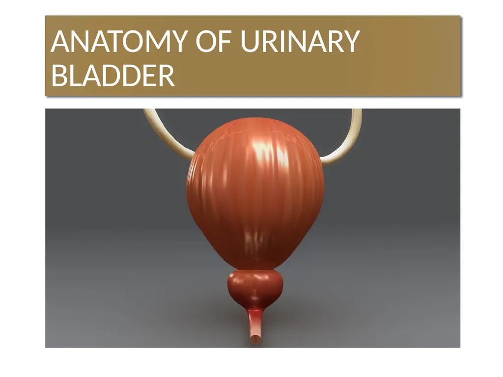
ANATOMY OF URINARY BLADDER<br>
02
Introduction The urinary bladder is a muscular organ which lies in the anterior part of pelvic cavity and helps in the storage of urine.
Shape, size and position of bladder varies according to the amount of urine contained in it.
An empty bladder lies entirely within the pelvis but as it fills it extends into abdominal cavity.<br>
03
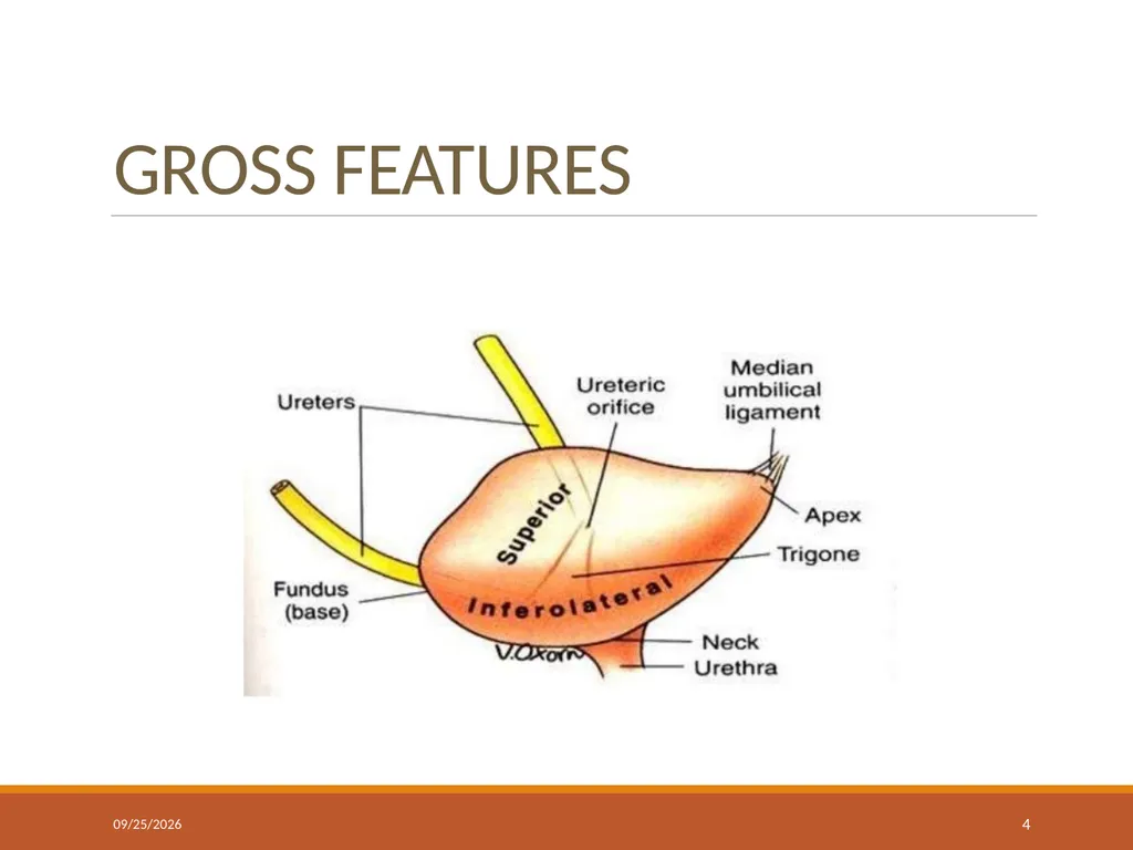
GROSS FEATURES An empty bladder is tetrahedral in shape & has:
Apex
Base or fundus
Neck
3 surfaces: superior, right & left inferolateral.
4 borders: anterior, posterior & 2 lateral lateral borders.<br>
04
GROSS FEATURES 4/24/2024 4<br>
05
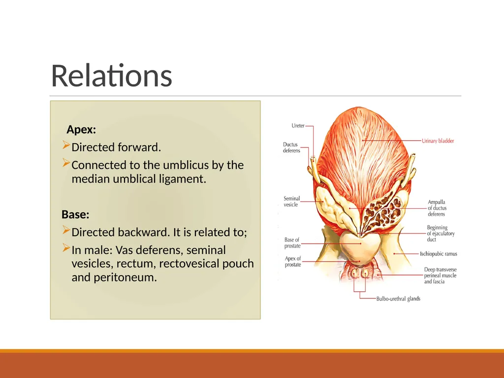
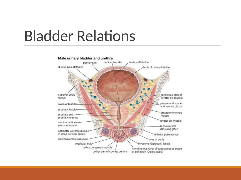
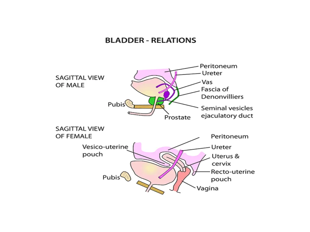
Relations Apex:
Directed forward.
Connected to the umblicus by the median umblical ligament.
Base:
Directed backward. It is related to;
In male: Vas deferens, seminal vesicles, rectum, rectovesical pouch and peritoneum.<br>
06
In female: Vagina and cervix of the uterus.
Neck:
Inferior and most fixed part.
In male: Related to prostate. Surrounded by smooth muscle fibres which form preprostatic sphincter.
In female: Related to pelvic fascia.<br>
07
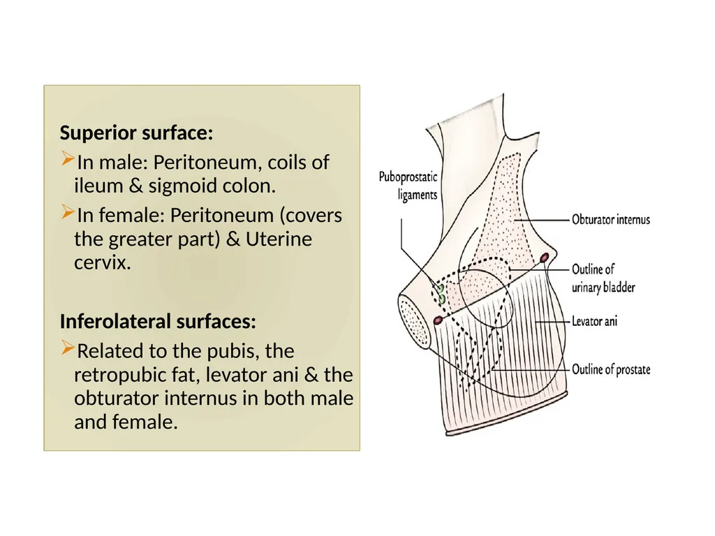
Superior surface:
In male: Peritoneum, coils of ileum & sigmoid colon.
In female: Peritoneum (covers the greater part) & Uterine cervix.
Inferolateral surfaces:
Related to the pubis, the retropubic fat, levator ani & the obturator internus in both male and female.<br>
08
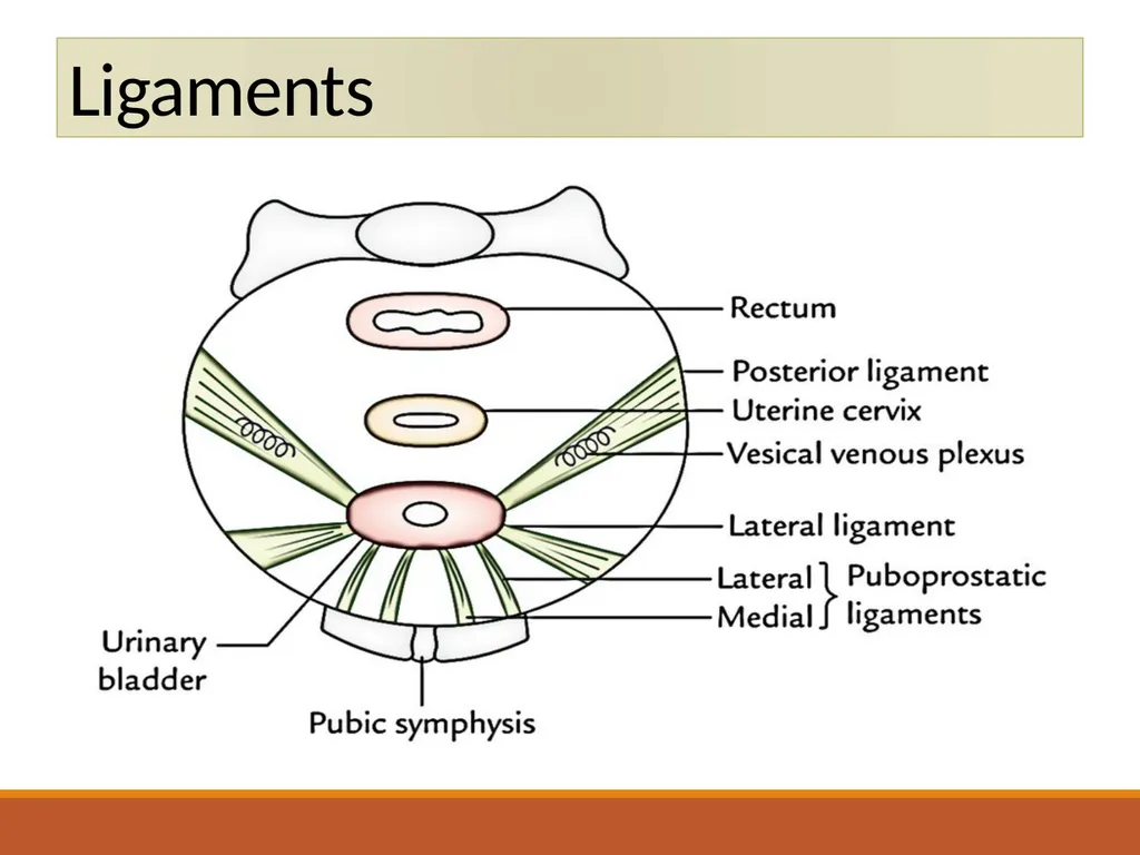
Puboprostatic ligaments in male & pubovesical ligaments in female.<br>
09
Bladder Relations<br>
10
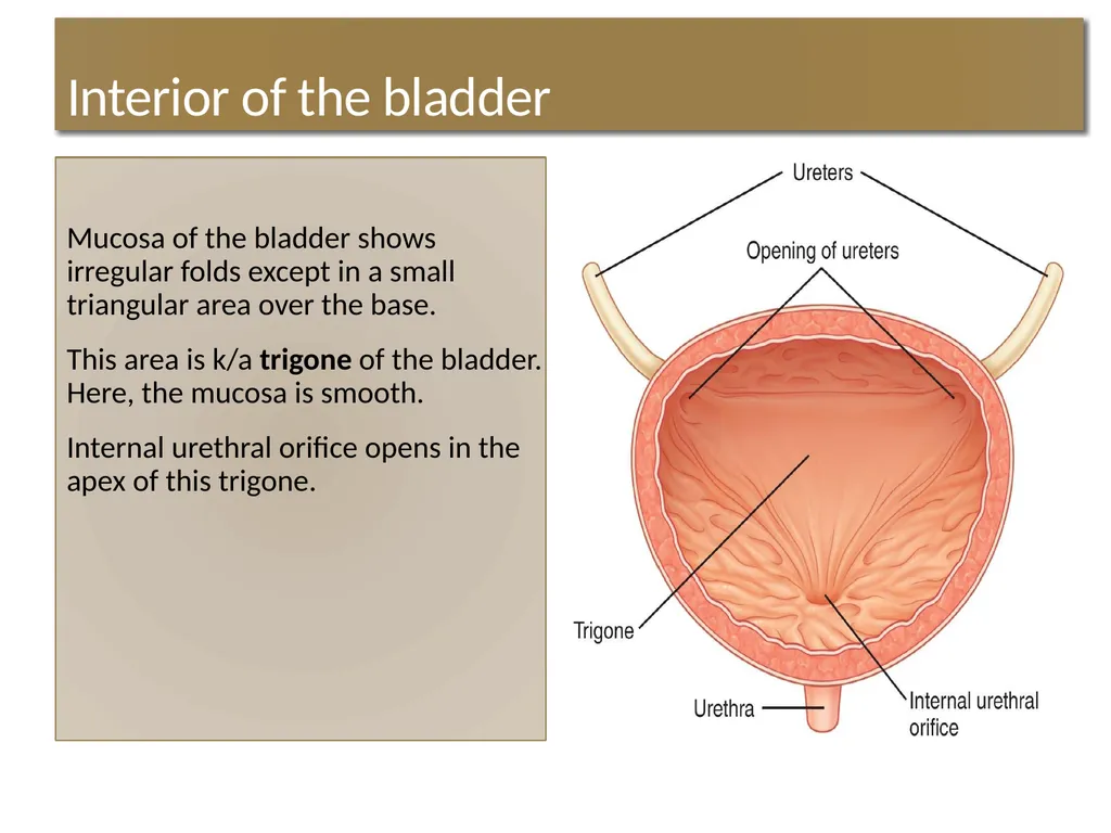
Interior of the bladder Mucosa of the bladder shows irregular folds except in a small triangular area over the base.
This area is k/a trigone of the bladder. Here, the mucosa is smooth.
Internal urethral orifice opens in the apex of this trigone.<br>
11
The ureters open at the posterolateral angle of the trigone.
Uvula vesicae: It is a small elevation on the trigone posterior to the urethral orifice produced by median lobe of prostate.<br>
12
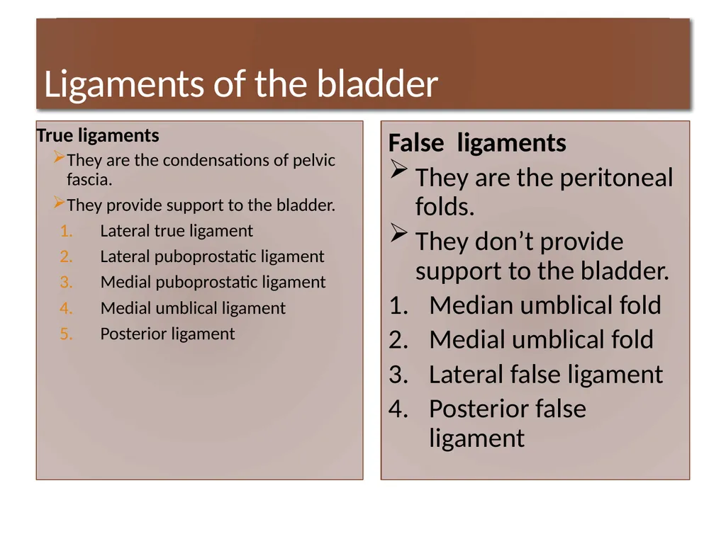
Ligaments of the bladder True ligaments
They are the condensations of pelvic fascia.
They provide support to the bladder.
Lateral true ligament
Lateral puboprostatic ligament
Medial puboprostatic ligament
Medial umblical ligament
Posterior ligament 4/24/2024 13 False ligaments
They are the peritoneal folds.
They don’t provide support to the bladder.
Median umblical fold
Medial umblical fold
Lateral false ligament
Posterior false ligament<br>
13
Ligaments<br>
14
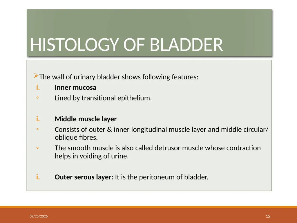
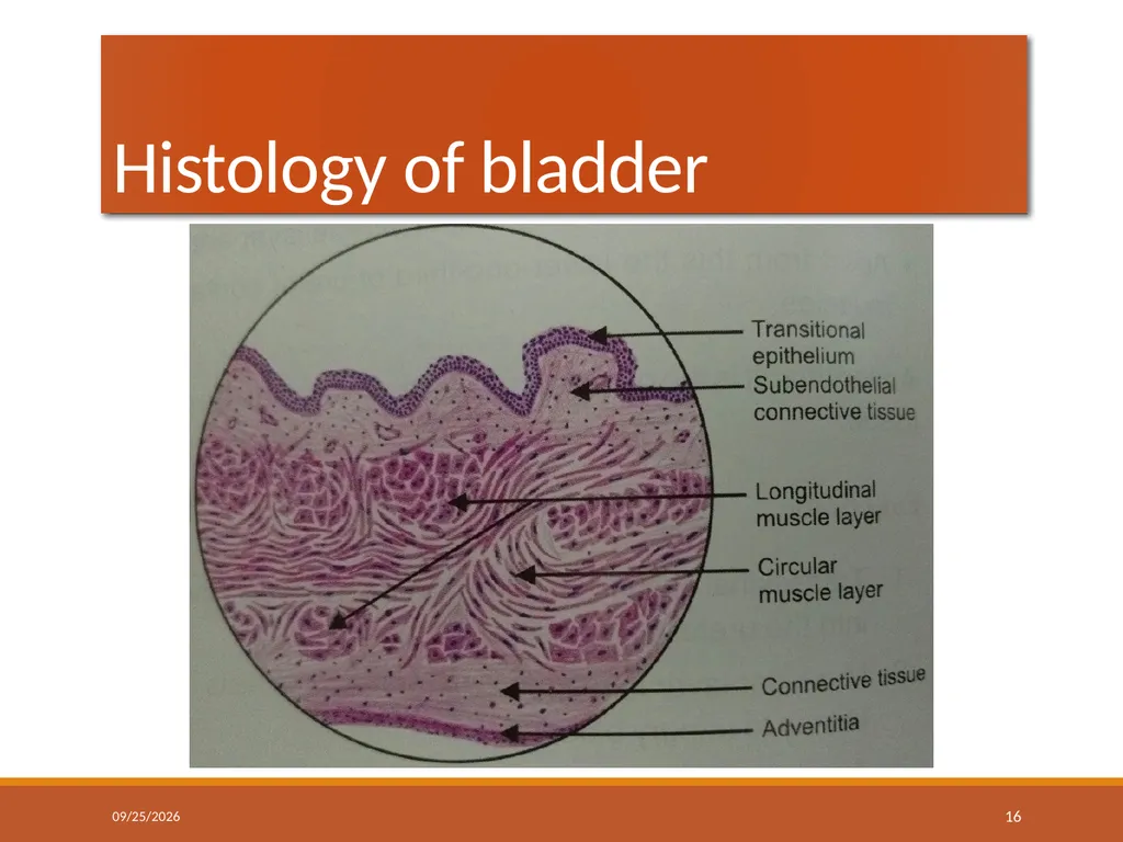
HISTOLOGY OF BLADDER The wall of urinary bladder shows following features:
Inner mucosa
Lined by transitional epithelium.
Middle muscle layer
Consists of outer & inner longitudinal muscle layer and middle circular/ oblique fibres.
The smooth muscle is also called detrusor muscle whose contraction helps in voiding of urine.
Outer serous layer: It is the peritoneum of bladder. 4/24/2024 15<br>