
Brain death declaration in children Dr Lokesh








Description: Brain death declaration in children Dr Lokesh Lingappa Consultant Paediatric Neurologist Rainbow Childrens Hospital and Perinatal Centre, Hyderabad outline Limitations of current guidelines Testing process Are there differences
Related Topics
Download Presentation
"Brain death declaration in children Dr Lokesh" is the property of its rightful owner. Permission is granted to download and print the materials on this website for personal, non-commercial use only, and to display it on your personal computer provided you do not modify the materials and that you retain all copyright notices contained in the materials. By downloading content from our website, you accept the terms of this agreement.
Presentation Transcript
slide1. Brain death declaration in children Dr Lokesh Lingappa
Consultant Paediatric Neurologist
Rainbow Children’s Hospital and Perinatal Centre, Hyderabad<br>
slide2. outline Limitations of current guidelines
Testing process
Are there differences adult/pediatric
Problems of newborn testing
Fallacies in intepretation of signs and testing results<br>
slide3. Key message The diagnosis of brain death should remain a clinical one to be made at the bedside by knowledgeable physicians who, in concert with grieving families, make the most agonizing of all life’s events (the death of a child) as bearable as possible for all concerned.
Freeman JM, Ferry PC. New Brain Death Guidelines in Children<br>
slide4. Understanding limitations of pediatric brain death guidelines Guidelines are 20 years old
Relied heavily upon EEG testing
Guidelines did not specifically address the trauma population
Guidelines were based upon limited clinical experience at the time of publication
Guidelines were based upon age criteria
No guidelines for neurologic death in neonates
Waiting times have never been validated<br>
slide5. History Determination of cause of death is necessary to ensure the absence of treatable or reversible conditions (ie, toxic or metabolic disorders, hypothermia, hypotension, or surgically remediable conditions).<br>
slide6. Primary requirement Irreversibility of brain function cessation is recognized
Cause of coma is established and is sufficient to account for the loss of brain function
Possibility of recovery of any brain function is excluded
Cessation of brain function persists for an appropriate period of observation or trial of therapy<br>
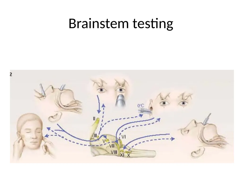
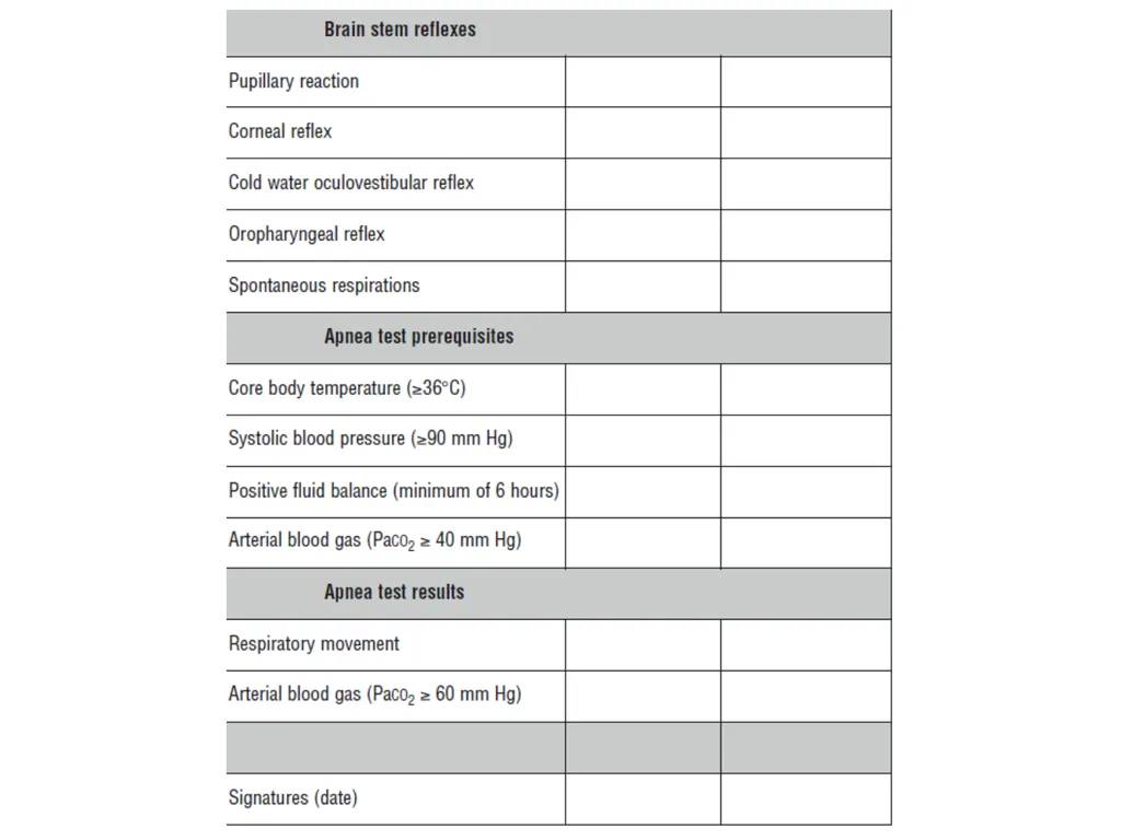
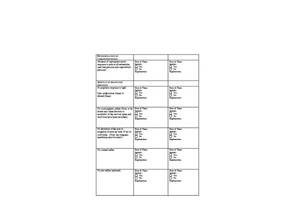
slide7. Diagnostic criteria Cessation of all brain function is recognized.
Cerebral functions are absent (ie, unresponsiveness)
Brainstem functions are absent:
Pupillary light reflex
Corneal reflex
Oculocephalic/oculovestibular reflex
Oropharyngeal reflex
Respiratory (apnea using an accepted apnea testing procedure)<br>
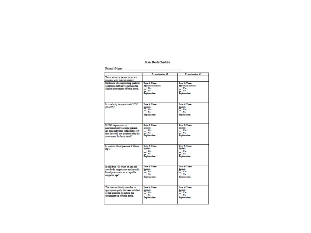
slide8. Confounding factors Complicating conditions are excluded
Drug and metabolic intoxication
Hypothermia
Circulatory shock
The patient has been monitored for an appropriate observation period<br>
slide9. Brainstem testing<br>
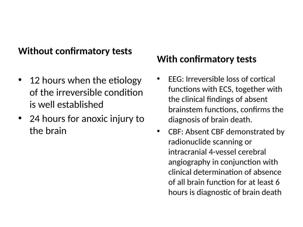
slide10. Without confirmatory tests 12 hours when the etiology of the irreversible condition is well established
24 hours for anoxic injury to the brain With confirmatory tests EEG: Irreversible loss of cortical functions with ECS, together with the clinical findings of absent brainstem functions, confirms the diagnosis of brain death.
CBF: Absent CBF demonstrated by radionuclide scanning or intracranial 4-vessel cerebral angiography in conjunction with clinical determination of absence of all brain function for at least 6 hours is diagnostic of brain death<br>
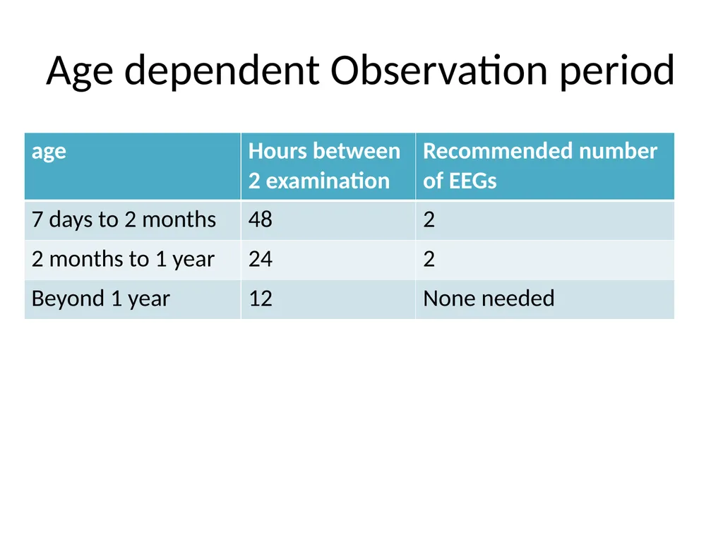
slide11. Age dependent Observation period<br>
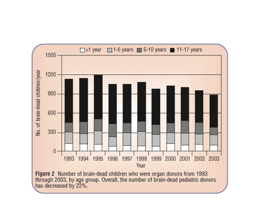
slide12. Brain death and organ donation<br>
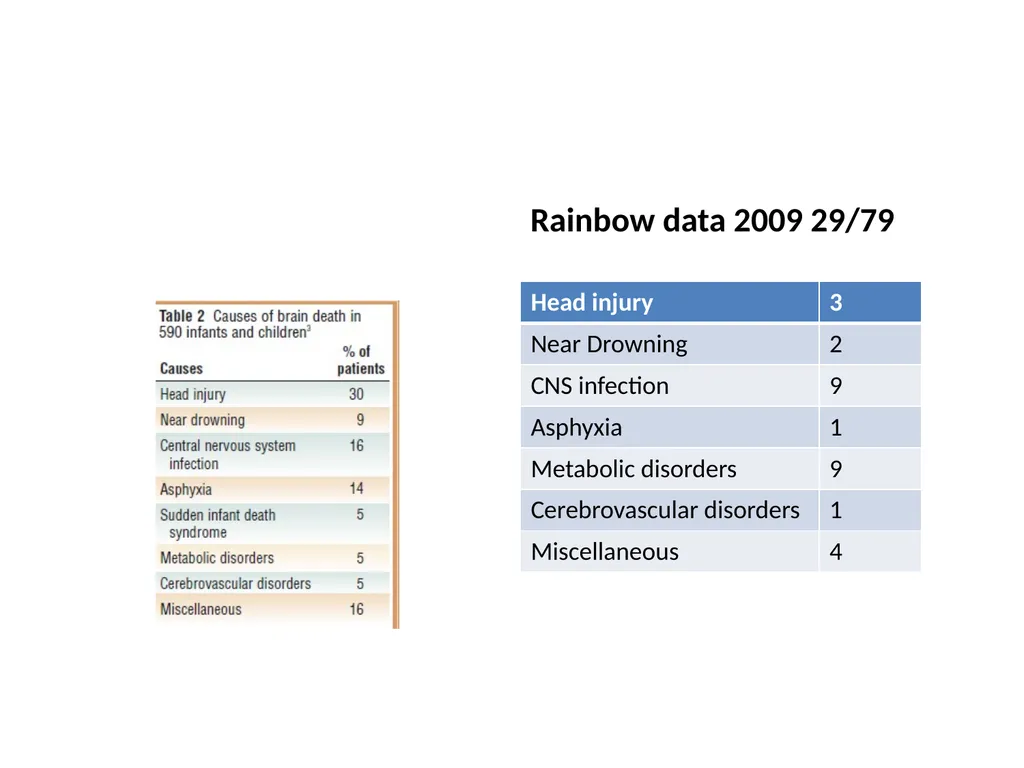
slide14. Rainbow data 2009 29/79<br>
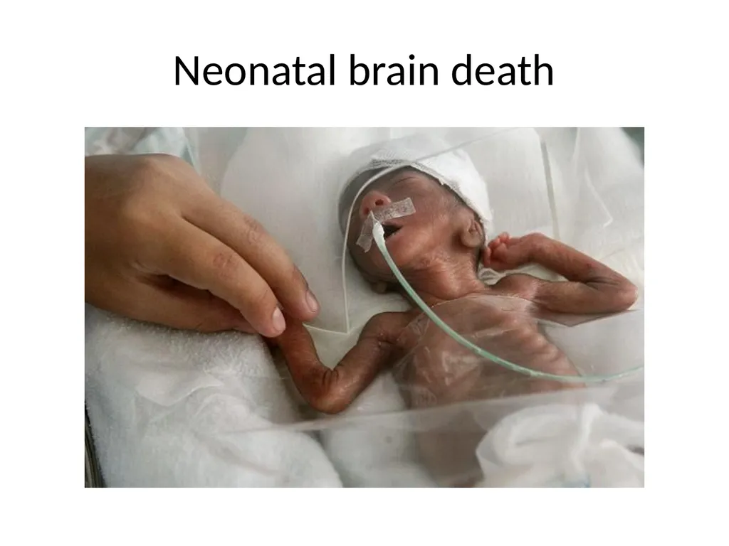
slide15. Neonatal brain death<br>
slide16. Neurologic death in the neonate “Brain death can be diagnosed in the term infant, even at less than 7 days of age. An observation period of 48 hours is recommended to confirm the diagnosis. If an EEG is isoelectric or if a CBF study shows no flow, then the observation period can be shortened to 24 hours.”
Ashwal. Brain death in the newborn. Clinics in Perinatology 1997;24:859-879<br>
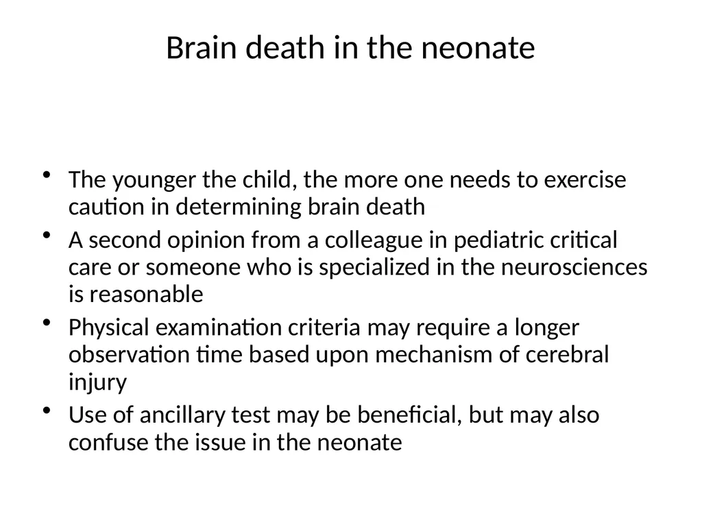
slide17. Brain death in the neonate The younger the child, the more one needs to exercise caution in determining brain death
A second opinion from a colleague in pediatric critical care or someone who is specialized in the neurosciences is reasonable
Physical examination criteria may require a longer observation time based upon mechanism of cerebral injury
Use of ancillary test may be beneficial, but may also confuse the issue in the neonate<br>
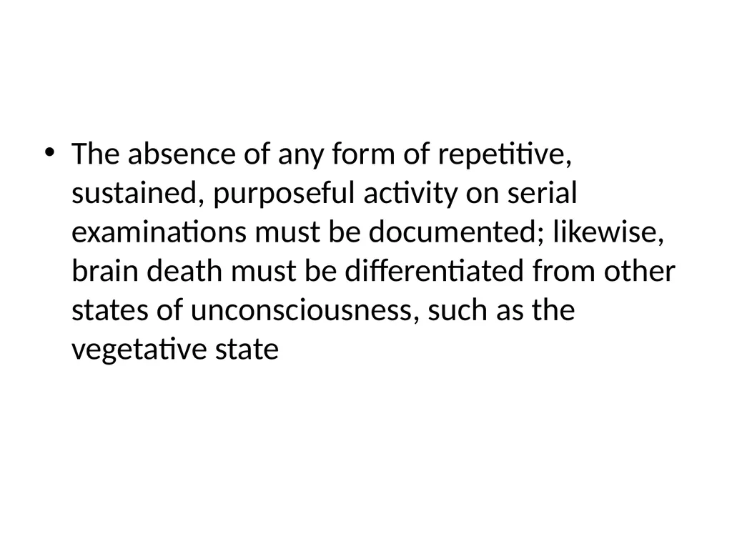
slide18. The absence of any form of repetitive, sustained, purposeful activity on serial examinations must be documented; likewise, brain death must be differentiated from other states of unconsciousness, such as the vegetative state<br>
slide19. preterm and term neonates several of the cranial nerve responses are not fully developed.
pupillary light reflex is absent before 29 to 30 weeks’ gestation,
oculocephalic reflex may not be elicited before 32 weeks’ gestation
Term and preterm infants are difficult to examine because their smallness makes it technically difficult to assess<br>
slide20. Preterm and neonate Assessment of pupillary reactivity can be compromised difficulties in gaining access –incubator, by corneal injury, retinal hemorrhages, and other anatomical factors (swelling or partial fusion of the eyelids)
smaller size of the pupils in newborns- make assessment of the loss of pupillary reactivity troublesome<br>
slide21. Preterm and neonate Assessing the caloric response adequately more difficult in neonates with a small external ear canal;
both the oculocephalic (doll’s eye) and oculovestibular (caloric) reflex should always be examined
corneal reflex- easiest brain stem reflex to examine in neonates and infants, it is often the least reliable
Contact irritation, dehydration and maceration of the cornea, use of lubricant drops, and use of analgesic medications often adversely affect tactile sensory information<br>
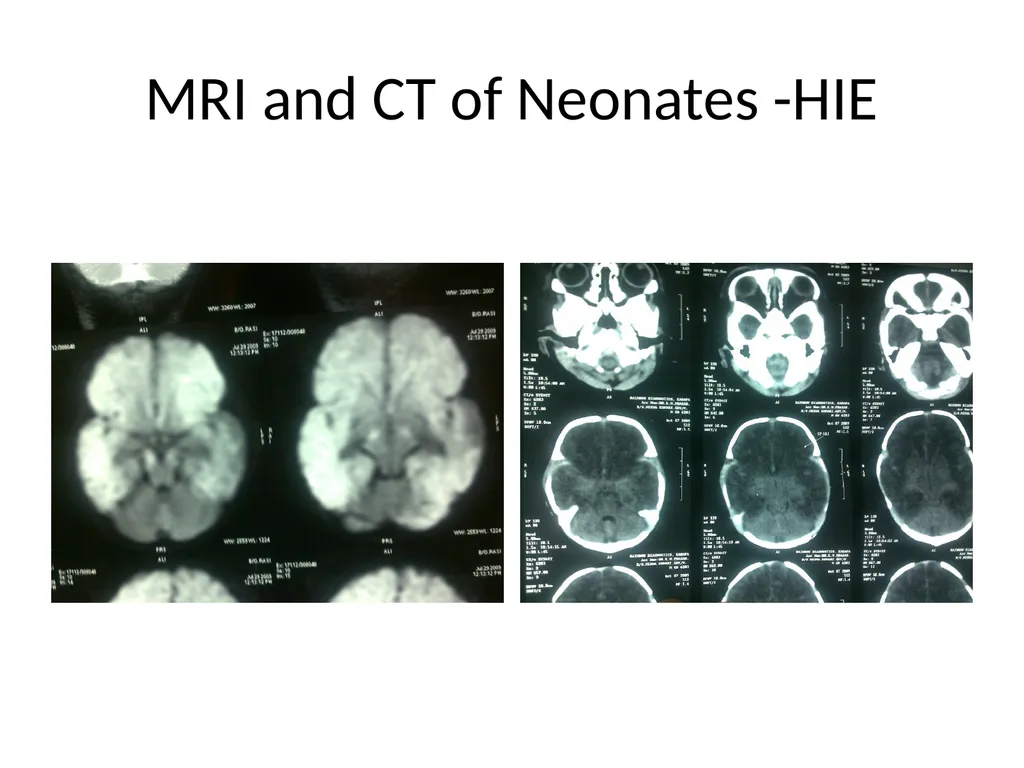
slide22. MRI and CT of Neonates -HIE<br>
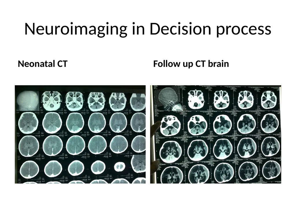
slide23. Neuroimaging in Decision process Neonatal CT Follow up CT brain<br>
slide24. Ancillary testing<br>
slide25. Considerations when diagnosing brain death in children Many times the cause of the child’s neurologic demise is known
Based upon presentation and examination many times we know that there will be no hope for survival or if the child does survive, the outcome will be dismal
The waiting period may be extended or decreased depending upon social and family related issues<br>
slide26. Ancillary testing Ancillary test that may aid in the diagnosis of brain death
EEG
Cerebral angiography
Radionuclide Scans
Brainstem evoked responses
Doppler sonography<br>
slide27. Ancillary testing Ancillary tests may aid in the diagnosis of brain death
Ancillary tests can provide additional information to help confirm brain death in situations where clinical examination and apnea testing are not feasible or cannot be completed because of undue circumstance
Facial injury
Acute lung injury
Cardiovascular instability<br>
slide28. Ancillary testing Ancillary tests are not mandatory
• Ancillary tests may provide another layer of comfort to the physician who is uncomfortable declaring brain death on clinical exam alone
• Ancillary tests may reduce observation periods thus increasing potential for retrieving viable transplant tissue
• Ancillary tests may also delay or prolong observation period<br>
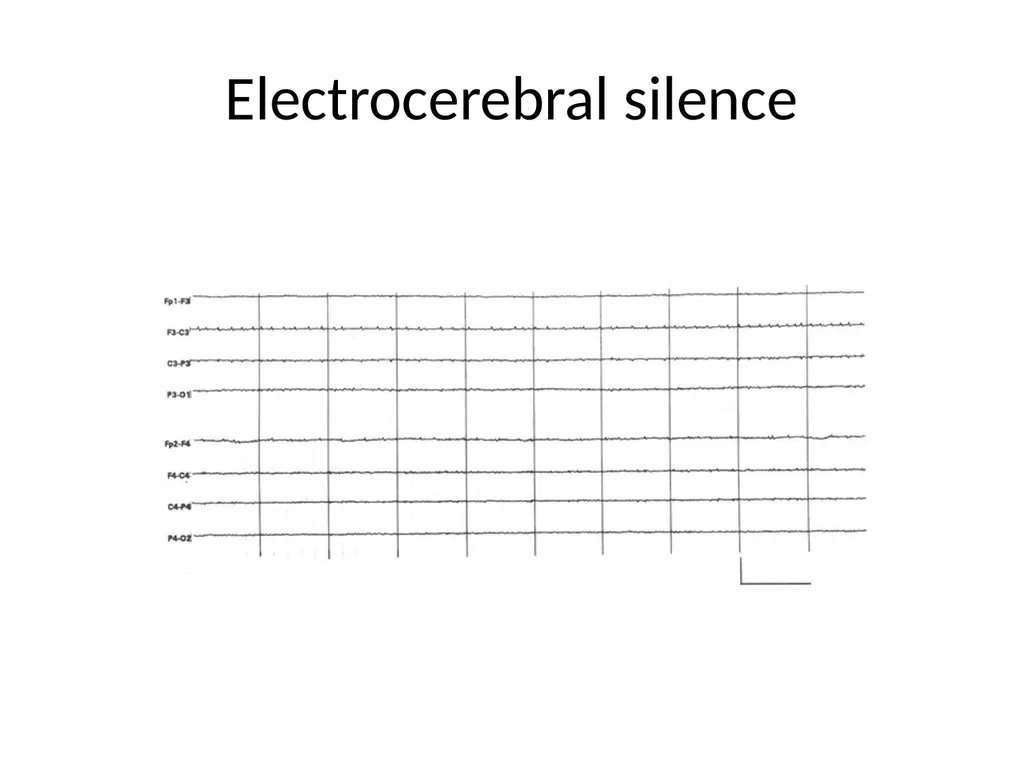
slide29. Recommendation for EEG the American Electroencephalographic Society retrospectively surveyed 1665 patients with electrocerebral silence (ECS), that is, no evidence of brain electrical activity greater than 2 µV between electrode pairs placed at a distance of 10 cm or more, who were in various levels of coma
Only 3 of the 1665 patients recovered cerebral function<br>
slide30. EEG in infants and children shorter interelectrode distances;
external artifacts in newborn ICUs and PICUs;
Distances between the heart and the brain, making the electrocardiographic contribution disproportionately large
reduced amplitude of cortical potentials in preterm and term neonates
longer duration of the effect of depressant medications
greater tendency for suppression burst patterns in infants with neurological disorders<br>
slide31. Need for EEG Two cases of acute inflammatory demyelinating polyradiculoneuropathy have -at satisfied the clinical criteria for brain death but had preserved EEG activity
EEG has an important role in the confirmation of brain death in such cases<br>
slide32. Electrocerebral silence<br>
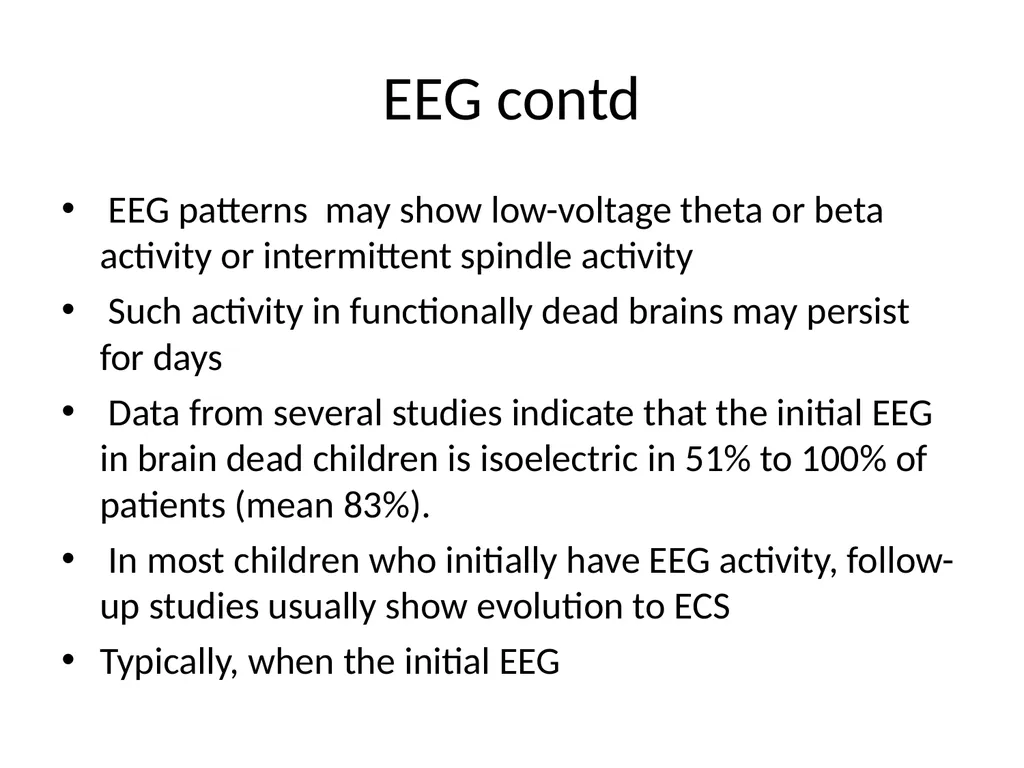
slide33. EEG contd EEG patterns may show low-voltage theta or beta activity or intermittent spindle activity
Such activity in functionally dead brains may persist for days
Data from several studies indicate that the initial EEG in brain dead children is isoelectric in 51% to 100% of patients (mean 83%).
In most children who initially have EEG activity, follow-up studies usually show evolution to ECS
Typically, when the initial EEG<br>
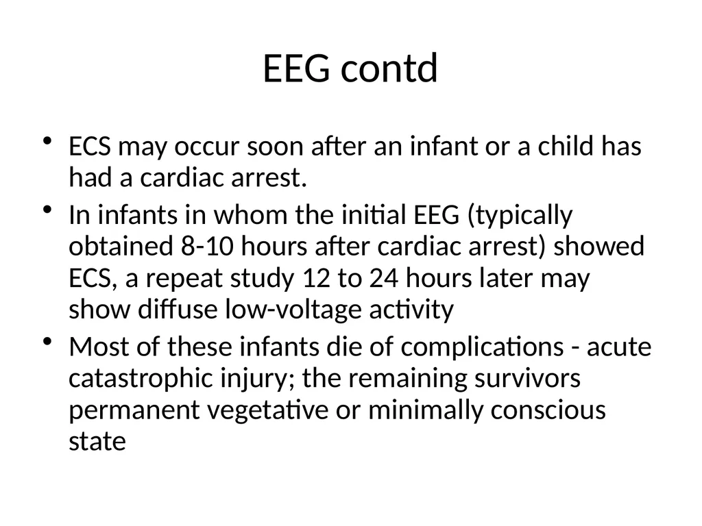
slide34. EEG contd ECS may occur soon after an infant or a child has had a cardiac arrest.
In infants in whom the initial EEG (typically obtained 8-10 hours after cardiac arrest) showed ECS, a repeat study 12 to 24 hours later may show diffuse low-voltage activity
Most of these infants die of complications - acute catastrophic injury; the remaining survivors permanent vegetative or minimally conscious state<br>
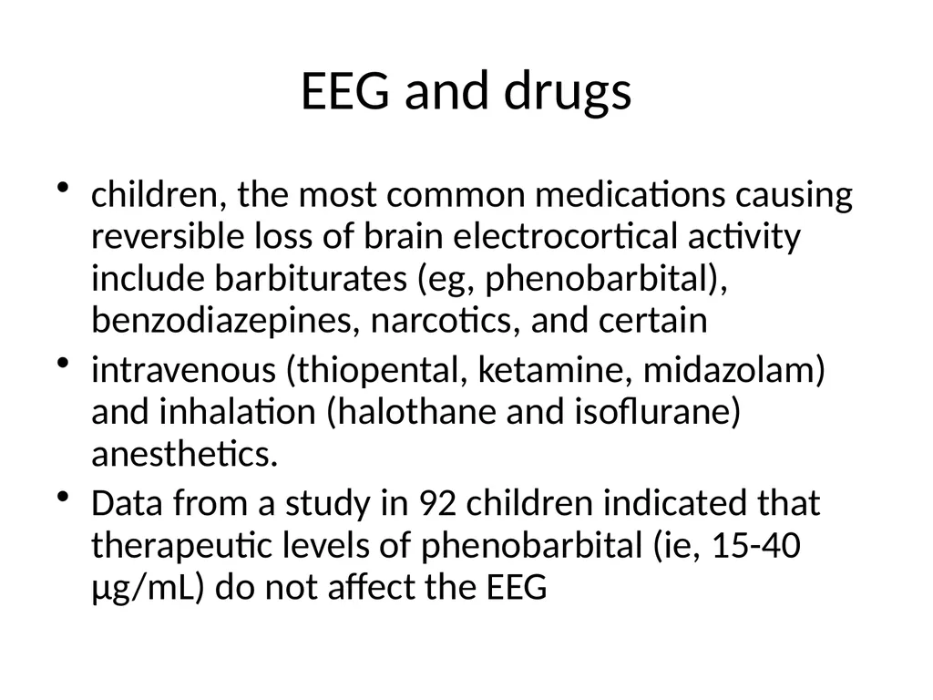
slide35. EEG and drugs children, the most common medications causing reversible loss of brain electrocortical activity include barbiturates (eg, phenobarbital), benzodiazepines, narcotics, and certain
intravenous (thiopental, ketamine, midazolam) and inhalation (halothane and isoflurane) anesthetics.
Data from a study in 92 children indicated that therapeutic levels of phenobarbital (ie, 15-40 μg/mL) do not affect the EEG<br>
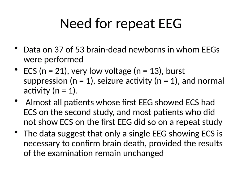
slide36. Need for repeat EEG Data on 37 of 53 brain-dead newborns in whom EEGs were performed
ECS (n = 21), very low voltage (n = 13), burst suppression (n = 1), seizure activity (n = 1), and normal activity (n = 1).
Almost all patients whose first EEG showed ECS had ECS on the second study, and most patients who did not show ECS on the first EEG did so on a repeat study
The data suggest that only a single EEG showing ECS is necessary to confirm brain death, provided the results of the examination remain unchanged<br>
slide37. Cerebral blood flow studies The absence of CBF in brain death is due primarily to low cerebral perfusion pressure and secondarily to release of vasoconstrictors
from vascular smooth muscle<br>
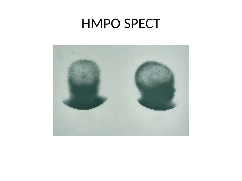
slide38. HMPO SPECT<br>
slide39. Cerebral blood flow-neonatal issues Newborns have patent sutures and an open fontanel, increases in ICP after acute injury are not significant
cascade of herniation from increased ICP and reduced cerebral perfusion is less likely to occur in newborns
Brain death can be diagnosed in newborns (even when younger than 7 days) if physician is aware of the limitations of the clinical examination and
laboratory testing<br>
slide40. Institutional Guidelines Does a policy regarding declaration of death exist in your institution? Policy should provide guidelines allowing flexibility and individuality for each child and their family
Decisions regarding determination of brain death should be left to the physician’s discretion within evolving standards of medical care
Who declares brain death in your institution?
Concentrate your efforts on educating these individuals and involve them in the establishment of institutional guidelines<br>
slide43. Take home messages Neurologic death occurs in children
• There are no unique legal issues in determining neurologic death in children
• Neurologic death is a clinical diagnosis
• Ancillary studies are not mandatory, however they can assist in determining neurologic death in certain situations
• Ancillary studies can reduce or prolong the recommended observation period
• Observation periods have never been validated and are meant to serve as guidelines only
• Neurologic death can occur and be diagnosed in infants less than 7 days of age<br>
slide44. Thank you<br>
slide45. One Class III study of 144 patients pronounced
brain dead found 55% (95% confidence interval
[CI] 47–63) of patients had retained plantar reflexes,
either flexion or “stimulation induced undulating toe
flexion.”22 Another study documented plantar flexion
and flexion synergy bilaterally that persisted for
32 hours after the determination of brain death.23<br>
slide46. ApneaTesting Absence of a breathing drive.
Prerequisites: 1) normotension, 2) normothermia,
3) euvolemia, 4) eucapnia
(PaCO2 35–45 mm Hg), 5) absence of
hypoxia, and
Procedure:
• Adjust vasopressors to a systolic blood
pressure 100 mm Hg.
Neurology 74 8, 2010<br>
slide47. Preoxygenate for at least 10 minutes with 100% oxygen to a PaO2 200 mm Hg
• 10 breaths per minute- eucapnia
• Reduce PEEP to 5 cm H2O (oxygen desaturation with decreasing PEEP may suggest difficulty with apnea testing).
• SpO2> 95%, obtain a baseline blood gas (PaO2, PaCO2, pH, bicarbonate, base excess)
• Disconnect the patient from the ventilator<br>
slide48. Preserve oxygenation (e.g., place an insufflation catheter through the endotracheal tube and close to the level of the carina and deliver 100% O2 at 6 L/min).
• Look for respiratory movements - 8–10 minutes.
• Abort if systolic blood pressure decreases to 90 mm Hg.
• Abort if oxygen saturation measured by pulse oximetry is 85% for 30 seconds.
Retry procedure with T-piece, CPAP 10 cm H2O, and 100% O2 12L/min<br>
slide49. If no respiratory drive- repeat blood gas after approximately 8 minutes.
• If respiratory movements are absent and arterial PCO2 is 60 mm Hg (or 20 mm Hg increase in arterial PCO2), the apnea test result is positive
• If test is inconclusive- patient is hemodynamically stable, it may be repeated for a longer period of time (10–15 minutes)
after the patient is again adequately preoxygenated.<br>
slide50. Take home messages Diagnosing brain death is not different in children as compared to adult
Newborn 34 week and above can be reliably daignosed to have brain death within first week of life
Most newborn withdrawal of care is based on future poor neurologic outcome rather than brain death
Most common ancillary testing required is EEG<br>
Consultant Paediatric Neurologist
Rainbow Children’s Hospital and Perinatal Centre, Hyderabad<br>
slide2. outline Limitations of current guidelines
Testing process
Are there differences adult/pediatric
Problems of newborn testing
Fallacies in intepretation of signs and testing results<br>
slide3. Key message The diagnosis of brain death should remain a clinical one to be made at the bedside by knowledgeable physicians who, in concert with grieving families, make the most agonizing of all life’s events (the death of a child) as bearable as possible for all concerned.
Freeman JM, Ferry PC. New Brain Death Guidelines in Children<br>
slide4. Understanding limitations of pediatric brain death guidelines Guidelines are 20 years old
Relied heavily upon EEG testing
Guidelines did not specifically address the trauma population
Guidelines were based upon limited clinical experience at the time of publication
Guidelines were based upon age criteria
No guidelines for neurologic death in neonates
Waiting times have never been validated<br>
slide5. History Determination of cause of death is necessary to ensure the absence of treatable or reversible conditions (ie, toxic or metabolic disorders, hypothermia, hypotension, or surgically remediable conditions).<br>
slide6. Primary requirement Irreversibility of brain function cessation is recognized
Cause of coma is established and is sufficient to account for the loss of brain function
Possibility of recovery of any brain function is excluded
Cessation of brain function persists for an appropriate period of observation or trial of therapy<br>
slide7. Diagnostic criteria Cessation of all brain function is recognized.
Cerebral functions are absent (ie, unresponsiveness)
Brainstem functions are absent:
Pupillary light reflex
Corneal reflex
Oculocephalic/oculovestibular reflex
Oropharyngeal reflex
Respiratory (apnea using an accepted apnea testing procedure)<br>
slide8. Confounding factors Complicating conditions are excluded
Drug and metabolic intoxication
Hypothermia
Circulatory shock
The patient has been monitored for an appropriate observation period<br>
slide9. Brainstem testing<br>
slide10. Without confirmatory tests 12 hours when the etiology of the irreversible condition is well established
24 hours for anoxic injury to the brain With confirmatory tests EEG: Irreversible loss of cortical functions with ECS, together with the clinical findings of absent brainstem functions, confirms the diagnosis of brain death.
CBF: Absent CBF demonstrated by radionuclide scanning or intracranial 4-vessel cerebral angiography in conjunction with clinical determination of absence of all brain function for at least 6 hours is diagnostic of brain death<br>
slide11. Age dependent Observation period<br>
slide12. Brain death and organ donation<br>
slide14. Rainbow data 2009 29/79<br>
slide15. Neonatal brain death<br>
slide16. Neurologic death in the neonate “Brain death can be diagnosed in the term infant, even at less than 7 days of age. An observation period of 48 hours is recommended to confirm the diagnosis. If an EEG is isoelectric or if a CBF study shows no flow, then the observation period can be shortened to 24 hours.”
Ashwal. Brain death in the newborn. Clinics in Perinatology 1997;24:859-879<br>
slide17. Brain death in the neonate The younger the child, the more one needs to exercise caution in determining brain death
A second opinion from a colleague in pediatric critical care or someone who is specialized in the neurosciences is reasonable
Physical examination criteria may require a longer observation time based upon mechanism of cerebral injury
Use of ancillary test may be beneficial, but may also confuse the issue in the neonate<br>
slide18. The absence of any form of repetitive, sustained, purposeful activity on serial examinations must be documented; likewise, brain death must be differentiated from other states of unconsciousness, such as the vegetative state<br>
slide19. preterm and term neonates several of the cranial nerve responses are not fully developed.
pupillary light reflex is absent before 29 to 30 weeks’ gestation,
oculocephalic reflex may not be elicited before 32 weeks’ gestation
Term and preterm infants are difficult to examine because their smallness makes it technically difficult to assess<br>
slide20. Preterm and neonate Assessment of pupillary reactivity can be compromised difficulties in gaining access –incubator, by corneal injury, retinal hemorrhages, and other anatomical factors (swelling or partial fusion of the eyelids)
smaller size of the pupils in newborns- make assessment of the loss of pupillary reactivity troublesome<br>
slide21. Preterm and neonate Assessing the caloric response adequately more difficult in neonates with a small external ear canal;
both the oculocephalic (doll’s eye) and oculovestibular (caloric) reflex should always be examined
corneal reflex- easiest brain stem reflex to examine in neonates and infants, it is often the least reliable
Contact irritation, dehydration and maceration of the cornea, use of lubricant drops, and use of analgesic medications often adversely affect tactile sensory information<br>
slide22. MRI and CT of Neonates -HIE<br>
slide23. Neuroimaging in Decision process Neonatal CT Follow up CT brain<br>
slide24. Ancillary testing<br>
slide25. Considerations when diagnosing brain death in children Many times the cause of the child’s neurologic demise is known
Based upon presentation and examination many times we know that there will be no hope for survival or if the child does survive, the outcome will be dismal
The waiting period may be extended or decreased depending upon social and family related issues<br>
slide26. Ancillary testing Ancillary test that may aid in the diagnosis of brain death
EEG
Cerebral angiography
Radionuclide Scans
Brainstem evoked responses
Doppler sonography<br>
slide27. Ancillary testing Ancillary tests may aid in the diagnosis of brain death
Ancillary tests can provide additional information to help confirm brain death in situations where clinical examination and apnea testing are not feasible or cannot be completed because of undue circumstance
Facial injury
Acute lung injury
Cardiovascular instability<br>
slide28. Ancillary testing Ancillary tests are not mandatory
• Ancillary tests may provide another layer of comfort to the physician who is uncomfortable declaring brain death on clinical exam alone
• Ancillary tests may reduce observation periods thus increasing potential for retrieving viable transplant tissue
• Ancillary tests may also delay or prolong observation period<br>
slide29. Recommendation for EEG the American Electroencephalographic Society retrospectively surveyed 1665 patients with electrocerebral silence (ECS), that is, no evidence of brain electrical activity greater than 2 µV between electrode pairs placed at a distance of 10 cm or more, who were in various levels of coma
Only 3 of the 1665 patients recovered cerebral function<br>
slide30. EEG in infants and children shorter interelectrode distances;
external artifacts in newborn ICUs and PICUs;
Distances between the heart and the brain, making the electrocardiographic contribution disproportionately large
reduced amplitude of cortical potentials in preterm and term neonates
longer duration of the effect of depressant medications
greater tendency for suppression burst patterns in infants with neurological disorders<br>
slide31. Need for EEG Two cases of acute inflammatory demyelinating polyradiculoneuropathy have -at satisfied the clinical criteria for brain death but had preserved EEG activity
EEG has an important role in the confirmation of brain death in such cases<br>
slide32. Electrocerebral silence<br>
slide33. EEG contd EEG patterns may show low-voltage theta or beta activity or intermittent spindle activity
Such activity in functionally dead brains may persist for days
Data from several studies indicate that the initial EEG in brain dead children is isoelectric in 51% to 100% of patients (mean 83%).
In most children who initially have EEG activity, follow-up studies usually show evolution to ECS
Typically, when the initial EEG<br>
slide34. EEG contd ECS may occur soon after an infant or a child has had a cardiac arrest.
In infants in whom the initial EEG (typically obtained 8-10 hours after cardiac arrest) showed ECS, a repeat study 12 to 24 hours later may show diffuse low-voltage activity
Most of these infants die of complications - acute catastrophic injury; the remaining survivors permanent vegetative or minimally conscious state<br>
slide35. EEG and drugs children, the most common medications causing reversible loss of brain electrocortical activity include barbiturates (eg, phenobarbital), benzodiazepines, narcotics, and certain
intravenous (thiopental, ketamine, midazolam) and inhalation (halothane and isoflurane) anesthetics.
Data from a study in 92 children indicated that therapeutic levels of phenobarbital (ie, 15-40 μg/mL) do not affect the EEG<br>
slide36. Need for repeat EEG Data on 37 of 53 brain-dead newborns in whom EEGs were performed
ECS (n = 21), very low voltage (n = 13), burst suppression (n = 1), seizure activity (n = 1), and normal activity (n = 1).
Almost all patients whose first EEG showed ECS had ECS on the second study, and most patients who did not show ECS on the first EEG did so on a repeat study
The data suggest that only a single EEG showing ECS is necessary to confirm brain death, provided the results of the examination remain unchanged<br>
slide37. Cerebral blood flow studies The absence of CBF in brain death is due primarily to low cerebral perfusion pressure and secondarily to release of vasoconstrictors
from vascular smooth muscle<br>
slide38. HMPO SPECT<br>
slide39. Cerebral blood flow-neonatal issues Newborns have patent sutures and an open fontanel, increases in ICP after acute injury are not significant
cascade of herniation from increased ICP and reduced cerebral perfusion is less likely to occur in newborns
Brain death can be diagnosed in newborns (even when younger than 7 days) if physician is aware of the limitations of the clinical examination and
laboratory testing<br>
slide40. Institutional Guidelines Does a policy regarding declaration of death exist in your institution? Policy should provide guidelines allowing flexibility and individuality for each child and their family
Decisions regarding determination of brain death should be left to the physician’s discretion within evolving standards of medical care
Who declares brain death in your institution?
Concentrate your efforts on educating these individuals and involve them in the establishment of institutional guidelines<br>
slide43. Take home messages Neurologic death occurs in children
• There are no unique legal issues in determining neurologic death in children
• Neurologic death is a clinical diagnosis
• Ancillary studies are not mandatory, however they can assist in determining neurologic death in certain situations
• Ancillary studies can reduce or prolong the recommended observation period
• Observation periods have never been validated and are meant to serve as guidelines only
• Neurologic death can occur and be diagnosed in infants less than 7 days of age<br>
slide44. Thank you<br>
slide45. One Class III study of 144 patients pronounced
brain dead found 55% (95% confidence interval
[CI] 47–63) of patients had retained plantar reflexes,
either flexion or “stimulation induced undulating toe
flexion.”22 Another study documented plantar flexion
and flexion synergy bilaterally that persisted for
32 hours after the determination of brain death.23<br>
slide46. ApneaTesting Absence of a breathing drive.
Prerequisites: 1) normotension, 2) normothermia,
3) euvolemia, 4) eucapnia
(PaCO2 35–45 mm Hg), 5) absence of
hypoxia, and
Procedure:
• Adjust vasopressors to a systolic blood
pressure 100 mm Hg.
Neurology 74 8, 2010<br>
slide47. Preoxygenate for at least 10 minutes with 100% oxygen to a PaO2 200 mm Hg
• 10 breaths per minute- eucapnia
• Reduce PEEP to 5 cm H2O (oxygen desaturation with decreasing PEEP may suggest difficulty with apnea testing).
• SpO2> 95%, obtain a baseline blood gas (PaO2, PaCO2, pH, bicarbonate, base excess)
• Disconnect the patient from the ventilator<br>
slide48. Preserve oxygenation (e.g., place an insufflation catheter through the endotracheal tube and close to the level of the carina and deliver 100% O2 at 6 L/min).
• Look for respiratory movements - 8–10 minutes.
• Abort if systolic blood pressure decreases to 90 mm Hg.
• Abort if oxygen saturation measured by pulse oximetry is 85% for 30 seconds.
Retry procedure with T-piece, CPAP 10 cm H2O, and 100% O2 12L/min<br>
slide49. If no respiratory drive- repeat blood gas after approximately 8 minutes.
• If respiratory movements are absent and arterial PCO2 is 60 mm Hg (or 20 mm Hg increase in arterial PCO2), the apnea test result is positive
• If test is inconclusive- patient is hemodynamically stable, it may be repeated for a longer period of time (10–15 minutes)
after the patient is again adequately preoxygenated.<br>
slide50. Take home messages Diagnosing brain death is not different in children as compared to adult
Newborn 34 week and above can be reliably daignosed to have brain death within first week of life
Most newborn withdrawal of care is based on future poor neurologic outcome rather than brain death
Most common ancillary testing required is EEG<br>