
Chapter 43: Neonatal Care CHAPTER 43 Introduction
JO
Published · 105 slides · 0 views

1 / 1



Description
Chapter 43: Neonatal Care CHAPTER 43 Introduction Newborn or neonate care must be tailored to meet the needs of the population. Newborn: Within the first few hours after birth Neonate: Within the first month after birth Supporting the needs
Related Topics
Share
Embed code
Download this presentation From Below
"Chapter 43: Neonatal Care CHAPTER 43 Introduction" is the property of its rightful owner. Permission is granted to download and print the materials on this website for personal, non-commercial use only, and to display it on your personal computer provided you do not modify the materials and that you retain all copyright notices contained in the materials. By downloading content from our website, you accept the terms of this agreement.
Presentation Transcript
01
Chapter 43: Neonatal Care CHAPTER 43<br>
02
Introduction Newborn or neonate care must be tailored to meet the needs of the population.
Newborn: Within the first few hours after birth
Neonate: Within the first month after birth
Supporting the needs of the newborn and parents or caregivers is important.<br>
Newborn: Within the first few hours after birth
Neonate: Within the first month after birth
Supporting the needs of the newborn and parents or caregivers is important.<br>
03
General Pathophysiology and Assessment Skilled care interventions to optimize cardiopulmonary function are required in about 1% of deliveries.
Approximately 8% of newborns delivered each year weigh less than 5.5 pounds.<br>
Approximately 8% of newborns delivered each year weigh less than 5.5 pounds.<br>
04
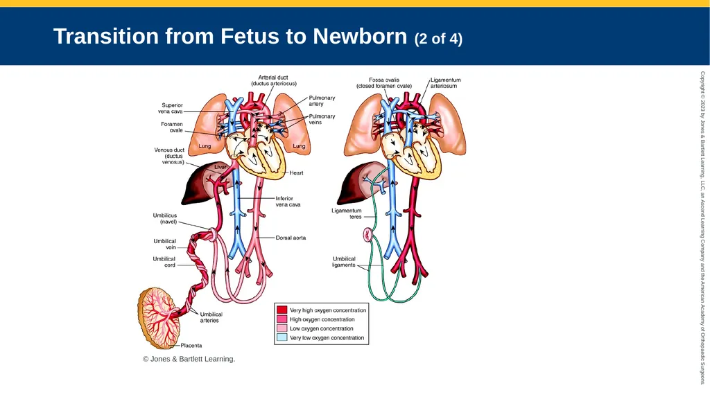
Transition from Fetus to Newborn (1 of 4) Fetal circulation has three major blood flow deviations or shunts:
Ductus venosus
Foramen ovale
Ductus arteriosus
These close off at birth<br>
Ductus venosus
Foramen ovale
Ductus arteriosus
These close off at birth<br>
05
Transition from Fetus to Newborn (2 of 4) © Jones & Bartlett Learning.<br>
06
Transition from Fetus to Newborn (3 of 4) First breath is triggered by mild hypoxia and hypercapnia.
Pulmonary vascular resistance drops as the lungs fill with air.
More blood flows to the lungs.<br>
Pulmonary vascular resistance drops as the lungs fill with air.
More blood flows to the lungs.<br>
07
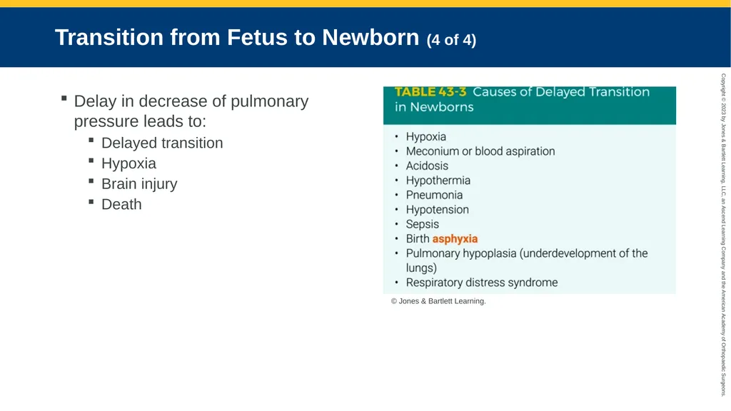
Transition from Fetus to Newborn (4 of 4) Delay in decrease of pulmonary pressure leads to:
Delayed transition
Hypoxia
Brain injury
Death © Jones & Bartlett Learning.<br>
Delayed transition
Hypoxia
Brain injury
Death © Jones & Bartlett Learning.<br>
08
Arrival of the Newborn (1 of 5) Obtain patient history and prepare environment.
Minimum needs:
Warm, dry blankets
Bulb syringe
Two small clamps or ties
Pair of clean scissors<br>
Minimum needs:
Warm, dry blankets
Bulb syringe
Two small clamps or ties
Pair of clean scissors<br>
09
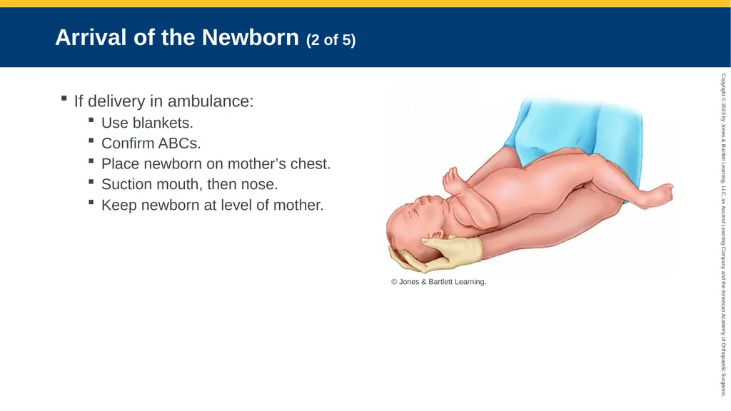
Arrival of the Newborn (2 of 5) If delivery in ambulance:
Use blankets.
Confirm ABCs.
Place newborn on mother’s chest.
Suction mouth, then nose.
Keep newborn at level of mother. © Jones & Bartlett Learning.<br>
Use blankets.
Confirm ABCs.
Place newborn on mother’s chest.
Suction mouth, then nose.
Keep newborn at level of mother. © Jones & Bartlett Learning.<br>
10
Arrival of the Newborn (3 of 5) Clamp and cut the umbilical cord.
Primary survey simultaneous with treatment interventions
Examine skin, head, and eyes for irregularities.
Inspect umbilical cord for abnormalities.
Note abdominal findings, such as hernias.<br>
Primary survey simultaneous with treatment interventions
Examine skin, head, and eyes for irregularities.
Inspect umbilical cord for abnormalities.
Note abdominal findings, such as hernias.<br>
11
Arrival of the Newborn (4 of 5) Newborns are at risk for hypothermia.
Place on prewarmed towels or radiant warmer.
Dry the head and body thoroughly.
Remove wet towels and cover with a dry towel.
Cover the head with a cap.<br>
Place on prewarmed towels or radiant warmer.
Dry the head and body thoroughly.
Remove wet towels and cover with a dry towel.
Cover the head with a cap.<br>
12
Arrival of the Newborn (5 of 5) All babies are cyanotic right after birth.
If the newborn remains vigorous and begins to turn pink in the first 5 minutes:
Maintain ongoing observation.
Continue thermoregulation with direct skin-to-skin contact with mother.<br>
If the newborn remains vigorous and begins to turn pink in the first 5 minutes:
Maintain ongoing observation.
Continue thermoregulation with direct skin-to-skin contact with mother.<br>
13
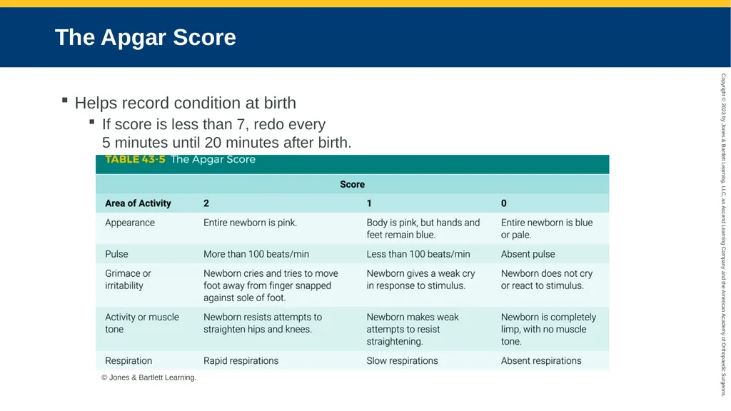
The Apgar Score Helps record condition at birth
If score is less than 7, redo every 5 minutes until 20 minutes after birth. © Jones & Bartlett Learning.<br>
If score is less than 7, redo every 5 minutes until 20 minutes after birth. © Jones & Bartlett Learning.<br>
14
Neonatal Resuscitation Algorithm (1 of 3) Follow current neonatal resuscitation guidelines.
First 60 seconds: Initial steps, reevaluation, and ventilation should occur.
Additional oxygen may be necessary if the target preductal oxygen saturation value has not been reached.<br>
First 60 seconds: Initial steps, reevaluation, and ventilation should occur.
Additional oxygen may be necessary if the target preductal oxygen saturation value has not been reached.<br>
15
Neonatal Resuscitation Algorithm (2 of 3) If the newborn’s pulse rate is apneic or less than 100 beats/min, begin PPV.
Begin chest compressions if the newborn’s pulse rate is less than 60 beats/min.
If ventilation and chest compression do not improve the bradycardia, administer epinephrine, preferably via IV line.<br>
Begin chest compressions if the newborn’s pulse rate is less than 60 beats/min.
If ventilation and chest compression do not improve the bradycardia, administer epinephrine, preferably via IV line.<br>
16
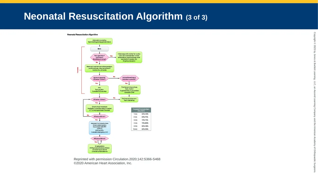
Neonatal Resuscitation Algorithm (3 of 3) Reprinted with permission Circulation.2020;142:S366-S468 ©2020 American Heart Association, Inc.<br>
17
Drying and Stimulation After ensuring airway patency, dry and stimulate newborn.
Position infant on back or side with neck in sniffing position.
Stimulate by drying newborn and flicking soles of feet.
Consider suctioning if PPV is needed or airway appears obstructed.<br>
Position infant on back or side with neck in sniffing position.
Stimulate by drying newborn and flicking soles of feet.
Consider suctioning if PPV is needed or airway appears obstructed.<br>
18
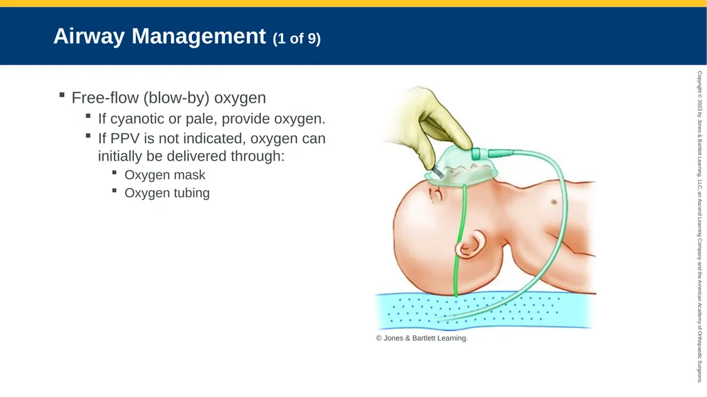
Airway Management (1 of 9) Free-flow (blow-by) oxygen
If cyanotic or pale, provide oxygen.
If PPV is not indicated, oxygen can initially be delivered through:
Oxygen mask
Oxygen tubing © Jones & Bartlett Learning.<br>
If cyanotic or pale, provide oxygen.
If PPV is not indicated, oxygen can initially be delivered through:
Oxygen mask
Oxygen tubing © Jones & Bartlett Learning.<br>
19
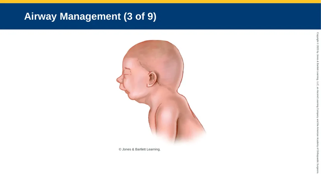
Airway Management (2 of 9) Oral airways
Conditions:
Bilateral choanal atresia
Pierre Robin sequence
Macroglossia
Craniofacial defects
In all these cases (except bilateral choanal atresia), an ET tube is inserted down a nostril.<br>
Conditions:
Bilateral choanal atresia
Pierre Robin sequence
Macroglossia
Craniofacial defects
In all these cases (except bilateral choanal atresia), an ET tube is inserted down a nostril.<br>
20
Airway Management (3 of 9) © Jones & Bartlett Learning.<br>
21
Airway Management (4 of 9) Bag-mask ventilation
Indicated if newborn:
Is apneic
Has inadequate respiratory effort
Has a pulse rate of less than 100 beats/min after the airway is cleared, airway is positioned, and stimulation is provided<br>
Indicated if newborn:
Is apneic
Has inadequate respiratory effort
Has a pulse rate of less than 100 beats/min after the airway is cleared, airway is positioned, and stimulation is provided<br>
22
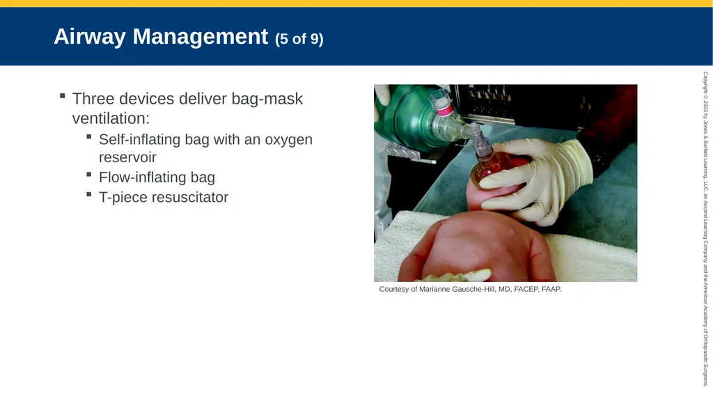
Airway Management (5 of 9) Three devices deliver bag-mask ventilation:
Self-inflating bag with an oxygen reservoir
Flow-inflating bag
T-piece resuscitator Courtesy of Marianne Gausche-Hill, MD, FACEP, FAAP.<br>
Self-inflating bag with an oxygen reservoir
Flow-inflating bag
T-piece resuscitator Courtesy of Marianne Gausche-Hill, MD, FACEP, FAAP.<br>
23
Airway Management (6 of 9) Bag-mask ventilation (cont’d)
Correct ventilation rate: 40 to 60 breaths/min
Causes of ineffective bag-mask ventilation:
Inadequate mask seal on the face
Incorrect head position
Copious secretions
Pneumothorax
Equipment malfunction<br>
Correct ventilation rate: 40 to 60 breaths/min
Causes of ineffective bag-mask ventilation:
Inadequate mask seal on the face
Incorrect head position
Copious secretions
Pneumothorax
Equipment malfunction<br>
24
Airway Management (7 of 9) Intubation
Indications:
Meconium-stained fluid, nonvigorous newborn
Congenital diaphragmatic hernia
Prolonged PPV needed
Craniofacial anomalies impede airway<br>
Indications:
Meconium-stained fluid, nonvigorous newborn
Congenital diaphragmatic hernia
Prolonged PPV needed
Craniofacial anomalies impede airway<br>
25
Airway Management (8 of 9) Intubation (cont’d)
Equipment needed:
Suction equipment
Laryngoscope
Blades
Shoulder roll and adhesive tape
ET tube and stylet<br>
Equipment needed:
Suction equipment
Laryngoscope
Blades
Shoulder roll and adhesive tape
ET tube and stylet<br>
26
Airway Management (9 of 9) Gastric decompression
Indications:
Prolonged bag-mask ventilation
Abdominal distention impeding ventilation
Diaphragmatic hernia or gastrointestinal congenital anomaly<br>
Indications:
Prolonged bag-mask ventilation
Abdominal distention impeding ventilation
Diaphragmatic hernia or gastrointestinal congenital anomaly<br>
27
Circulation (1 of 6) Chest compressions
Indicated if pulse rate remains at less than 60 beats/min after resuscitation efforts
Two people are needed for effective chest compressions and ventilation.<br>
Indicated if pulse rate remains at less than 60 beats/min after resuscitation efforts
Two people are needed for effective chest compressions and ventilation.<br>
28
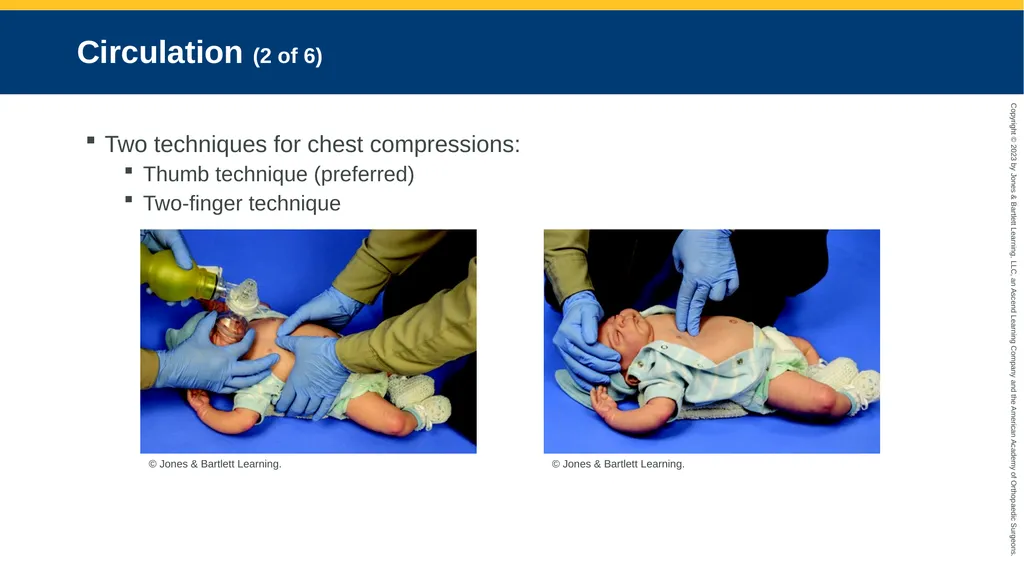
Circulation (2 of 6) Two techniques for chest compressions:
Thumb technique (preferred)
Two-finger technique © Jones & Bartlett Learning. © Jones & Bartlett Learning.<br>
Thumb technique (preferred)
Two-finger technique © Jones & Bartlett Learning. © Jones & Bartlett Learning.<br>
29
Circulation (3 of 6) Chest compressions (cont’d)
Depth: One-third of the anteroposterior diameter
Do not deliver simultaneously with artificial ventilation.
Coordinate 90 compressions and 30 breaths/min.<br>
Depth: One-third of the anteroposterior diameter
Do not deliver simultaneously with artificial ventilation.
Coordinate 90 compressions and 30 breaths/min.<br>
30
Circulation (4 of 6) Chest compressions (cont’d)
Reassess for the presence of a pulse after 60 seconds of well-coordinated chest compressions and ventilations.
If the pulse rate is above 60 beats/min, stop compressions and recheck pulse.
If rate goes above 100 beats/min, gradually slow the rate and decrease PPV pressure.<br>
Reassess for the presence of a pulse after 60 seconds of well-coordinated chest compressions and ventilations.
If the pulse rate is above 60 beats/min, stop compressions and recheck pulse.
If rate goes above 100 beats/min, gradually slow the rate and decrease PPV pressure.<br>
31
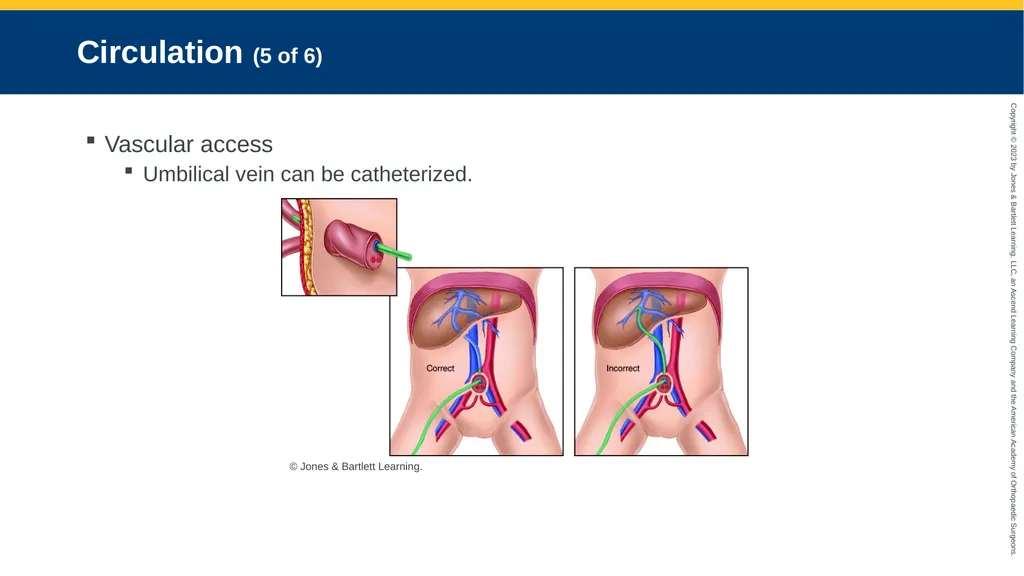
Circulation (5 of 6) Vascular access
Umbilical vein can be catheterized. © Jones & Bartlett Learning.<br>
Umbilical vein can be catheterized. © Jones & Bartlett Learning.<br>
32
Circulation (6 of 6) Vascular access (cont’d)
IV and IO access are more common in prehospital setting.
Seek medical director’s guidance to determine if IO option is available.<br>
IV and IO access are more common in prehospital setting.
Seek medical director’s guidance to determine if IO option is available.<br>
33
Pharmacologic Interventions Rarely needed in newborn resuscitation
Medication dosages based on weight<br>
Medication dosages based on weight<br>
34
Family and Transport Considerations (1 of 2) Transport to nearest facility once the newborn is stabilized as much as possible.
Communicate with the family.
Monitor the newborn and assess status.<br>
Communicate with the family.
Monitor the newborn and assess status.<br>
35
Family and Transport Considerations (2 of 2) Transport of a high-risk newborn
Physician at referring hospital initiates request.
Mode of transportation is chosen.
Transport team is mobilized and equipment assembled.
On arrival, transport team continues to stabilize the newborn.<br>
Physician at referring hospital initiates request.
Mode of transportation is chosen.
Transport team is mobilized and equipment assembled.
On arrival, transport team continues to stabilize the newborn.<br>
36
Apnea (1 of 2) Respiratory pause of 20 seconds or longer
Can lead to hypoxemia and bradycardia
Often follows hypoxia or hypothermia
Newborn needs respiratory support to minimize brain and organ damage.<br>
Can lead to hypoxemia and bradycardia
Often follows hypoxia or hypothermia
Newborn needs respiratory support to minimize brain and organ damage.<br>
37
Apnea (2 of 2) Assessment and management
Careful history to find etiologic risk factors
Perform physical exam.
Differentiate between:
Primary apnea
Secondary apnea<br>
Careful history to find etiologic risk factors
Perform physical exam.
Differentiate between:
Primary apnea
Secondary apnea<br>
38
Bradycardia (1 of 2) Most frequently is a result of hypoxia
Often responds to effective PPV
Some causes include hypovolemia, pneumothorax, and head injury.
Therapeutic interventions can also cause bradycardia.<br>
Often responds to effective PPV
Some causes include hypovolemia, pneumothorax, and head injury.
Therapeutic interventions can also cause bradycardia.<br>
39
Bradycardia (2 of 2) Assessment and management
Heart rate less than 100 beats/min:
Provide PPV.
If still less than 60 beats/min:
Begin chest compressions.
If still less than 60 beats/min:
Administer epinephrine.
Check pulse after 1 minute.<br>
Heart rate less than 100 beats/min:
Provide PPV.
If still less than 60 beats/min:
Begin chest compressions.
If still less than 60 beats/min:
Administer epinephrine.
Check pulse after 1 minute.<br>
40
Acidosis (1 of 2) Suspect metabolic acidosis if bradycardia persists after:
Adequate ventilation
Chest compressions
Volume expansion<br>
Adequate ventilation
Chest compressions
Volume expansion<br>
41
Acidosis (2 of 2) Consider administering a bolus of normal saline to aid in improved perfusion and clearance of acid.
Best treatment is to identify and correct the underlying cause.<br>
Best treatment is to identify and correct the underlying cause.<br>
42
Pneumothorax Decompression (1 of 2) A pneumothorax can occur if:
Infant inhales meconium.
Lung is weakened by infection.
PPV is too aggressive.
Signs of significant pneumothorax:
Severe respiratory distress unresponsive to PPV
Unilateral decreased breath sounds<br>
Infant inhales meconium.
Lung is weakened by infection.
PPV is too aggressive.
Signs of significant pneumothorax:
Severe respiratory distress unresponsive to PPV
Unilateral decreased breath sounds<br>
43
Pneumothorax Decompression (2 of 2) Assessment and management
Clean area with alcohol.
Prepare equipment.
Insert needle above upper edge of second rib.
If symptomatic ongoing air leak, insert a 22-gauge angiocatheter in a similar location.
During transport, monitor for reaccumulation of the pneumothorax.<br>
Clean area with alcohol.
Prepare equipment.
Insert needle above upper edge of second rib.
If symptomatic ongoing air leak, insert a 22-gauge angiocatheter in a similar location.
During transport, monitor for reaccumulation of the pneumothorax.<br>
44
Meconium-Stained Amniotic Fluid (1 of 4) Carries a high risk of morbidity
If newborns inhale meconium-stained amniotic fluid, airway may become plugged.
Atelectasis
Persistent pulmonary hypertension
Hypoxemia
Aspiration pneumonitis
Pneumothorax<br>
If newborns inhale meconium-stained amniotic fluid, airway may become plugged.
Atelectasis
Persistent pulmonary hypertension
Hypoxemia
Aspiration pneumonitis
Pneumothorax<br>
45
Meconium-Stained Amniotic Fluid (2 of 4) To decrease risk of persistent pulmonary hypertension:
Ensure a clear airway.
Keep newborn warm.
Minimize stimulation.
Administer supplemental oxygen when necessary.<br>
Ensure a clear airway.
Keep newborn warm.
Minimize stimulation.
Administer supplemental oxygen when necessary.<br>
46
Meconium-Stained Amniotic Fluid (3 of 4) Assessment and management
If depressed, start PPV.
Intubation and suctioning may be required to remove an obstruction.<br>
If depressed, start PPV.
Intubation and suctioning may be required to remove an obstruction.<br>
47
Meconium-Stained Amniotic Fluid (4 of 4) Assessment and management (cont’d)
If the newborn is not responding well to the care outlined in the neonatal resuscitation algorithm:
Suspect airway occlusion or pneumothorax.
Minimize hypothermia.
Reassess frequently.<br>
If the newborn is not responding well to the care outlined in the neonatal resuscitation algorithm:
Suspect airway occlusion or pneumothorax.
Minimize hypothermia.
Reassess frequently.<br>
48
Low Blood Volume (1 of 2) Fluid resuscitation may be needed if the newborn has significant depletion due to:
Abruptio placentae
Twin-to-twin transfusion
Placenta previa
Septic shock<br>
Abruptio placentae
Twin-to-twin transfusion
Placenta previa
Septic shock<br>
49
Low Blood Volume (2 of 2) Signs of hypovolemia include:
Pallor
Persistently low pulse rate
Weak pulse
No improvement in circulatory status
Consider administering a fluid bolus.<br>
Pallor
Persistently low pulse rate
Weak pulse
No improvement in circulatory status
Consider administering a fluid bolus.<br>
50
Diaphragmatic Hernia (1 of 2) An abnormal opening in the diaphragm
Postnatal signs and symptoms include:
Respiratory distress
Heart sounds shifted to the right
Bowel sounds heard in the chest<br>
Postnatal signs and symptoms include:
Respiratory distress
Heart sounds shifted to the right
Bowel sounds heard in the chest<br>
51
Diaphragmatic Hernia (2 of 2) Assessment and management
May be few or no symptoms or severe hypoxia
Resuscitate with supplemental oxygen.
Monitor heart rate continuously.
Ultimately requires surgical correction<br>
May be few or no symptoms or severe hypoxia
Resuscitate with supplemental oxygen.
Monitor heart rate continuously.
Ultimately requires surgical correction<br>
52
Respiratory Distress and Cyanosis (1 of 3) Single most common cause is prematurity
Respiratory causes
Other causes:
CNS depression
Septic shock and severe metabolic acidosis
Cardiac anomalies<br>
Respiratory causes
Other causes:
CNS depression
Septic shock and severe metabolic acidosis
Cardiac anomalies<br>
53
Respiratory Distress and Cyanosis (2 of 3) Assessment and management
Ensure patent airway.
Check breathing is adequate.
Check pulse is present.
Assess respirations.
Ask about increased symptoms with feeding.<br>
Ensure patent airway.
Check breathing is adequate.
Check pulse is present.
Assess respirations.
Ask about increased symptoms with feeding.<br>
54
Respiratory Distress and Cyanosis (3 of 3) Assessment and management (cont’d)
Treatment includes establishing:
Patent airway
Adequate oxygen delivery
Effective ventilation
Adequate circulation<br>
Treatment includes establishing:
Patent airway
Adequate oxygen delivery
Effective ventilation
Adequate circulation<br>
55
Respiratory Depression Secondary to Opioids (1 of 3) Administration of naloxone (Narcan) to the newborn of a mother with opioid use disorder:
May precipitate seizures that can potentially cause death
No longer recommended as a first-line drug in resuscitation<br>
May precipitate seizures that can potentially cause death
No longer recommended as a first-line drug in resuscitation<br>
56
Respiratory Depression Secondary to Opioids (2 of 3) For respiratory depression from the mother’s chronic use of opioids:
Provide ventilatory support.
Transport immediately.<br>
Provide ventilatory support.
Transport immediately.<br>
57
Respiratory Depression Secondary to Opioids (3 of 3) If respiratory depression is the result of the mother being treated acutely with opioids:
Naloxone may be administered to the newborn via the IV (preferred) or intramuscular route to reverse the opioid effect.<br>
Naloxone may be administered to the newborn via the IV (preferred) or intramuscular route to reverse the opioid effect.<br>
58
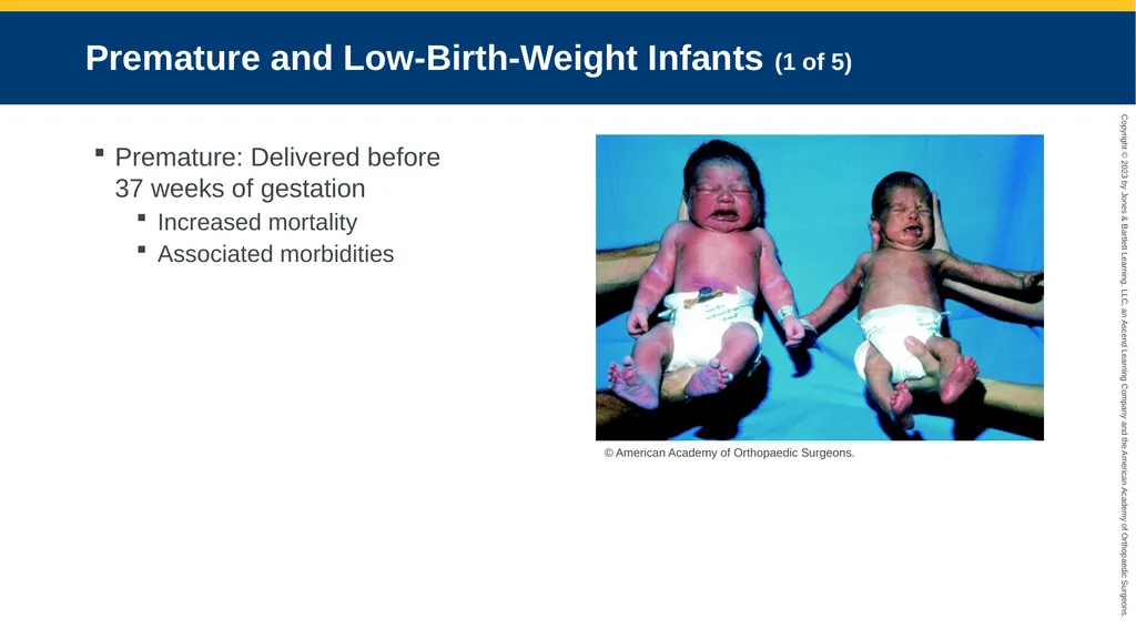
Premature and Low-Birth-Weight Infants (1 of 5) Premature: Delivered before
37 weeks of gestation
Increased mortality
Associated morbidities © American Academy of Orthopaedic Surgeons.<br>
Increased mortality
Associated morbidities © American Academy of Orthopaedic Surgeons.<br>
59
Premature and Low-Birth-Weight Infants (2 of 5) Low birth weight—newborns weighing less than 5.5 lb (2,500 g)
Morbidity and mortality are related to degree of prematurity.<br>
Morbidity and mortality are related to degree of prematurity.<br>
60
Premature and Low-Birth-Weight Infants (3 of 5) To determine prematurity, rely on:
Physical features
Information from family about gestational dating
Information related to maternal or fetal complications<br>
Physical features
Information from family about gestational dating
Information related to maternal or fetal complications<br>
61
Premature and Low-Birth-Weight Infants (4 of 5) To optimize survival in the field:
Provide cardiorespiratory support.
Provide thermoneutral environment.
Use only minimum pressure necessary to move chest when providing PPV.<br>
Provide cardiorespiratory support.
Provide thermoneutral environment.
Use only minimum pressure necessary to move chest when providing PPV.<br>
62
Premature and Low-Birth-Weight Infants (5 of 5) Assessment and management
Management focuses on:
Clearing airway
Gentle stimulation
Administering supplemental oxygen and PPV if needed
Providing chest compressions
Maintaining a warm environment<br>
Management focuses on:
Clearing airway
Gentle stimulation
Administering supplemental oxygen and PPV if needed
Providing chest compressions
Maintaining a warm environment<br>
63
Seizures in the Newborn (1 of 4) Suggest the presence of neurologic disorder
Usually related to an underlying abnormality
Prolonged seizures may cause brain injury.<br>
Usually related to an underlying abnormality
Prolonged seizures may cause brain injury.<br>
64
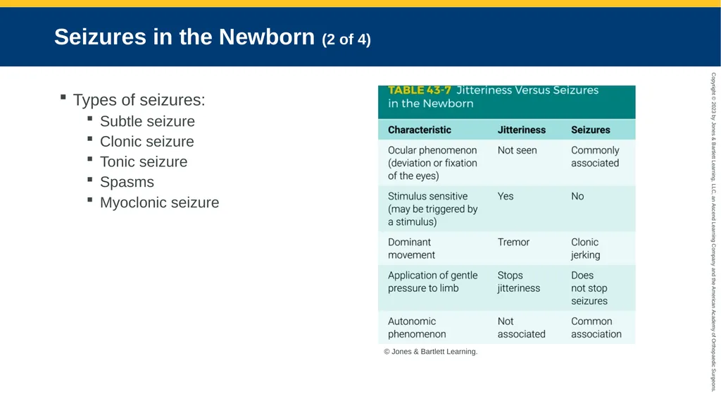
Seizures in the Newborn (2 of 4) Types of seizures:
Subtle seizure
Clonic seizure
Tonic seizure
Spasms
Myoclonic seizure © Jones & Bartlett Learning.<br>
Subtle seizure
Clonic seizure
Tonic seizure
Spasms
Myoclonic seizure © Jones & Bartlett Learning.<br>
65
Seizures in the Newborn (3 of 4) Assessment and management
Evaluate prenatal and birth history.
Perform a careful physical exam.
Obtain vital signs and oxygen saturation.
Provide additional oxygen, assisted ventilation, blood pressure evaluation, and IV access.<br>
Evaluate prenatal and birth history.
Perform a careful physical exam.
Obtain vital signs and oxygen saturation.
Provide additional oxygen, assisted ventilation, blood pressure evaluation, and IV access.<br>
66
Seizures in the Newborn (4 of 4) Assessment and management (cont’d)
If blood glucose level is less than 40 mg/dL, give an IV bolus of 10% dextrose solution.
Monitor respiratory status and oxygen saturation.
Maintain normal body temperature.
Keep family informed as you transport.<br>
If blood glucose level is less than 40 mg/dL, give an IV bolus of 10% dextrose solution.
Monitor respiratory status and oxygen saturation.
Maintain normal body temperature.
Keep family informed as you transport.<br>
67
Hypoglycemia (1 of 3) Blood glucose level of less than 45 mg/dL
May result in:
Seizures
Brain damage<br>
May result in:
Seizures
Brain damage<br>
68
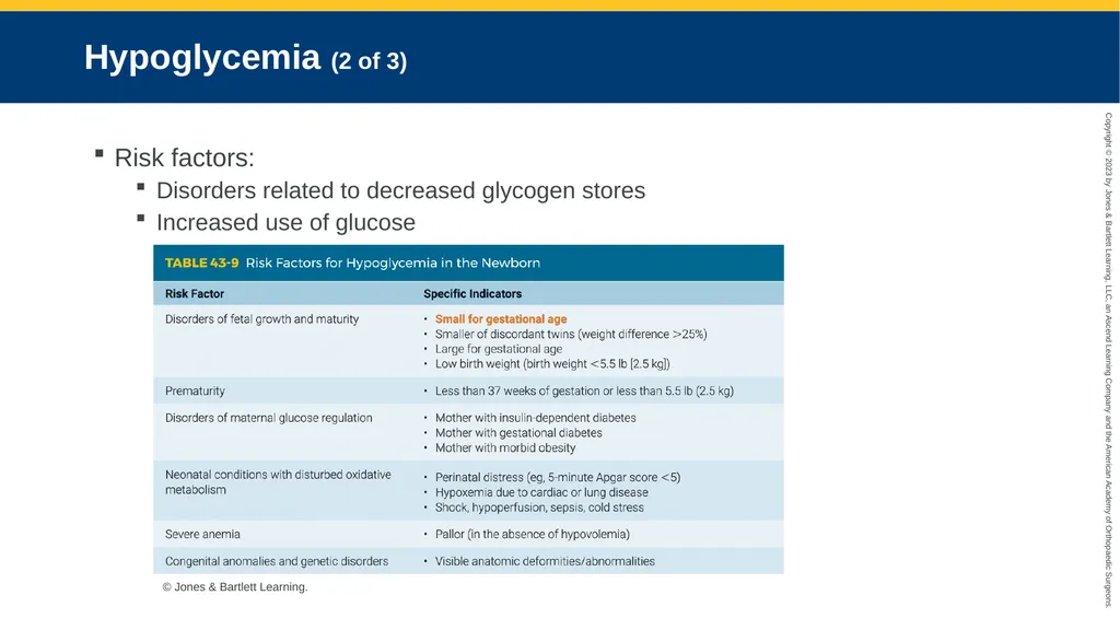
Hypoglycemia (2 of 3) Risk factors:
Disorders related to decreased glycogen stores
Increased use of glucose © Jones & Bartlett Learning.<br>
Disorders related to decreased glycogen stores
Increased use of glucose © Jones & Bartlett Learning.<br>
69
Hypoglycemia (3 of 3) Assessment and management
Symptoms vary.
Check blood glucose level and vital signs.
Manage hypoglycemia after establishing good oxygen, ventilation, and circulation.
Maintain normal body temperature.<br>
Symptoms vary.
Check blood glucose level and vital signs.
Manage hypoglycemia after establishing good oxygen, ventilation, and circulation.
Maintain normal body temperature.<br>
70
Vomiting (1 of 5) Common in newborns
Persistent or bilious vomiting may indicate:
Upper digestive tract obstruction
Increased intracranial pressure
Vomitus aspiration may cause respiratory complications, including airway obstruction.<br>
Persistent or bilious vomiting may indicate:
Upper digestive tract obstruction
Increased intracranial pressure
Vomitus aspiration may cause respiratory complications, including airway obstruction.<br>
71
Vomiting (2 of 5) Causes of vomiting
Esophageal atresia
Gastroesophageal reflux
Infantile hypertrophic pyloric stenosis (IHPS)
Intestinal malrotation
Congenital conditions
Hirschsprung disease<br>
Esophageal atresia
Gastroesophageal reflux
Infantile hypertrophic pyloric stenosis (IHPS)
Intestinal malrotation
Congenital conditions
Hirschsprung disease<br>
72
Vomiting (3 of 5) Sudden, unexpected, and forceful vomiting may occur in conjunction with:
Asphyxia
Meningitis
Hydrocephalus
Withdrawal syndromes<br>
Asphyxia
Meningitis
Hydrocephalus
Withdrawal syndromes<br>
73
Vomiting (4 of 5) Assessment and management
Stomach may be distended.
Suspect infection if newborn has fever or history of contact with people who are ill.
May also note:
Temperature instability
Apnea/bradycardia
Abdominal tenderness/guarding
Minimal/absent bowel sounds<br>
Stomach may be distended.
Suspect infection if newborn has fever or history of contact with people who are ill.
May also note:
Temperature instability
Apnea/bradycardia
Abdominal tenderness/guarding
Minimal/absent bowel sounds<br>
74
Vomiting (5 of 5) Assessment and management (cont’d)
Start management with ABCs.
Consider decompressing the stomach.
Do not administer antiemetics.
May need fluid resuscitation if dehydrated
Place newborn on their side when transporting.<br>
Start management with ABCs.
Consider decompressing the stomach.
Do not administer antiemetics.
May need fluid resuscitation if dehydrated
Place newborn on their side when transporting.<br>
75
Diarrhea (1 of 2) Common cause for hospitalization in neonatal and pediatric populations
Causes include:
Poisoning
Gastroenteritis
Lactose intolerance
Neonatal abstinence syndrome<br>
Causes include:
Poisoning
Gastroenteritis
Lactose intolerance
Neonatal abstinence syndrome<br>
76
Diarrhea (2 of 2) Assessment and management
Estimate:
Number and volume of loose stools
Decreased urinary output
Degree of dehydration
Patient management begins with ABCs.<br>
Estimate:
Number and volume of loose stools
Decreased urinary output
Degree of dehydration
Patient management begins with ABCs.<br>
77
Neonatal Jaundice (1 of 2) Results from immaturity of the liver to conjugate bilirubin in the first week
Can result from:
Hemolysis
Red blood cell disorders
Polycythemia
Excessive bruising<br>
Can result from:
Hemolysis
Red blood cell disorders
Polycythemia
Excessive bruising<br>
78
Neonatal Jaundice (2 of 2) Assessment and management
Transport is essential.
Start on IV fluids if neonate shows significant clinical jaundice.
Communicate with medical control.
Phototherapy is treatment in most cases.<br>
Transport is essential.
Start on IV fluids if neonate shows significant clinical jaundice.
Communicate with medical control.
Phototherapy is treatment in most cases.<br>
79
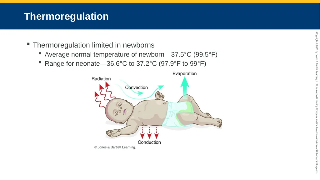
Thermoregulation Thermoregulation limited in newborns
Average normal temperature of newborn—37.5°C (99.5°F)
Range for neonate—36.6°C to 37.2°C (97.9°F to 99°F) © Jones & Bartlett Learning.<br>
Average normal temperature of newborn—37.5°C (99.5°F)
Range for neonate—36.6°C to 37.2°C (97.9°F to 99°F) © Jones & Bartlett Learning.<br>
80
Fever (1 of 4) Rectal temperature greater than 38°C (100.4°F)
Newborn may not always present with fever in an illness or infection.
May be caused by overheating or dehydration<br>
Newborn may not always present with fever in an illness or infection.
May be caused by overheating or dehydration<br>
81
Fever (2 of 4) Limited ability to control body temperature.
Signs and symptoms include:
Irritability
Somnolence
Decreased feeding
Warm to touch<br>
Signs and symptoms include:
Irritability
Somnolence
Decreased feeding
Warm to touch<br>
82
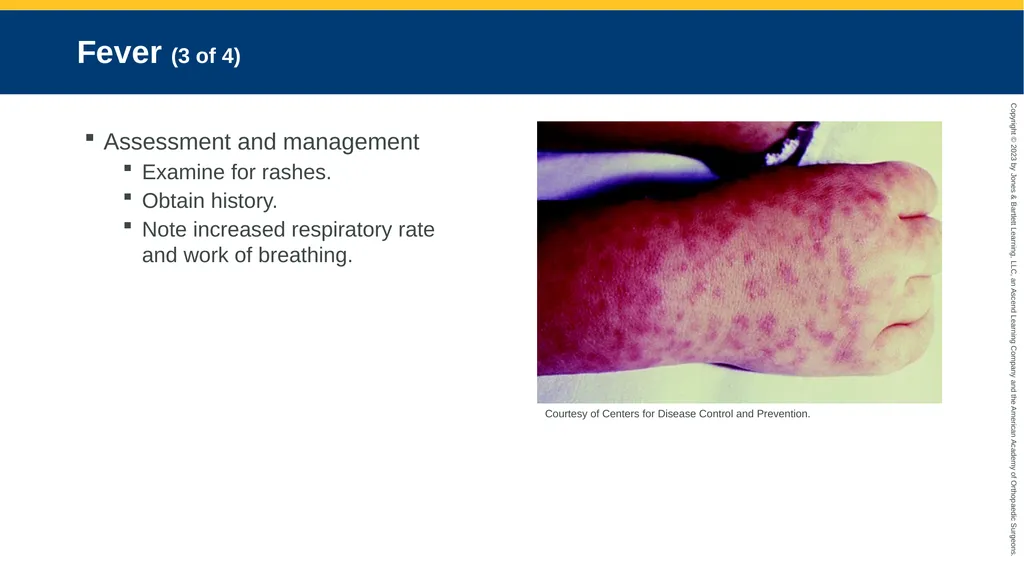
Fever (3 of 4) Assessment and management
Examine for rashes.
Obtain history.
Note increased respiratory rate and work of breathing. Courtesy of Centers for Disease Control and Prevention.<br>
Examine for rashes.
Obtain history.
Note increased respiratory rate and work of breathing. Courtesy of Centers for Disease Control and Prevention.<br>
83
Fever (4 of 4) Assessment and management (cont’d)
Obtain vital signs and ensure adequate oxygenation and ventilation.
Perform chest compressions, if indicated.
Cool the patient.<br>
Obtain vital signs and ensure adequate oxygenation and ventilation.
Perform chest compressions, if indicated.
Cool the patient.<br>
84
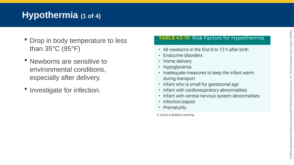
Hypothermia (1 of 4) Drop in body temperature to less than 35°C (95°F)
Newborns are sensitive to environmental conditions, especially after delivery.
Investigate for infection. © Jones & Bartlett Learning.<br>
Newborns are sensitive to environmental conditions, especially after delivery.
Investigate for infection. © Jones & Bartlett Learning.<br>
85
Hypothermia (2 of 4) Assessment and management
Hypothermic newborns may be:
Cool to the touch
Pale with acrocyanosis
Presentation may include:
Apnea
Bradycardia
Central cyanosis<br>
Hypothermic newborns may be:
Cool to the touch
Pale with acrocyanosis
Presentation may include:
Apnea
Bradycardia
Central cyanosis<br>
86
Hypothermia (3 of 4) Preventive measures include:
Warming hands before touching the newborn
Drying thoroughly after birth
Placing a prewarmed cap on the head
Placing the newborn skin-to-skin with mother<br>
Warming hands before touching the newborn
Drying thoroughly after birth
Placing a prewarmed cap on the head
Placing the newborn skin-to-skin with mother<br>
87
Hypothermia (4 of 4) Treatment includes:
Administration of D10W if newborn is hypoglycemic
Once stabilized:
Cover newborn with warm blankets and place on mother’s chest.
Turn up the heat in the ambulance.
Consider heat packs, but never place directly on skin.<br>
Administration of D10W if newborn is hypoglycemic
Once stabilized:
Cover newborn with warm blankets and place on mother’s chest.
Turn up the heat in the ambulance.
Consider heat packs, but never place directly on skin.<br>
88
Common Birth Injuries in the Newborn (1 of 5) Most are self-limiting
Newborn injuries can occur because of:
Newborn size
Position during labor and delivery<br>
Newborn injuries can occur because of:
Newborn size
Position during labor and delivery<br>
89
Common Birth Injuries in the Newborn (2 of 5) Birth trauma injuries include:
Those involving instruments during delivery
Excessive molding of the head
Caput succedaneum
Cephalohematoma
Linear skull fractures<br>
Those involving instruments during delivery
Excessive molding of the head
Caput succedaneum
Cephalohematoma
Linear skull fractures<br>
90
Common Birth Injuries in the Newborn (3 of 5) Birth trauma injuries include (cont’d):
Brachial plexus injuries
Facial nerve palsy
Diaphragmatic paralysis
Laryngeal nerve injury
Spinal cord injury<br>
Brachial plexus injuries
Facial nerve palsy
Diaphragmatic paralysis
Laryngeal nerve injury
Spinal cord injury<br>
91
Common Birth Injuries in the Newborn (4 of 5) Clavicle—most frequently fractured bone
Examination will show:
Crepitus
Palpable bony irregularity
Possible lack of arm movement on affected side<br>
Examination will show:
Crepitus
Palpable bony irregularity
Possible lack of arm movement on affected side<br>
92
Common Birth Injuries in the Newborn (5 of 5) Long bone fracture may present as loss of spontaneous arm or leg movement.
Intra-abdominal injury is uncommon.
Hypoxia and shock could be caused by birth trauma.<br>
Intra-abdominal injury is uncommon.
Hypoxia and shock could be caused by birth trauma.<br>
93
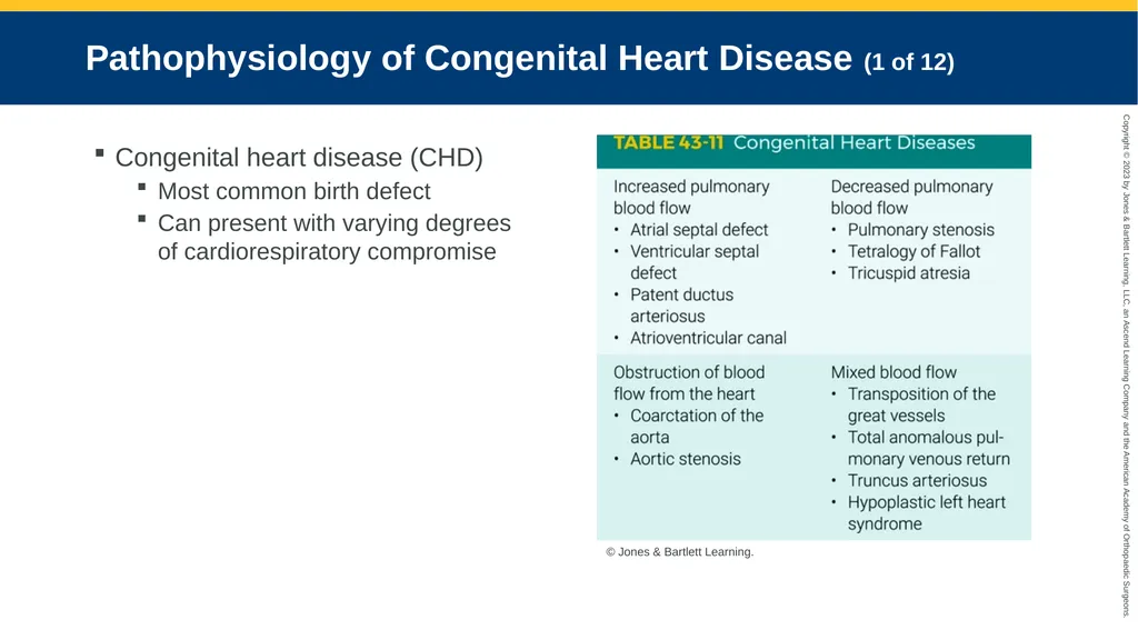
Pathophysiology of Congenital Heart Disease (1 of 12) Congenital heart disease (CHD)
Most common birth defect
Can present with varying degrees of cardiorespiratory compromise © Jones & Bartlett Learning.<br>
Most common birth defect
Can present with varying degrees of cardiorespiratory compromise © Jones & Bartlett Learning.<br>
94
Pathophysiology of Congenital Heart Disease (2 of 12) Noncyanotic disease
Oxygenated blood is shunted from the left side of the heart to the right side.
Called a left-to-right shunt<br>
Oxygenated blood is shunted from the left side of the heart to the right side.
Called a left-to-right shunt<br>
95
Pathophysiology of Congenital Heart Disease (3 of 12) Atrial septal defect
Abnormal opening exists in the wall separating the atrial chambers of the heart.
Ventricular septal defect:
Abnormal opening exists in the wall separating the right and left ventricles.<br>
Abnormal opening exists in the wall separating the atrial chambers of the heart.
Ventricular septal defect:
Abnormal opening exists in the wall separating the right and left ventricles.<br>
96
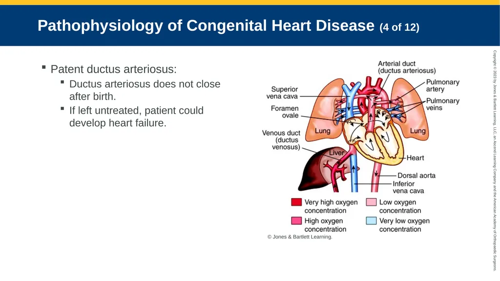
Pathophysiology of Congenital Heart Disease (4 of 12) Patent ductus arteriosus:
Ductus arteriosus does not close after birth.
If left untreated, patient could develop heart failure. © Jones & Bartlett Learning.<br>
Ductus arteriosus does not close after birth.
If left untreated, patient could develop heart failure. © Jones & Bartlett Learning.<br>
97
Pathophysiology of Congenital Heart Disease (5 of 12) Coarctation of the aorta (CoA)
Narrowing of the aorta
May be associated with other cardiac defects
Often asymptomatic until later in childhood<br>
Narrowing of the aorta
May be associated with other cardiac defects
Often asymptomatic until later in childhood<br>
98
Pathophysiology of Congenital Heart Disease (6 of 12) Cyanotic disease
Deoxygenated blood from the right side of the heart mixes with the left side.
Known as a right-to-left shunt<br>
Deoxygenated blood from the right side of the heart mixes with the left side.
Known as a right-to-left shunt<br>
99
Pathophysiology of Congenital Heart Disease (7 of 12) Pulmonary stenosis
Pulmonic valve near right ventricle becomes damaged.
Patient will present with:
Jugular vein distention
Cyanosis
Right ventricular hypertrophy<br>
Pulmonic valve near right ventricle becomes damaged.
Patient will present with:
Jugular vein distention
Cyanosis
Right ventricular hypertrophy<br>
100
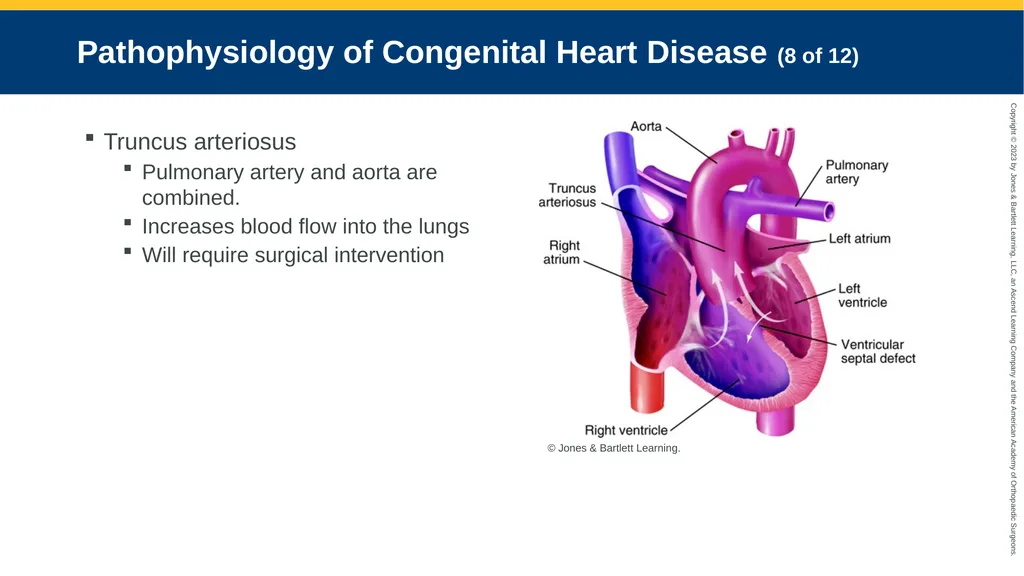
Pathophysiology of Congenital Heart Disease (8 of 12) Truncus arteriosus
Pulmonary artery and aorta are combined.
Increases blood flow into the lungs
Will require surgical intervention © Jones & Bartlett Learning.<br>
Pulmonary artery and aorta are combined.
Increases blood flow into the lungs
Will require surgical intervention © Jones & Bartlett Learning.<br>
101
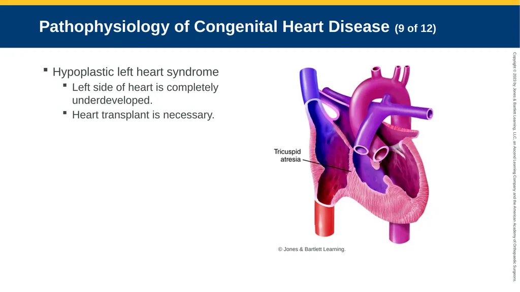
Pathophysiology of Congenital Heart Disease (9 of 12) Hypoplastic left heart syndrome
Left side of heart is completely underdeveloped.
Heart transplant is necessary. © Jones & Bartlett Learning.<br>
Left side of heart is completely underdeveloped.
Heart transplant is necessary. © Jones & Bartlett Learning.<br>
102
Pathophysiology of Congenital Heart Disease (10 of 12) Tetralogy of Fallot (ToF)
Combination of four heart defects:
Ventricular septal defect
Pulmonary stenosis
Right ventricular hypertrophy
Overriding aorta
Open heart surgery is required.<br>
Combination of four heart defects:
Ventricular septal defect
Pulmonary stenosis
Right ventricular hypertrophy
Overriding aorta
Open heart surgery is required.<br>
103
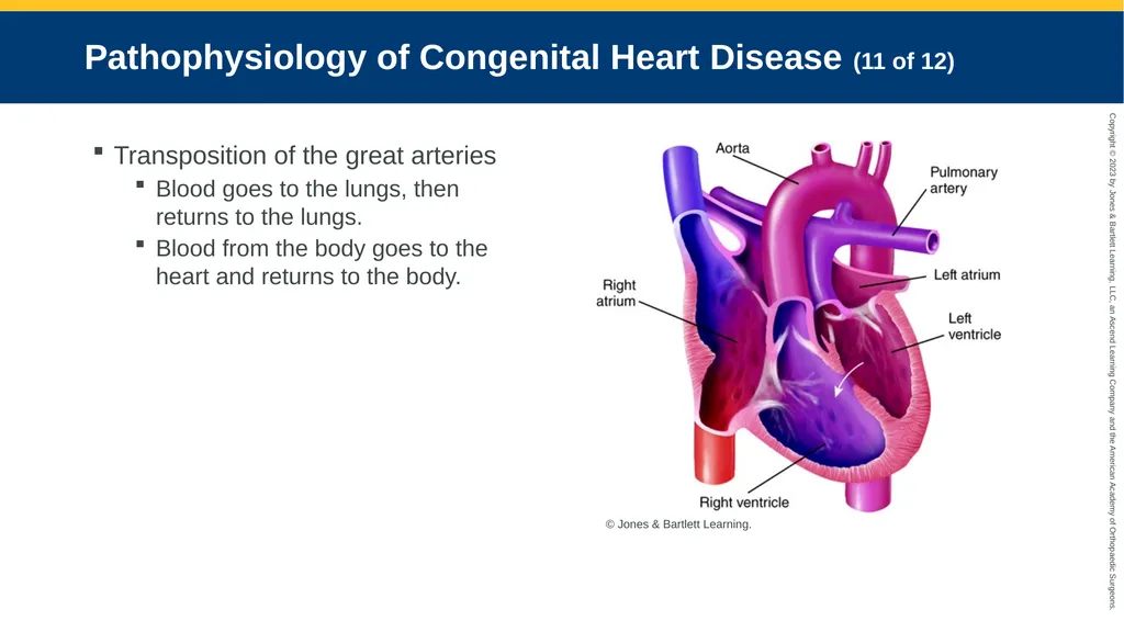
Pathophysiology of Congenital Heart Disease (11 of 12) Transposition of the great arteries
Blood goes to the lungs, then returns to the lungs.
Blood from the body goes to the heart and returns to the body. © Jones & Bartlett Learning.<br>
Blood goes to the lungs, then returns to the lungs.
Blood from the body goes to the heart and returns to the body. © Jones & Bartlett Learning.<br>
104
Pathophysiology of Congenital Heart Disease (12 of 12) Total anomalous pulmonary venous return
Four pulmonary veins connect to the right atrium instead of the left atrium.
Results in pulmonary overcirculation due to excessive blood return to right side of heart.<br>
Four pulmonary veins connect to the right atrium instead of the left atrium.
Results in pulmonary overcirculation due to excessive blood return to right side of heart.<br>
105
Assessment and Management of Congenital Heart Disease Rapid detection and transport are mandatory.
Communication with medical control is crucial.<br>
Communication with medical control is crucial.<br>