
D DIARRHOEA Dr. Anil Kumar Asst. Prof. VCC, BVC,




Description: D DIARRHOEA Dr. Anil Kumar Asst. Prof. VCC, BVC, Patna VCP-II, 4th Professional Diarrhoea Diarrhoea is defined as increased volume or fluidity of faeces or increased frequency of defecation. Normal physiologic functions of the small
Related Topics
Download Presentation
"D DIARRHOEA Dr. Anil Kumar Asst. Prof. VCC, BVC," is the property of its rightful owner. Permission is granted to download and print the materials on this website for personal, non-commercial use only, and to display it on your personal computer provided you do not modify the materials and that you retain all copyright notices contained in the materials. By downloading content from our website, you accept the terms of this agreement.
Presentation Transcript
slide1. D
DIARRHOEA
Dr. Anil Kumar
Asst. Prof. VCC, BVC, Patna
VCP-II, 4th Professional<br>
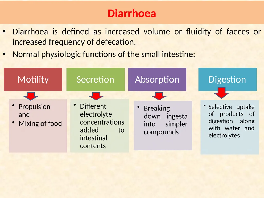
slide2. Diarrhoea Diarrhoea is defined as increased volume or fluidity of faeces or increased frequency of defecation.
Normal physiologic functions of the small intestine:<br>
slide3. Food enters the stomach leads to the production of semj-fluid chyme with the help of combination of an acid environment, pepsin proteolytic activity and active gastric motility.
Then it passed to duodenum in controlled quantities and also receives intestinal and pancreatic secretions and bile, which are mixed with chyme.
Pancreatic secretions (sodium bicarbonate)neutralizes gastric acid and also releases lipase, amylase and trypsin
Bile contains bile salts and bile pigments, but the bile salts are important for the emulsification of undigested fats into micelles, which greatly increases exposure to the fat digesting enzyme lipase and produce free fatty acids and monoglycerides
Carbohydrate digestion is initiated by salivary amylase and contiJ1ued by pancreatic amylase.<br>
slide4. The produced disaccharides and oligosaccharides, which are further digested by disaccharidase (lactase and sucrase) and oligosaccharidase enzymes in the small intestinal brush border.
Monosaccbarides are absorbed into the mucosal cell by co-transport with sodium (glucose, galactose) or by facilitated diffusion (fructose).
Protein digestion is initiated by pepsin in the stomach and continued by pancreatic proteolytic enzymes (including trypsin and chymotrypsin) and by peptidases of the intestinal brush border.
Amino acids are absorbed into the mucosal cell by various carriers and enter the blood stream
Mixing of ingesta and secreted digestive components in the small intestine is facilitated by segmental contraction of the bowel wall, which promotes digestion and absorption at the brush border by maximizing contact with luminal contents.<br>
slide5. lngesta is passed distally along the intestine by peristaltic waves and finally this material enters the colon via the ileo-colic valve
The storage and co-ordinated elimination of faecal material and dehydration of faeces (by absorbing approximately 90% of the water entering)—through Large intestine
The colon only accounts for approximately 10-15% of total water absorption in the gastrointestinal tract (jejunun and ileum)
The net flux of water through the gastrointestinal tract is at least 0.15 1 kg-1 bodyweight day·
The major solutes in diarrhea fluid are sodium, chloride, organic anions, and potassium.
During diarrheal diseases the most important source of potassium loss is via urine, mediated by aldosterone released in response to extracellular fluid volume depletion
Mild metabolic acidosis and hypokalemia are the most common acid-base and electrolyte alterations observed in patients with acute small intestinal disease and diarrhea.<br>
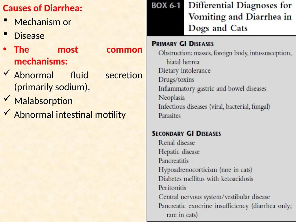
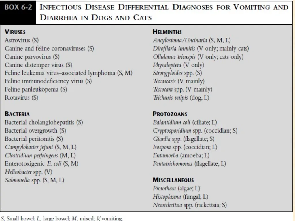
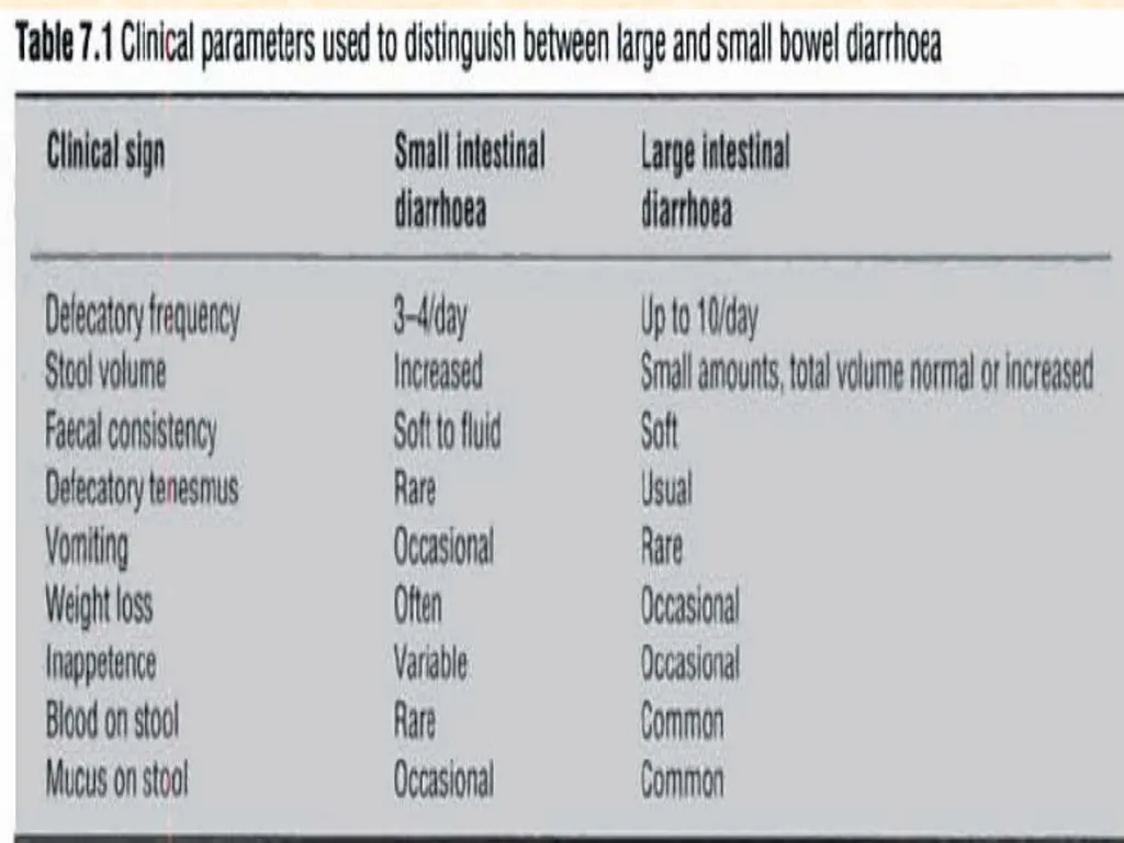
slide6. Causes of Diarrhea:
Mechanism or
Disease
The most common mechanisms:
Abnormal fluid secretion (primarily sodium),
Malabsorption
Abnormal intestinal motility<br>
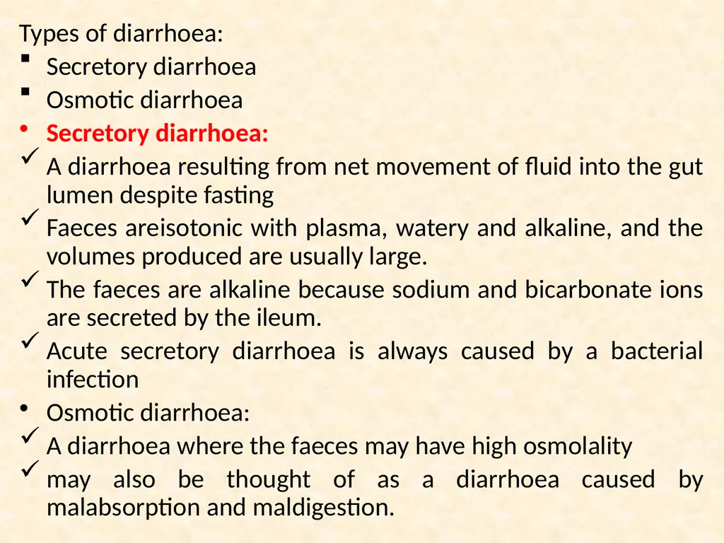
slide9. Types of diarrhoea:
Secretory diarrhoea
Osmotic diarrhoea
Secretory diarrhoea:
A diarrhoea resulting from net movement of fluid into the gut lumen despite fasting
Faeces areisotonic with plasma, watery and alkaline, and the volumes produced are usually large.
The faeces are alkaline because sodium and bicarbonate ions are secreted by the ileum.
Acute secretory diarrhoea is always caused by a bacterial infection
Osmotic diarrhoea:
A diarrhoea where the faeces may have high osmolality
may also be thought of as a diarrhoea caused by malabsorption and maldigestion.<br>
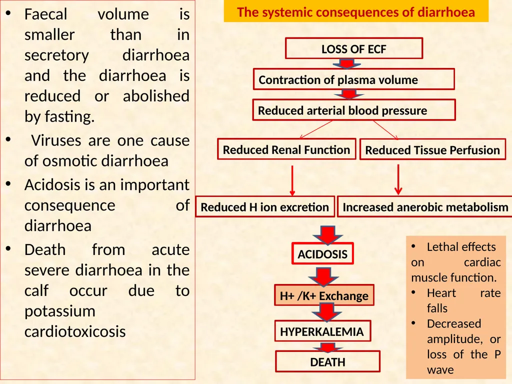
slide10. Faecal volume is smaller than in secretory diarrhoea and the diarrhoea is reduced or abolished by fasting.
Viruses are one cause of osmotic diarrhoea
Acidosis is an important consequence of diarrhoea
Death from acute severe diarrhoea in the calf occur due to potassium cardiotoxicosis LOSS OF ECF Contraction of plasma volume Reduced arterial blood pressure Reduced Renal Function Reduced Tissue Perfusion Reduced H ion excretion Increased anerobic metabolism ACIDOSIS H+ /K+ Exchange HYPERKALEMIA DEATH The systemic consequences of diarrhoea Lethal effects
on cardiac muscle function.
Heart rate falls
Decreased amplitude, or loss of the P wave<br>
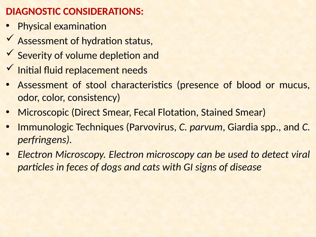
slide11. DIAGNOSTIC CONSIDERATIONS:
Physical examination
Assessment of hydration status,
Severity of volume depletion and
Initial fluid replacement needs
Assessment of stool characteristics (presence of blood or mucus, odor, color, consistency)
Microscopic (Direct Smear, Fecal Flotation, Stained Smear)
Immunologic Techniques (Parvovirus, C. parvum, Giardia spp., and C. perfringens).
Electron Microscopy. Electron microscopy can be used to detect viral particles in feces of dogs and cats with GI signs of disease<br>
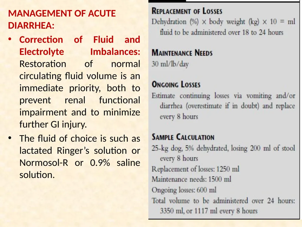
slide12. MANAGEMENT OF ACUTE DIARRHEA:
Correction of Fluid and Electrolyte Imbalances: Restoration of normal circulating fluid volume is an immediate priority, both to prevent renal functional impairment and to minimize further GI injury.
The fluid of choice is such as lactated Ringer’s solution or Normosol-R or 0.9% saline solution.<br>
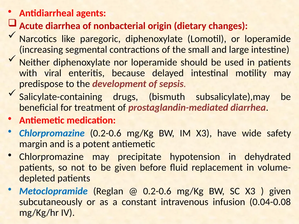
slide13. Antidiarrheal agents:
Acute diarrhea of nonbacterial origin (dietary changes):
Narcotics like paregoric, diphenoxylate (Lomotil), or loperamide (increasing segmental contractions of the small and large intestine)
Neither diphenoxylate nor loperamide should be used in patients with viral enteritis, because delayed intestinal motility may predispose to the development of sepsis.
Salicylate-containing drugs, (bismuth subsalicylate),may be beneficial for treatment of prostaglandin-mediated diarrhea.
Antiemetic medication:
Chlorpromazine (0.2-0.6 mg/Kg BW, IM X3), have wide safety margin and is a potent antiemetic
Chlorpromazine may precipitate hypotension in dehydrated patients, so not to be given before fluid replacement in volume-depleted patients
Metoclopramide (Reglan @ 0.2-0.6 mg/Kg BW, SC X3 ) given subcutaneously or as a constant intravenous infusion (0.04-0.08 mg/Kg/hr IV).<br>
slide14. Ondansetron @0.1-0.2 mg/Kg BW slow IV two to three times a day<br>
slide15. Source:
Handbook of Small Animal Gastroenterology, 2nd Edn. Elsevier Science (USA).
Bovine Medicine Diseases and Husbandry of Cattle Second edition THANKS<br>
DIARRHOEA
Dr. Anil Kumar
Asst. Prof. VCC, BVC, Patna
VCP-II, 4th Professional<br>
slide2. Diarrhoea Diarrhoea is defined as increased volume or fluidity of faeces or increased frequency of defecation.
Normal physiologic functions of the small intestine:<br>
slide3. Food enters the stomach leads to the production of semj-fluid chyme with the help of combination of an acid environment, pepsin proteolytic activity and active gastric motility.
Then it passed to duodenum in controlled quantities and also receives intestinal and pancreatic secretions and bile, which are mixed with chyme.
Pancreatic secretions (sodium bicarbonate)neutralizes gastric acid and also releases lipase, amylase and trypsin
Bile contains bile salts and bile pigments, but the bile salts are important for the emulsification of undigested fats into micelles, which greatly increases exposure to the fat digesting enzyme lipase and produce free fatty acids and monoglycerides
Carbohydrate digestion is initiated by salivary amylase and contiJ1ued by pancreatic amylase.<br>
slide4. The produced disaccharides and oligosaccharides, which are further digested by disaccharidase (lactase and sucrase) and oligosaccharidase enzymes in the small intestinal brush border.
Monosaccbarides are absorbed into the mucosal cell by co-transport with sodium (glucose, galactose) or by facilitated diffusion (fructose).
Protein digestion is initiated by pepsin in the stomach and continued by pancreatic proteolytic enzymes (including trypsin and chymotrypsin) and by peptidases of the intestinal brush border.
Amino acids are absorbed into the mucosal cell by various carriers and enter the blood stream
Mixing of ingesta and secreted digestive components in the small intestine is facilitated by segmental contraction of the bowel wall, which promotes digestion and absorption at the brush border by maximizing contact with luminal contents.<br>
slide5. lngesta is passed distally along the intestine by peristaltic waves and finally this material enters the colon via the ileo-colic valve
The storage and co-ordinated elimination of faecal material and dehydration of faeces (by absorbing approximately 90% of the water entering)—through Large intestine
The colon only accounts for approximately 10-15% of total water absorption in the gastrointestinal tract (jejunun and ileum)
The net flux of water through the gastrointestinal tract is at least 0.15 1 kg-1 bodyweight day·
The major solutes in diarrhea fluid are sodium, chloride, organic anions, and potassium.
During diarrheal diseases the most important source of potassium loss is via urine, mediated by aldosterone released in response to extracellular fluid volume depletion
Mild metabolic acidosis and hypokalemia are the most common acid-base and electrolyte alterations observed in patients with acute small intestinal disease and diarrhea.<br>
slide6. Causes of Diarrhea:
Mechanism or
Disease
The most common mechanisms:
Abnormal fluid secretion (primarily sodium),
Malabsorption
Abnormal intestinal motility<br>
slide9. Types of diarrhoea:
Secretory diarrhoea
Osmotic diarrhoea
Secretory diarrhoea:
A diarrhoea resulting from net movement of fluid into the gut lumen despite fasting
Faeces areisotonic with plasma, watery and alkaline, and the volumes produced are usually large.
The faeces are alkaline because sodium and bicarbonate ions are secreted by the ileum.
Acute secretory diarrhoea is always caused by a bacterial infection
Osmotic diarrhoea:
A diarrhoea where the faeces may have high osmolality
may also be thought of as a diarrhoea caused by malabsorption and maldigestion.<br>
slide10. Faecal volume is smaller than in secretory diarrhoea and the diarrhoea is reduced or abolished by fasting.
Viruses are one cause of osmotic diarrhoea
Acidosis is an important consequence of diarrhoea
Death from acute severe diarrhoea in the calf occur due to potassium cardiotoxicosis LOSS OF ECF Contraction of plasma volume Reduced arterial blood pressure Reduced Renal Function Reduced Tissue Perfusion Reduced H ion excretion Increased anerobic metabolism ACIDOSIS H+ /K+ Exchange HYPERKALEMIA DEATH The systemic consequences of diarrhoea Lethal effects
on cardiac muscle function.
Heart rate falls
Decreased amplitude, or loss of the P wave<br>
slide11. DIAGNOSTIC CONSIDERATIONS:
Physical examination
Assessment of hydration status,
Severity of volume depletion and
Initial fluid replacement needs
Assessment of stool characteristics (presence of blood or mucus, odor, color, consistency)
Microscopic (Direct Smear, Fecal Flotation, Stained Smear)
Immunologic Techniques (Parvovirus, C. parvum, Giardia spp., and C. perfringens).
Electron Microscopy. Electron microscopy can be used to detect viral particles in feces of dogs and cats with GI signs of disease<br>
slide12. MANAGEMENT OF ACUTE DIARRHEA:
Correction of Fluid and Electrolyte Imbalances: Restoration of normal circulating fluid volume is an immediate priority, both to prevent renal functional impairment and to minimize further GI injury.
The fluid of choice is such as lactated Ringer’s solution or Normosol-R or 0.9% saline solution.<br>
slide13. Antidiarrheal agents:
Acute diarrhea of nonbacterial origin (dietary changes):
Narcotics like paregoric, diphenoxylate (Lomotil), or loperamide (increasing segmental contractions of the small and large intestine)
Neither diphenoxylate nor loperamide should be used in patients with viral enteritis, because delayed intestinal motility may predispose to the development of sepsis.
Salicylate-containing drugs, (bismuth subsalicylate),may be beneficial for treatment of prostaglandin-mediated diarrhea.
Antiemetic medication:
Chlorpromazine (0.2-0.6 mg/Kg BW, IM X3), have wide safety margin and is a potent antiemetic
Chlorpromazine may precipitate hypotension in dehydrated patients, so not to be given before fluid replacement in volume-depleted patients
Metoclopramide (Reglan @ 0.2-0.6 mg/Kg BW, SC X3 ) given subcutaneously or as a constant intravenous infusion (0.04-0.08 mg/Kg/hr IV).<br>
slide14. Ondansetron @0.1-0.2 mg/Kg BW slow IV two to three times a day<br>
slide15. Source:
Handbook of Small Animal Gastroenterology, 2nd Edn. Elsevier Science (USA).
Bovine Medicine Diseases and Husbandry of Cattle Second edition THANKS<br>