
Deflazacort Scientific Communication Platform
LS
Published · 75 slides · 0 views

1 / 1



Description
Deflazacort Scientific Communication Platform ENTER Scientific Communication Platform Pillars Pillar 1: Pathophysiology of Dystrophinopathy Pillar 2: Early Screening, Diagnosis, and Treatment Pillar 3: Dystrophinopathy Treatment and Unmet
Related Topics
Share
Embed code
Download this presentation From Below
"Deflazacort Scientific Communication Platform" is the property of its rightful owner. Permission is granted to download and print the materials on this website for personal, non-commercial use only, and to display it on your personal computer provided you do not modify the materials and that you retain all copyright notices contained in the materials. By downloading content from our website, you accept the terms of this agreement.
Presentation Transcript
01
Deflazacort
Scientific Communication Platform ENTER ►<br>
02
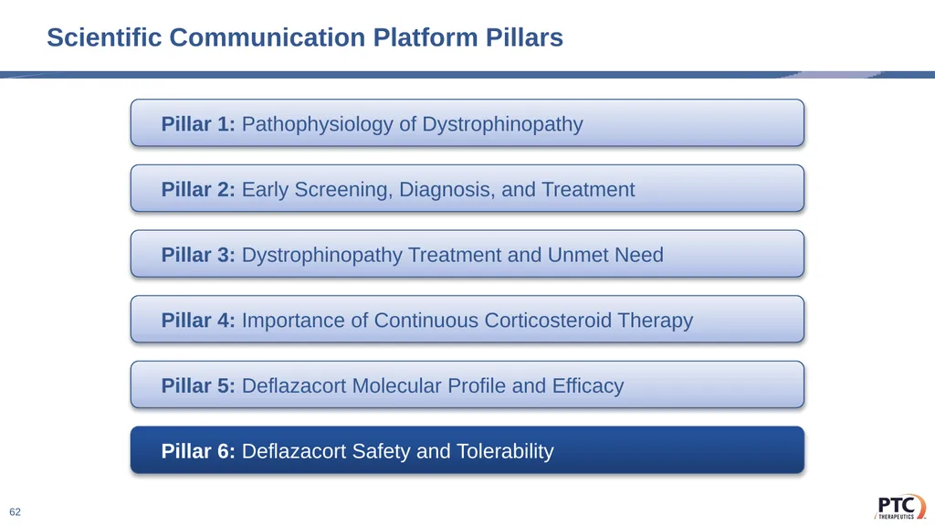
Scientific Communication Platform Pillars Pillar 1: Pathophysiology of Dystrophinopathy Pillar 2: Early Screening, Diagnosis, and Treatment Pillar 3: Dystrophinopathy Treatment and Unmet Need Pillar 4: Importance of Continuous Corticosteroid Therapy Pillar 5: Deflazacort Molecular Profile and Efficacy Pillar 6: Deflazacort Safety and Tolerability 2<br>
03
Scientific Communication Platform Pillars Pillar 1: Pathophysiology of Dystrophinopathy Pillar 2: Early Screening, Diagnosis, and Treatment Pillar 3: Dystrophinopathy Treatment and Unmet Need Pillar 4: Importance of Continuous Corticosteroid Therapy Pillar 5: Deflazacort Molecular Profile and Efficacy Pillar 6: Deflazacort Safety and Tolerability 3<br>
04
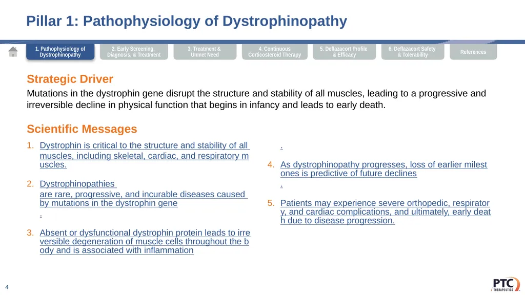
Pillar 1: Pathophysiology of Dystrophinopathy Strategic Driver
Mutations in the dystrophin gene disrupt the structure and stability of all muscles, leading to a progressive and irreversible decline in physical function that begins in infancy and leads to early death. 4 Scientific Messages Dystrophin is critical to the structure and stability of all muscles, including skeletal, cardiac, and respiratory muscles.
Dystrophinopathies are rare, progressive, and incurable diseases caused by mutations in the dystrophin gene.
Absent or dysfunctional dystrophin protein leads to irreversible degeneration of muscle cells throughout the body and is associated with inflammation.
As dystrophinopathy progresses, loss of earlier milestones is predictive of future declines.
Patients may experience severe orthopedic, respiratory, and cardiac complications, and ultimately, early death due to disease progression. 1. Pathophysiology of Dystrophinopathy<br>
Mutations in the dystrophin gene disrupt the structure and stability of all muscles, leading to a progressive and irreversible decline in physical function that begins in infancy and leads to early death. 4 Scientific Messages Dystrophin is critical to the structure and stability of all muscles, including skeletal, cardiac, and respiratory muscles.
Dystrophinopathies are rare, progressive, and incurable diseases caused by mutations in the dystrophin gene.
Absent or dysfunctional dystrophin protein leads to irreversible degeneration of muscle cells throughout the body and is associated with inflammation.
As dystrophinopathy progresses, loss of earlier milestones is predictive of future declines.
Patients may experience severe orthopedic, respiratory, and cardiac complications, and ultimately, early death due to disease progression. 1. Pathophysiology of Dystrophinopathy<br>
05
Pillar 1: Scientific Statements 5 Scientific Message 1
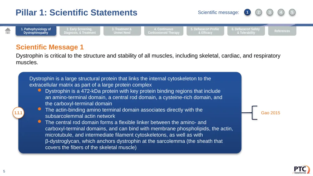
Dystrophin is critical to the structure and stability of all muscles, including skeletal, cardiac, and respiratory muscles. Dystrophin is a large structural protein that links the internal cytoskeleton to the extracellular matrix as part of a large protein complex
Dystrophin is a 472-kDa protein with key protein binding regions that include an amino-terminal domain, a central rod domain, a cysteine-rich domain, and the carboxyl-terminal domain
The actin-binding amino terminal domain associates directly with the subsarcolemmal actin network
The central rod domain forms a flexible linker between the amino- and carboxyl-terminal domains, and can bind with membrane phospholipids, the actin, microtubule, and intermediate filament cytoskeletons, as well as with β-dystroglycan, which anchors dystrophin at the sarcolemma (the sheath that covers the fibers of the skeletal muscle) 1.1.1 Gao 2015 1 3 2 4 5 Scientific message: 1. Pathophysiology of Dystrophinopathy<br>
Dystrophin is critical to the structure and stability of all muscles, including skeletal, cardiac, and respiratory muscles. Dystrophin is a large structural protein that links the internal cytoskeleton to the extracellular matrix as part of a large protein complex
Dystrophin is a 472-kDa protein with key protein binding regions that include an amino-terminal domain, a central rod domain, a cysteine-rich domain, and the carboxyl-terminal domain
The actin-binding amino terminal domain associates directly with the subsarcolemmal actin network
The central rod domain forms a flexible linker between the amino- and carboxyl-terminal domains, and can bind with membrane phospholipids, the actin, microtubule, and intermediate filament cytoskeletons, as well as with β-dystroglycan, which anchors dystrophin at the sarcolemma (the sheath that covers the fibers of the skeletal muscle) 1.1.1 Gao 2015 1 3 2 4 5 Scientific message: 1. Pathophysiology of Dystrophinopathy<br>
06
Pillar 1: Scientific Statements 6 Scientific Message 1
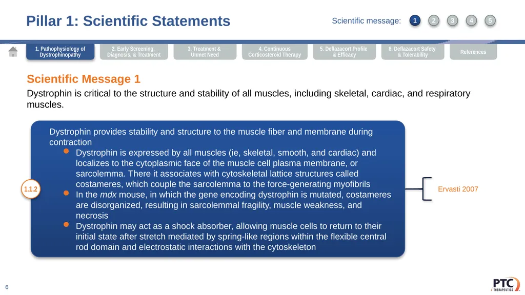
Dystrophin is critical to the structure and stability of all muscles, including skeletal, cardiac, and respiratory muscles. Dystrophin provides stability and structure to the muscle fiber and membrane during contraction
Dystrophin is expressed by all muscles (ie, skeletal, smooth, and cardiac) and localizes to the cytoplasmic face of the muscle cell plasma membrane, or sarcolemma. There it associates with cytoskeletal lattice structures called costameres, which couple the sarcolemma to the force-generating myofibrils
In the mdx mouse, in which the gene encoding dystrophin is mutated, costameres are disorganized, resulting in sarcolemmal fragility, muscle weakness, and necrosis
Dystrophin may act as a shock absorber, allowing muscle cells to return to their initial state after stretch mediated by spring-like regions within the flexible central rod domain and electrostatic interactions with the cytoskeleton 1.1.2 Ervasti 2007 1. Pathophysiology of Dystrophinopathy 1 3 2 4 5 Scientific message:<br>
Dystrophin is critical to the structure and stability of all muscles, including skeletal, cardiac, and respiratory muscles. Dystrophin provides stability and structure to the muscle fiber and membrane during contraction
Dystrophin is expressed by all muscles (ie, skeletal, smooth, and cardiac) and localizes to the cytoplasmic face of the muscle cell plasma membrane, or sarcolemma. There it associates with cytoskeletal lattice structures called costameres, which couple the sarcolemma to the force-generating myofibrils
In the mdx mouse, in which the gene encoding dystrophin is mutated, costameres are disorganized, resulting in sarcolemmal fragility, muscle weakness, and necrosis
Dystrophin may act as a shock absorber, allowing muscle cells to return to their initial state after stretch mediated by spring-like regions within the flexible central rod domain and electrostatic interactions with the cytoskeleton 1.1.2 Ervasti 2007 1. Pathophysiology of Dystrophinopathy 1 3 2 4 5 Scientific message:<br>
07
Pillar 1: Scientific Statements 7 Scientific Message 2
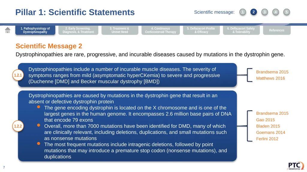
Dystrophinopathies are rare, progressive, and incurable diseases caused by mutations in the dystrophin gene. Dystrophinopathies include a number of incurable muscle diseases. The severity of symptoms ranges from mild (asymptomatic hyperCKemia) to severe and progressive (Duchenne [DMD] and Becker muscular dystrophy [BMD]) 1.2.1 Brandsema 2015
Matthews 2016 Dystrophinopathies are caused by mutations in the dystrophin gene that result in an absent or defective dystrophin protein
The gene encoding dystrophin is located on the X chromosome and is one of the largest genes in the human genome. It encompasses 2.6 million base pairs of DNA that encode 79 exons
Overall, more than 7000 mutations have been identified for DMD, many of which are clinically relevant, including deletions, duplications, and small mutations such as nonsense mutations
The most frequent mutations include intragenic deletions, followed by point mutations that may introduce a premature stop codon (nonsense mutations), and duplications 1.2.2 Brandsema 2015
Gao 2015
Bladen 2015
Goemans 2014
Ferlini 2012 1. Pathophysiology of Dystrophinopathy 1 3 2 4 5 Scientific message:<br>
Dystrophinopathies are rare, progressive, and incurable diseases caused by mutations in the dystrophin gene. Dystrophinopathies include a number of incurable muscle diseases. The severity of symptoms ranges from mild (asymptomatic hyperCKemia) to severe and progressive (Duchenne [DMD] and Becker muscular dystrophy [BMD]) 1.2.1 Brandsema 2015
Matthews 2016 Dystrophinopathies are caused by mutations in the dystrophin gene that result in an absent or defective dystrophin protein
The gene encoding dystrophin is located on the X chromosome and is one of the largest genes in the human genome. It encompasses 2.6 million base pairs of DNA that encode 79 exons
Overall, more than 7000 mutations have been identified for DMD, many of which are clinically relevant, including deletions, duplications, and small mutations such as nonsense mutations
The most frequent mutations include intragenic deletions, followed by point mutations that may introduce a premature stop codon (nonsense mutations), and duplications 1.2.2 Brandsema 2015
Gao 2015
Bladen 2015
Goemans 2014
Ferlini 2012 1. Pathophysiology of Dystrophinopathy 1 3 2 4 5 Scientific message:<br>
08
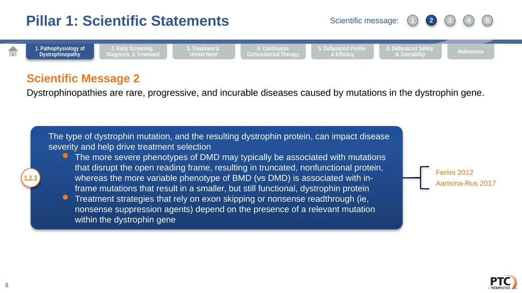
The type of dystrophin mutation, and the resulting dystrophin protein, can impact disease severity and help drive treatment selection
The more severe phenotypes of DMD may typically be associated with mutations that disrupt the open reading frame, resulting in truncated, nonfunctional protein, whereas the more variable phenotype of BMD (vs DMD) is associated with in-frame mutations that result in a smaller, but still functional, dystrophin protein
Treatment strategies that rely on exon skipping or nonsense readthrough (ie, nonsense suppression agents) depend on the presence of a relevant mutation within the dystrophin gene Pillar 1: Scientific Statements 8 Scientific Message 2
Dystrophinopathies are rare, progressive, and incurable diseases caused by mutations in the dystrophin gene. 1.2.3 Ferlini 2012
Aartsma-Rus 2017 1. Pathophysiology of Dystrophinopathy 1 3 2 4 5 Scientific message:<br>
The more severe phenotypes of DMD may typically be associated with mutations that disrupt the open reading frame, resulting in truncated, nonfunctional protein, whereas the more variable phenotype of BMD (vs DMD) is associated with in-frame mutations that result in a smaller, but still functional, dystrophin protein
Treatment strategies that rely on exon skipping or nonsense readthrough (ie, nonsense suppression agents) depend on the presence of a relevant mutation within the dystrophin gene Pillar 1: Scientific Statements 8 Scientific Message 2
Dystrophinopathies are rare, progressive, and incurable diseases caused by mutations in the dystrophin gene. 1.2.3 Ferlini 2012
Aartsma-Rus 2017 1. Pathophysiology of Dystrophinopathy 1 3 2 4 5 Scientific message:<br>
09
Pillar 1: Scientific Statements 9 Scientific Message 2
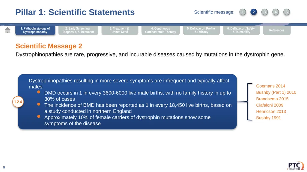
Dystrophinopathies are rare, progressive, and incurable diseases caused by mutations in the dystrophin gene. Dystrophinopathies resulting in more severe symptoms are infrequent and typically affect males
DMD occurs in 1 in every 3600-6000 live male births, with no family history in up to 30% of cases
The incidence of BMD has been reported as 1 in every 18,450 live births, based on a study conducted in northern England
Approximately 10% of female carriers of dystrophin mutations show some symptoms of the disease 1.2.4 Goemans 2014
Bushby (Part 1) 2010
Brandsema 2015
Ciafaloni 2009
Henricson 2013
Bushby 1991 1. Pathophysiology of Dystrophinopathy 1 3 2 4 5 Scientific message:<br>
Dystrophinopathies are rare, progressive, and incurable diseases caused by mutations in the dystrophin gene. Dystrophinopathies resulting in more severe symptoms are infrequent and typically affect males
DMD occurs in 1 in every 3600-6000 live male births, with no family history in up to 30% of cases
The incidence of BMD has been reported as 1 in every 18,450 live births, based on a study conducted in northern England
Approximately 10% of female carriers of dystrophin mutations show some symptoms of the disease 1.2.4 Goemans 2014
Bushby (Part 1) 2010
Brandsema 2015
Ciafaloni 2009
Henricson 2013
Bushby 1991 1. Pathophysiology of Dystrophinopathy 1 3 2 4 5 Scientific message:<br>
10
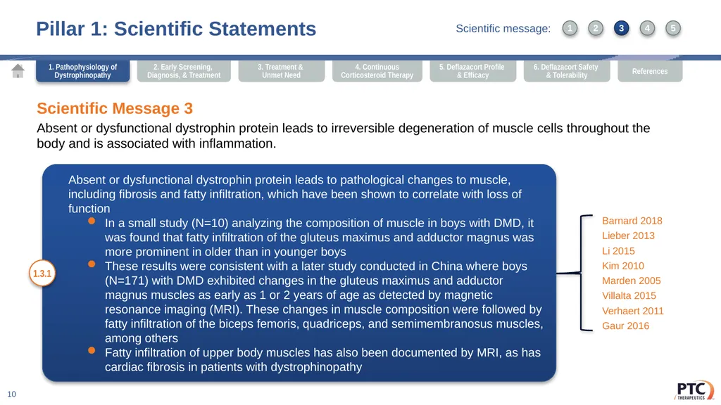
Pillar 1: Scientific Statements 10 Scientific Message 3
Absent or dysfunctional dystrophin protein leads to irreversible degeneration of muscle cells throughout the body and is associated with inflammation. Absent or dysfunctional dystrophin protein leads to pathological changes to muscle, including fibrosis and fatty infiltration, which have been shown to correlate with loss of function
In a small study (N=10) analyzing the composition of muscle in boys with DMD, it was found that fatty infiltration of the gluteus maximus and adductor magnus was more prominent in older than in younger boys
These results were consistent with a later study conducted in China where boys (N=171) with DMD exhibited changes in the gluteus maximus and adductor magnus muscles as early as 1 or 2 years of age as detected by magnetic resonance imaging (MRI). These changes in muscle composition were followed by fatty infiltration of the biceps femoris, quadriceps, and semimembranosus muscles, among others
Fatty infiltration of upper body muscles has also been documented by MRI, as has cardiac fibrosis in patients with dystrophinopathy 1.3.1 Barnard 2018
Lieber 2013
Li 2015
Kim 2010
Marden 2005
Villalta 2015
Verhaert 2011
Gaur 2016 1. Pathophysiology of Dystrophinopathy 1 3 2 4 5 Scientific message:<br>
Absent or dysfunctional dystrophin protein leads to irreversible degeneration of muscle cells throughout the body and is associated with inflammation. Absent or dysfunctional dystrophin protein leads to pathological changes to muscle, including fibrosis and fatty infiltration, which have been shown to correlate with loss of function
In a small study (N=10) analyzing the composition of muscle in boys with DMD, it was found that fatty infiltration of the gluteus maximus and adductor magnus was more prominent in older than in younger boys
These results were consistent with a later study conducted in China where boys (N=171) with DMD exhibited changes in the gluteus maximus and adductor magnus muscles as early as 1 or 2 years of age as detected by magnetic resonance imaging (MRI). These changes in muscle composition were followed by fatty infiltration of the biceps femoris, quadriceps, and semimembranosus muscles, among others
Fatty infiltration of upper body muscles has also been documented by MRI, as has cardiac fibrosis in patients with dystrophinopathy 1.3.1 Barnard 2018
Lieber 2013
Li 2015
Kim 2010
Marden 2005
Villalta 2015
Verhaert 2011
Gaur 2016 1. Pathophysiology of Dystrophinopathy 1 3 2 4 5 Scientific message:<br>
11
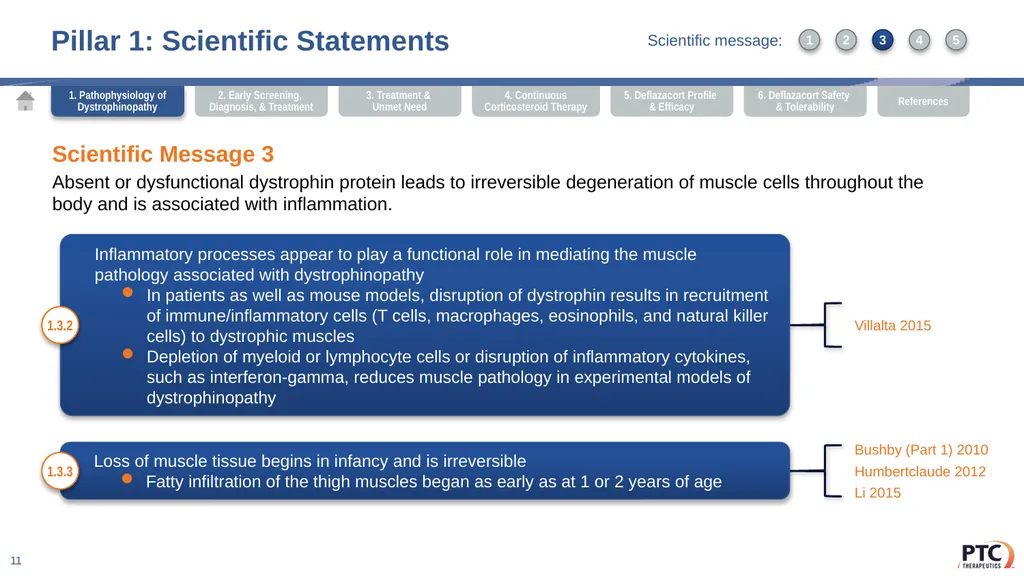
Pillar 1: Scientific Statements 11 Scientific Message 3
Absent or dysfunctional dystrophin protein leads to irreversible degeneration of muscle cells throughout the body and is associated with inflammation. Inflammatory processes appear to play a functional role in mediating the muscle pathology associated with dystrophinopathy
In patients as well as mouse models, disruption of dystrophin results in recruitment of immune/inflammatory cells (T cells, macrophages, eosinophils, and natural killer cells) to dystrophic muscles
Depletion of myeloid or lymphocyte cells or disruption of inflammatory cytokines, such as interferon-gamma, reduces muscle pathology in experimental models of dystrophinopathy 1.3.2 Villalta 2015 Loss of muscle tissue begins in infancy and is irreversible
Fatty infiltration of the thigh muscles began as early as at 1 or 2 years of age 1.3.3 Bushby (Part 1) 2010
Humbertclaude 2012
Li 2015 1. Pathophysiology of Dystrophinopathy 1 3 2 4 5 Scientific message:<br>
Absent or dysfunctional dystrophin protein leads to irreversible degeneration of muscle cells throughout the body and is associated with inflammation. Inflammatory processes appear to play a functional role in mediating the muscle pathology associated with dystrophinopathy
In patients as well as mouse models, disruption of dystrophin results in recruitment of immune/inflammatory cells (T cells, macrophages, eosinophils, and natural killer cells) to dystrophic muscles
Depletion of myeloid or lymphocyte cells or disruption of inflammatory cytokines, such as interferon-gamma, reduces muscle pathology in experimental models of dystrophinopathy 1.3.2 Villalta 2015 Loss of muscle tissue begins in infancy and is irreversible
Fatty infiltration of the thigh muscles began as early as at 1 or 2 years of age 1.3.3 Bushby (Part 1) 2010
Humbertclaude 2012
Li 2015 1. Pathophysiology of Dystrophinopathy 1 3 2 4 5 Scientific message:<br>
12
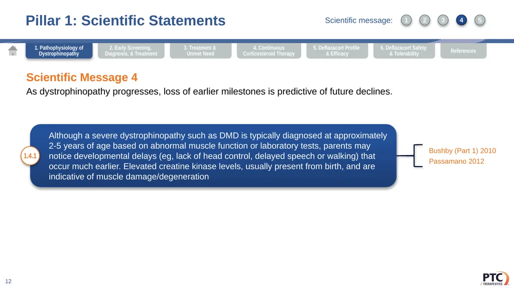
Pillar 1: Scientific Statements 12 Scientific Message 4
As dystrophinopathy progresses, loss of earlier milestones is predictive of future declines. Although a severe dystrophinopathy such as DMD is typically diagnosed at approximately 2-5 years of age based on abnormal muscle function or laboratory tests, parents may notice developmental delays (eg, lack of head control, delayed speech or walking) that occur much earlier. Elevated creatine kinase levels, usually present from birth, and are indicative of muscle damage/degeneration 1.4.1 Bushby (Part 1) 2010
Passamano 2012 1. Pathophysiology of Dystrophinopathy 1 3 2 4 5 Scientific message:<br>
As dystrophinopathy progresses, loss of earlier milestones is predictive of future declines. Although a severe dystrophinopathy such as DMD is typically diagnosed at approximately 2-5 years of age based on abnormal muscle function or laboratory tests, parents may notice developmental delays (eg, lack of head control, delayed speech or walking) that occur much earlier. Elevated creatine kinase levels, usually present from birth, and are indicative of muscle damage/degeneration 1.4.1 Bushby (Part 1) 2010
Passamano 2012 1. Pathophysiology of Dystrophinopathy 1 3 2 4 5 Scientific message:<br>
13
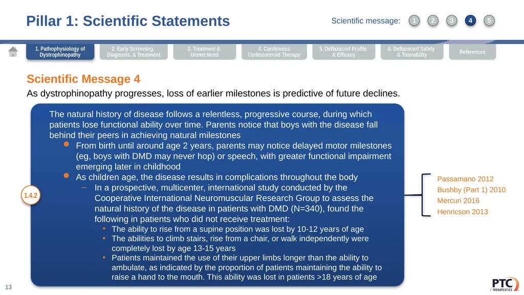
Pillar 1: Scientific Statements 13 Scientific Message 4
As dystrophinopathy progresses, loss of earlier milestones is predictive of future declines. The natural history of disease follows a relentless, progressive course, during which patients lose functional ability over time. Parents notice that boys with the disease fall behind their peers in achieving natural milestones
From birth until around age 2 years, parents may notice delayed motor milestones (eg, boys with DMD may never hop) or speech, with greater functional impairment emerging later in childhood
As children age, the disease results in complications throughout the body
In a prospective, multicenter, international study conducted by the Cooperative International Neuromuscular Research Group to assess the natural history of the disease in patients with DMD (N=340), found the following in patients who did not receive treatment:
The ability to rise from a supine position was lost by 10-12 years of age
The abilities to climb stairs, rise from a chair, or walk independently were completely lost by age 13-15 years
Patients maintained the use of their upper limbs longer than the ability to ambulate, as indicated by the proportion of patients maintaining the ability to raise a hand to the mouth. This ability was lost in patients >18 years of age 1.4.2 Passamano 2012
Bushby (Part 1) 2010
Mercuri 2016
Henricson 2013 1. Pathophysiology of Dystrophinopathy 1 3 2 4 5 Scientific message:<br>
As dystrophinopathy progresses, loss of earlier milestones is predictive of future declines. The natural history of disease follows a relentless, progressive course, during which patients lose functional ability over time. Parents notice that boys with the disease fall behind their peers in achieving natural milestones
From birth until around age 2 years, parents may notice delayed motor milestones (eg, boys with DMD may never hop) or speech, with greater functional impairment emerging later in childhood
As children age, the disease results in complications throughout the body
In a prospective, multicenter, international study conducted by the Cooperative International Neuromuscular Research Group to assess the natural history of the disease in patients with DMD (N=340), found the following in patients who did not receive treatment:
The ability to rise from a supine position was lost by 10-12 years of age
The abilities to climb stairs, rise from a chair, or walk independently were completely lost by age 13-15 years
Patients maintained the use of their upper limbs longer than the ability to ambulate, as indicated by the proportion of patients maintaining the ability to raise a hand to the mouth. This ability was lost in patients >18 years of age 1.4.2 Passamano 2012
Bushby (Part 1) 2010
Mercuri 2016
Henricson 2013 1. Pathophysiology of Dystrophinopathy 1 3 2 4 5 Scientific message:<br>
14
Pillar 1: Scientific Statements 14 Scientific Message 4
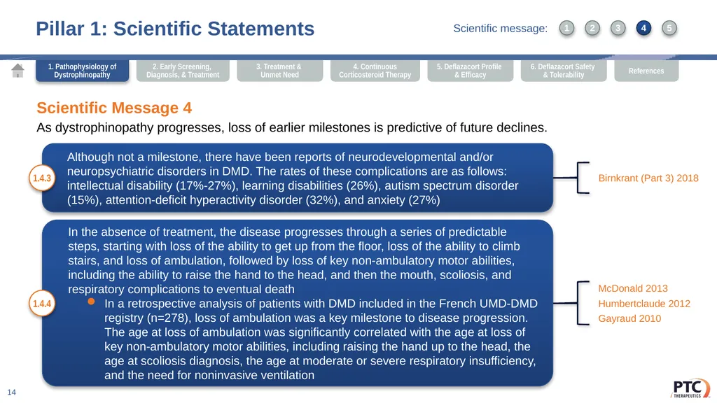
As dystrophinopathy progresses, loss of earlier milestones is predictive of future declines. Although not a milestone, there have been reports of neurodevelopmental and/or neuropsychiatric disorders in DMD. The rates of these complications are as follows: intellectual disability (17%-27%), learning disabilities (26%), autism spectrum disorder (15%), attention-deficit hyperactivity disorder (32%), and anxiety (27%) 1.4.3 Birnkrant (Part 3) 2018 In the absence of treatment, the disease progresses through a series of predictable steps, starting with loss of the ability to get up from the floor, loss of the ability to climb stairs, and loss of ambulation, followed by loss of key non-ambulatory motor abilities, including the ability to raise the hand to the head, and then the mouth, scoliosis, and respiratory complications to eventual death
In a retrospective analysis of patients with DMD included in the French UMD-DMD registry (n=278), loss of ambulation was a key milestone to disease progression. The age at loss of ambulation was significantly correlated with the age at loss of key non-ambulatory motor abilities, including raising the hand up to the head, the age at scoliosis diagnosis, the age at moderate or severe respiratory insufficiency, and the need for noninvasive ventilation 1.4.4 McDonald 2013
Humbertclaude 2012
Gayraud 2010 1. Pathophysiology of Dystrophinopathy 1 3 2 4 5 Scientific message:<br>
As dystrophinopathy progresses, loss of earlier milestones is predictive of future declines. Although not a milestone, there have been reports of neurodevelopmental and/or neuropsychiatric disorders in DMD. The rates of these complications are as follows: intellectual disability (17%-27%), learning disabilities (26%), autism spectrum disorder (15%), attention-deficit hyperactivity disorder (32%), and anxiety (27%) 1.4.3 Birnkrant (Part 3) 2018 In the absence of treatment, the disease progresses through a series of predictable steps, starting with loss of the ability to get up from the floor, loss of the ability to climb stairs, and loss of ambulation, followed by loss of key non-ambulatory motor abilities, including the ability to raise the hand to the head, and then the mouth, scoliosis, and respiratory complications to eventual death
In a retrospective analysis of patients with DMD included in the French UMD-DMD registry (n=278), loss of ambulation was a key milestone to disease progression. The age at loss of ambulation was significantly correlated with the age at loss of key non-ambulatory motor abilities, including raising the hand up to the head, the age at scoliosis diagnosis, the age at moderate or severe respiratory insufficiency, and the need for noninvasive ventilation 1.4.4 McDonald 2013
Humbertclaude 2012
Gayraud 2010 1. Pathophysiology of Dystrophinopathy 1 3 2 4 5 Scientific message:<br>
15
Pillar 1: Scientific Statements 15 Scientific Message 5
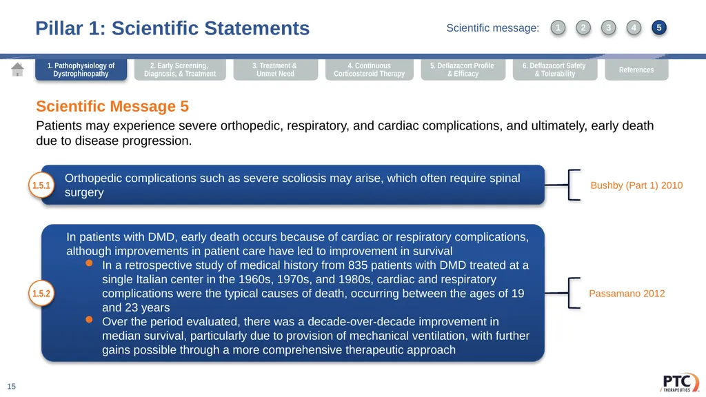
Patients may experience severe orthopedic, respiratory, and cardiac complications, and ultimately, early death due to disease progression. Orthopedic complications such as severe scoliosis may arise, which often require spinal surgery 1.5.1 Bushby (Part 1) 2010 In patients with DMD, early death occurs because of cardiac or respiratory complications, although improvements in patient care have led to improvement in survival
In a retrospective study of medical history from 835 patients with DMD treated at a single Italian center in the 1960s, 1970s, and 1980s, cardiac and respiratory complications were the typical causes of death, occurring between the ages of 19 and 23 years
Over the period evaluated, there was a decade-over-decade improvement in median survival, particularly due to provision of mechanical ventilation, with further gains possible through a more comprehensive therapeutic approach 1.5.2 Passamano 2012 1. Pathophysiology of Dystrophinopathy 1 3 2 4 5 Scientific message:<br>
Patients may experience severe orthopedic, respiratory, and cardiac complications, and ultimately, early death due to disease progression. Orthopedic complications such as severe scoliosis may arise, which often require spinal surgery 1.5.1 Bushby (Part 1) 2010 In patients with DMD, early death occurs because of cardiac or respiratory complications, although improvements in patient care have led to improvement in survival
In a retrospective study of medical history from 835 patients with DMD treated at a single Italian center in the 1960s, 1970s, and 1980s, cardiac and respiratory complications were the typical causes of death, occurring between the ages of 19 and 23 years
Over the period evaluated, there was a decade-over-decade improvement in median survival, particularly due to provision of mechanical ventilation, with further gains possible through a more comprehensive therapeutic approach 1.5.2 Passamano 2012 1. Pathophysiology of Dystrophinopathy 1 3 2 4 5 Scientific message:<br>
16
Pillar 1: Scientific Statements 16 Scientific Message 5
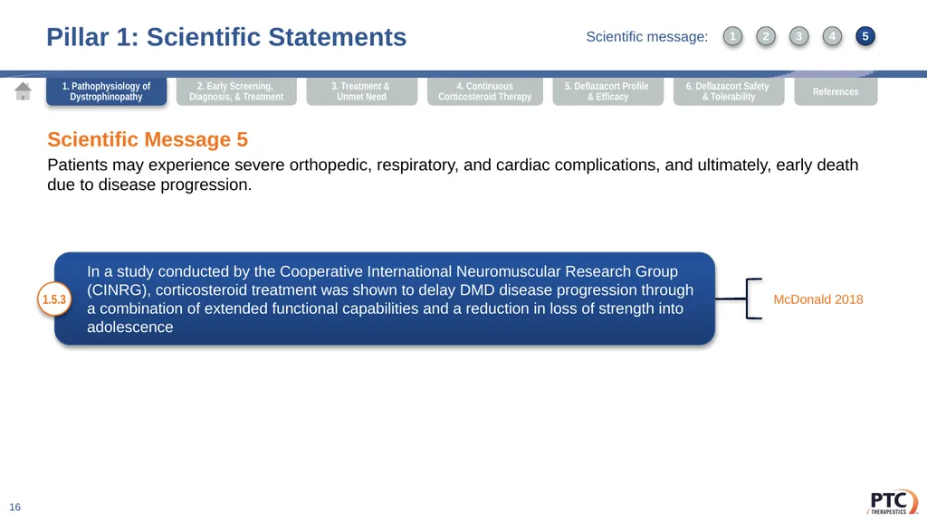
Patients may experience severe orthopedic, respiratory, and cardiac complications, and ultimately, early death due to disease progression. In a study conducted by the Cooperative International Neuromuscular Research Group (CINRG), corticosteroid treatment was shown to delay DMD disease progression through a combination of extended functional capabilities and a reduction in loss of strength into adolescence 1.5.3 McDonald 2018 1. Pathophysiology of Dystrophinopathy 1 3 2 4 5 Scientific message:<br>
Patients may experience severe orthopedic, respiratory, and cardiac complications, and ultimately, early death due to disease progression. In a study conducted by the Cooperative International Neuromuscular Research Group (CINRG), corticosteroid treatment was shown to delay DMD disease progression through a combination of extended functional capabilities and a reduction in loss of strength into adolescence 1.5.3 McDonald 2018 1. Pathophysiology of Dystrophinopathy 1 3 2 4 5 Scientific message:<br>
17
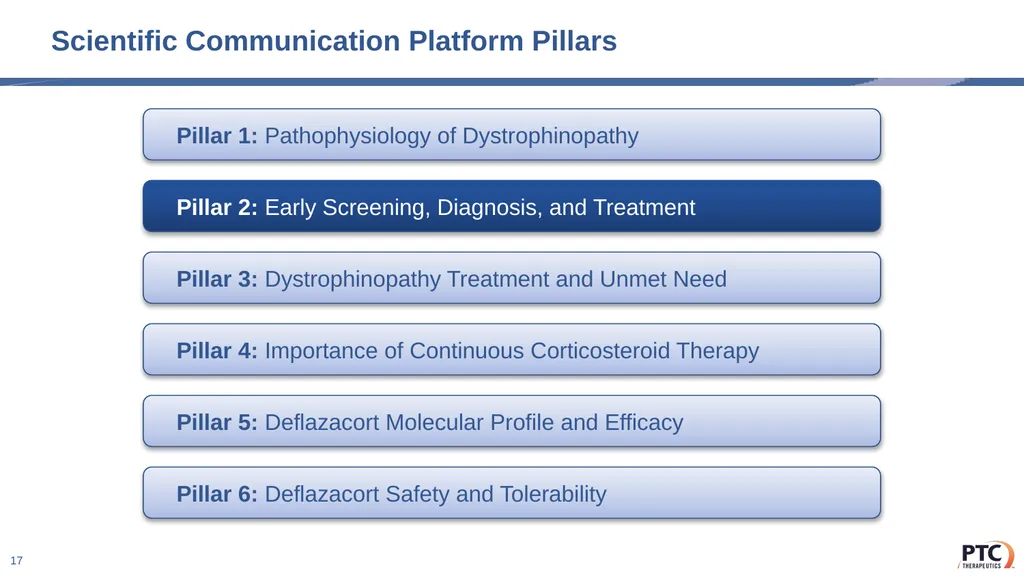
Scientific Communication Platform Pillars 17 Pillar 1: Pathophysiology of Dystrophinopathy Pillar 2: Early Screening, Diagnosis, and Treatment Pillar 3: Dystrophinopathy Treatment and Unmet Need Pillar 4: Importance of Continuous Corticosteroid Therapy Pillar 5: Deflazacort Molecular Profile and Efficacy Pillar 6: Deflazacort Safety and Tolerability<br>
18
Pillar 2: Early Screening, Diagnosis, and Treatment 18 Strategic Driver
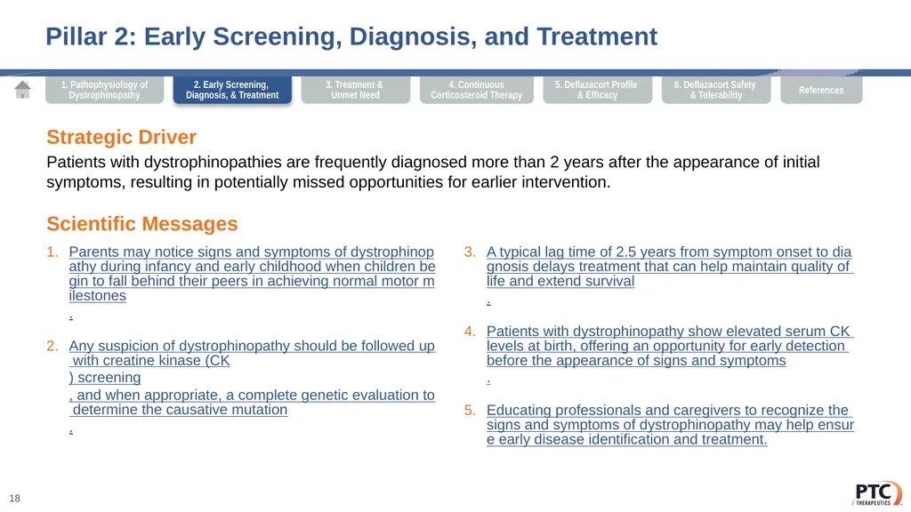
Patients with dystrophinopathies are frequently diagnosed more than 2 years after the appearance of initial symptoms, resulting in potentially missed opportunities for earlier intervention. Scientific Messages Parents may notice signs and symptoms of dystrophinopathy during infancy and early childhood when children begin to fall behind their peers in achieving normal motor milestones.
Any suspicion of dystrophinopathy should be followed up with creatine kinase (CK) screening, and when appropriate, a complete genetic evaluation to determine the causative mutation.
A typical lag time of 2.5 years from symptom onset to diagnosis delays treatment that can help maintain quality of life and extend survival.
Patients with dystrophinopathy show elevated serum CK levels at birth, offering an opportunity for early detection before the appearance of signs and symptoms.
Educating professionals and caregivers to recognize the signs and symptoms of dystrophinopathy may help ensure early disease identification and treatment. 2. Early Screening, Diagnosis, & Treatment<br>
Patients with dystrophinopathies are frequently diagnosed more than 2 years after the appearance of initial symptoms, resulting in potentially missed opportunities for earlier intervention. Scientific Messages Parents may notice signs and symptoms of dystrophinopathy during infancy and early childhood when children begin to fall behind their peers in achieving normal motor milestones.
Any suspicion of dystrophinopathy should be followed up with creatine kinase (CK) screening, and when appropriate, a complete genetic evaluation to determine the causative mutation.
A typical lag time of 2.5 years from symptom onset to diagnosis delays treatment that can help maintain quality of life and extend survival.
Patients with dystrophinopathy show elevated serum CK levels at birth, offering an opportunity for early detection before the appearance of signs and symptoms.
Educating professionals and caregivers to recognize the signs and symptoms of dystrophinopathy may help ensure early disease identification and treatment. 2. Early Screening, Diagnosis, & Treatment<br>
19
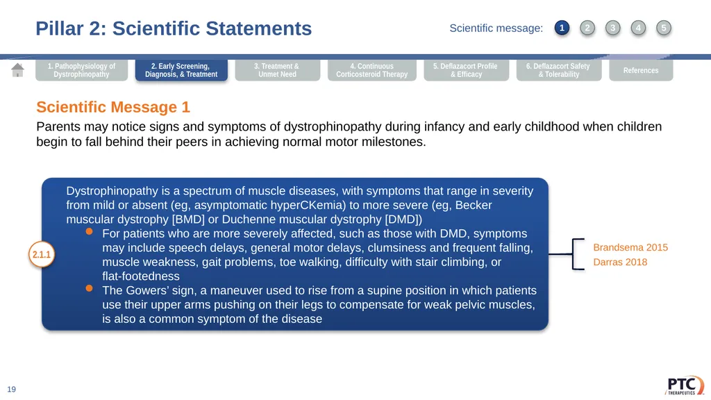
Pillar 2: Scientific Statements 19 Dystrophinopathy is a spectrum of muscle diseases, with symptoms that range in severity from mild or absent (eg, asymptomatic hyperCKemia) to more severe (eg, Becker muscular dystrophy [BMD] or Duchenne muscular dystrophy [DMD])
For patients who are more severely affected, such as those with DMD, symptoms may include speech delays, general motor delays, clumsiness and frequent falling, muscle weakness, gait problems, toe walking, difficulty with stair climbing, or flat-footedness
The Gowers’ sign, a maneuver used to rise from a supine position in which patients use their upper arms pushing on their legs to compensate for weak pelvic muscles, is also a common symptom of the disease 2.1.1 Brandsema 2015
Darras 2018 Scientific Message 1
Parents may notice signs and symptoms of dystrophinopathy during infancy and early childhood when children begin to fall behind their peers in achieving normal motor milestones. 1 3 2 4 5 2. Early Screening, Diagnosis, & Treatment Scientific message:<br>
For patients who are more severely affected, such as those with DMD, symptoms may include speech delays, general motor delays, clumsiness and frequent falling, muscle weakness, gait problems, toe walking, difficulty with stair climbing, or flat-footedness
The Gowers’ sign, a maneuver used to rise from a supine position in which patients use their upper arms pushing on their legs to compensate for weak pelvic muscles, is also a common symptom of the disease 2.1.1 Brandsema 2015
Darras 2018 Scientific Message 1
Parents may notice signs and symptoms of dystrophinopathy during infancy and early childhood when children begin to fall behind their peers in achieving normal motor milestones. 1 3 2 4 5 2. Early Screening, Diagnosis, & Treatment Scientific message:<br>
20
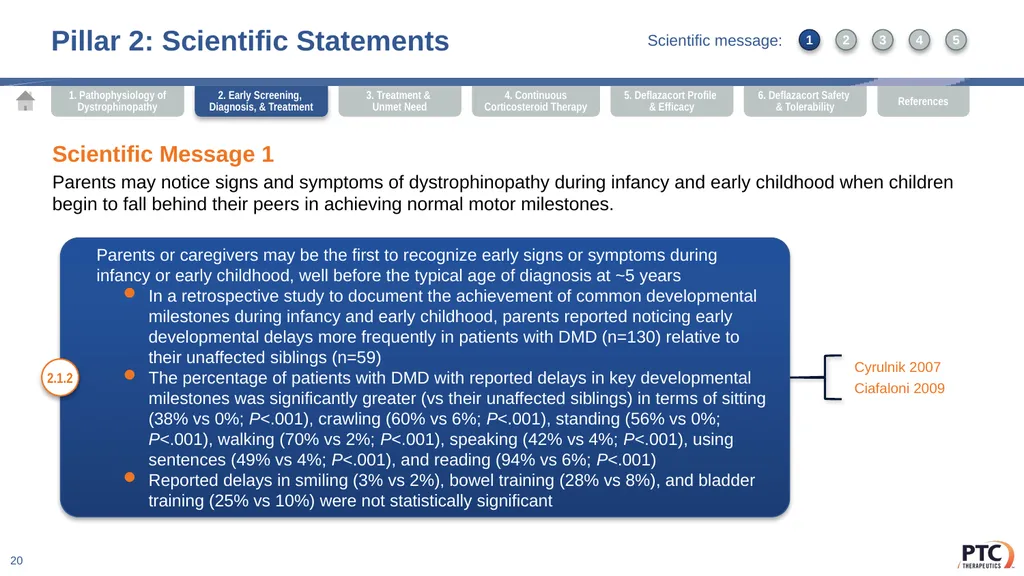
Pillar 2: Scientific Statements 20 Parents or caregivers may be the first to recognize early signs or symptoms during infancy or early childhood, well before the typical age of diagnosis at ~5 years
In a retrospective study to document the achievement of common developmental milestones during infancy and early childhood, parents reported noticing early developmental delays more frequently in patients with DMD (n=130) relative to their unaffected siblings (n=59)
The percentage of patients with DMD with reported delays in key developmental milestones was significantly greater (vs their unaffected siblings) in terms of sitting (38% vs 0%; P<.001), crawling (60% vs 6%; P<.001), standing (56% vs 0%; P<.001), walking (70% vs 2%; P<.001), speaking (42% vs 4%; P<.001), using sentences (49% vs 4%; P<.001), and reading (94% vs 6%; P<.001)
Reported delays in smiling (3% vs 2%), bowel training (28% vs 8%), and bladder training (25% vs 10%) were not statistically significant 2.1.2 Cyrulnik 2007
Ciafaloni 2009 Scientific Message 1
Parents may notice signs and symptoms of dystrophinopathy during infancy and early childhood when children begin to fall behind their peers in achieving normal motor milestones. 2. Early Screening, Diagnosis, & Treatment 1 3 2 4 5 Scientific message:<br>
In a retrospective study to document the achievement of common developmental milestones during infancy and early childhood, parents reported noticing early developmental delays more frequently in patients with DMD (n=130) relative to their unaffected siblings (n=59)
The percentage of patients with DMD with reported delays in key developmental milestones was significantly greater (vs their unaffected siblings) in terms of sitting (38% vs 0%; P<.001), crawling (60% vs 6%; P<.001), standing (56% vs 0%; P<.001), walking (70% vs 2%; P<.001), speaking (42% vs 4%; P<.001), using sentences (49% vs 4%; P<.001), and reading (94% vs 6%; P<.001)
Reported delays in smiling (3% vs 2%), bowel training (28% vs 8%), and bladder training (25% vs 10%) were not statistically significant 2.1.2 Cyrulnik 2007
Ciafaloni 2009 Scientific Message 1
Parents may notice signs and symptoms of dystrophinopathy during infancy and early childhood when children begin to fall behind their peers in achieving normal motor milestones. 2. Early Screening, Diagnosis, & Treatment 1 3 2 4 5 Scientific message:<br>
21
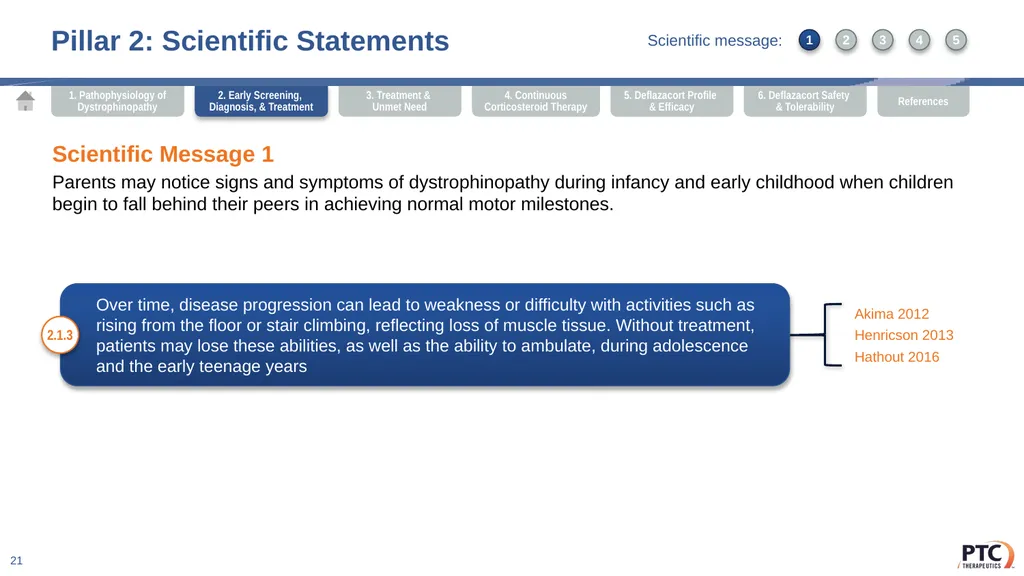
Pillar 2: Scientific Statements 21 Over time, disease progression can lead to weakness or difficulty with activities such as rising from the floor or stair climbing, reflecting loss of muscle tissue. Without treatment, patients may lose these abilities, as well as the ability to ambulate, during adolescence and the early teenage years 2.1.3 Akima 2012
Henricson 2013
Hathout 2016 Scientific Message 1
Parents may notice signs and symptoms of dystrophinopathy during infancy and early childhood when children begin to fall behind their peers in achieving normal motor milestones. 2. Early Screening, Diagnosis, & Treatment 1 3 2 4 5 Scientific message:<br>
Henricson 2013
Hathout 2016 Scientific Message 1
Parents may notice signs and symptoms of dystrophinopathy during infancy and early childhood when children begin to fall behind their peers in achieving normal motor milestones. 2. Early Screening, Diagnosis, & Treatment 1 3 2 4 5 Scientific message:<br>
22
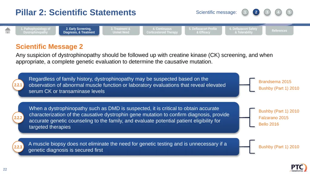
Pillar 2: Scientific Statements 22 Regardless of family history, dystrophinopathy may be suspected based on the observation of abnormal muscle function or laboratory evaluations that reveal elevated serum CK or transaminase levels 2.2.1 Brandsema 2015
Bushby (Part 1) 2010 Scientific Message 2
Any suspicion of dystrophinopathy should be followed up with creatine kinase (CK) screening, and when appropriate, a complete genetic evaluation to determine the causative mutation. When a dystrophinopathy such as DMD is suspected, it is critical to obtain accurate characterization of the causative dystrophin gene mutation to confirm diagnosis, provide accurate genetic counseling to the family, and evaluate potential patient eligibility for targeted therapies 2.2.2 Bushby (Part 1) 2010
Falzarano 2015
Bello 2016 A muscle biopsy does not eliminate the need for genetic testing and is unnecessary if a genetic diagnosis is secured first 2.2.3 Bushby (Part 1) 2010 2. Early Screening, Diagnosis, & Treatment 1 3 2 4 5 Scientific message:<br>
Bushby (Part 1) 2010 Scientific Message 2
Any suspicion of dystrophinopathy should be followed up with creatine kinase (CK) screening, and when appropriate, a complete genetic evaluation to determine the causative mutation. When a dystrophinopathy such as DMD is suspected, it is critical to obtain accurate characterization of the causative dystrophin gene mutation to confirm diagnosis, provide accurate genetic counseling to the family, and evaluate potential patient eligibility for targeted therapies 2.2.2 Bushby (Part 1) 2010
Falzarano 2015
Bello 2016 A muscle biopsy does not eliminate the need for genetic testing and is unnecessary if a genetic diagnosis is secured first 2.2.3 Bushby (Part 1) 2010 2. Early Screening, Diagnosis, & Treatment 1 3 2 4 5 Scientific message:<br>
23
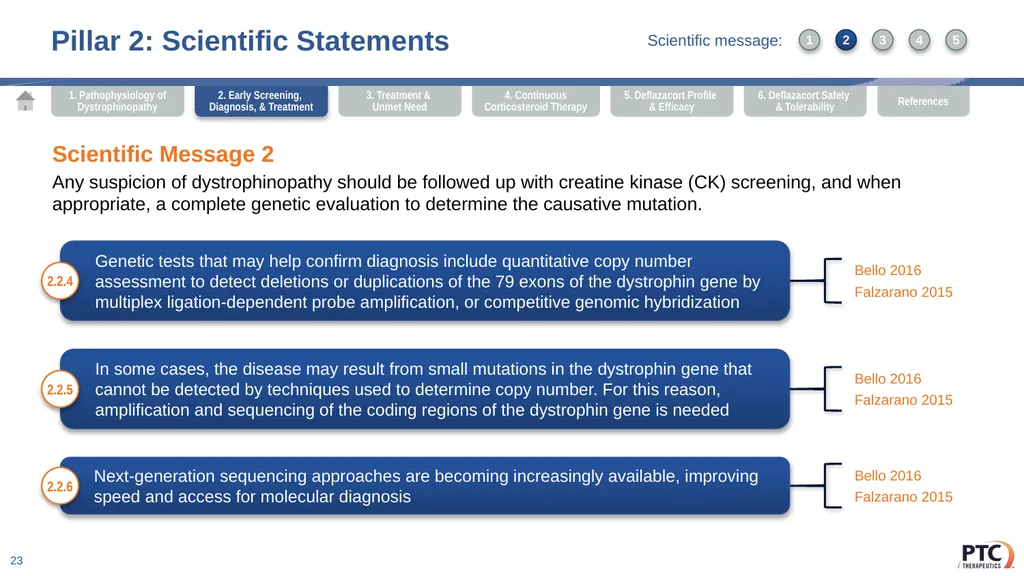
Pillar 2: Scientific Statements 23 Genetic tests that may help confirm diagnosis include quantitative copy number assessment to detect deletions or duplications of the 79 exons of the dystrophin gene by multiplex ligation-dependent probe amplification, or competitive genomic hybridization 2.2.4 Bello 2016
Falzarano 2015 Scientific Message 2
Any suspicion of dystrophinopathy should be followed up with creatine kinase (CK) screening, and when appropriate, a complete genetic evaluation to determine the causative mutation. In some cases, the disease may result from small mutations in the dystrophin gene that cannot be detected by techniques used to determine copy number. For this reason, amplification and sequencing of the coding regions of the dystrophin gene is needed 2.2.5 Bello 2016
Falzarano 2015 Next-generation sequencing approaches are becoming increasingly available, improving speed and access for molecular diagnosis 2.2.6 Bello 2016
Falzarano 2015 2. Early Screening, Diagnosis, & Treatment 1 3 2 4 5 Scientific message:<br>
Falzarano 2015 Scientific Message 2
Any suspicion of dystrophinopathy should be followed up with creatine kinase (CK) screening, and when appropriate, a complete genetic evaluation to determine the causative mutation. In some cases, the disease may result from small mutations in the dystrophin gene that cannot be detected by techniques used to determine copy number. For this reason, amplification and sequencing of the coding regions of the dystrophin gene is needed 2.2.5 Bello 2016
Falzarano 2015 Next-generation sequencing approaches are becoming increasingly available, improving speed and access for molecular diagnosis 2.2.6 Bello 2016
Falzarano 2015 2. Early Screening, Diagnosis, & Treatment 1 3 2 4 5 Scientific message:<br>
24
Pillar 2: Scientific Statements 24 Scientific Message 3
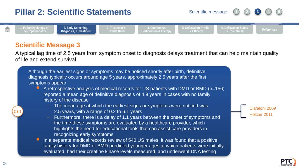
A typical lag time of 2.5 years from symptom onset to diagnosis delays treatment that can help maintain quality of life and extend survival. Although the earliest signs or symptoms may be noticed shortly after birth, definitive diagnosis typically occurs around age 5 years, approximately 2.5 years after the first symptoms appear
A retrospective analysis of medical records for US patients with DMD or BMD (n=156) reported a mean age of definitive diagnosis of 4.9 years in cases with no family history of the disease
The mean age at which the earliest signs or symptoms were noticed was 2.5 years, with a range of 0.2 to 6.1 years
Furthermore, there is a delay of 1.1 years between the onset of symptoms and the time these symptoms are evaluated by a healthcare provider, which highlights the need for educational tools that can assist care providers in recognizing early symptoms
In a separate medical records review of 540 US males, it was found that a positive family history for DMD or BMD predicted younger ages at which patients were initially evaluated, had their creatine kinase levels measured, and underwent DNA testing 2.3.1 Ciafaloni 2009
Holtzer 2011 2. Early Screening, Diagnosis, & Treatment 1 3 2 4 5 Scientific message:<br>
A typical lag time of 2.5 years from symptom onset to diagnosis delays treatment that can help maintain quality of life and extend survival. Although the earliest signs or symptoms may be noticed shortly after birth, definitive diagnosis typically occurs around age 5 years, approximately 2.5 years after the first symptoms appear
A retrospective analysis of medical records for US patients with DMD or BMD (n=156) reported a mean age of definitive diagnosis of 4.9 years in cases with no family history of the disease
The mean age at which the earliest signs or symptoms were noticed was 2.5 years, with a range of 0.2 to 6.1 years
Furthermore, there is a delay of 1.1 years between the onset of symptoms and the time these symptoms are evaluated by a healthcare provider, which highlights the need for educational tools that can assist care providers in recognizing early symptoms
In a separate medical records review of 540 US males, it was found that a positive family history for DMD or BMD predicted younger ages at which patients were initially evaluated, had their creatine kinase levels measured, and underwent DNA testing 2.3.1 Ciafaloni 2009
Holtzer 2011 2. Early Screening, Diagnosis, & Treatment 1 3 2 4 5 Scientific message:<br>
25
Pillar 2: Scientific Statements 25 Scientific Message 3
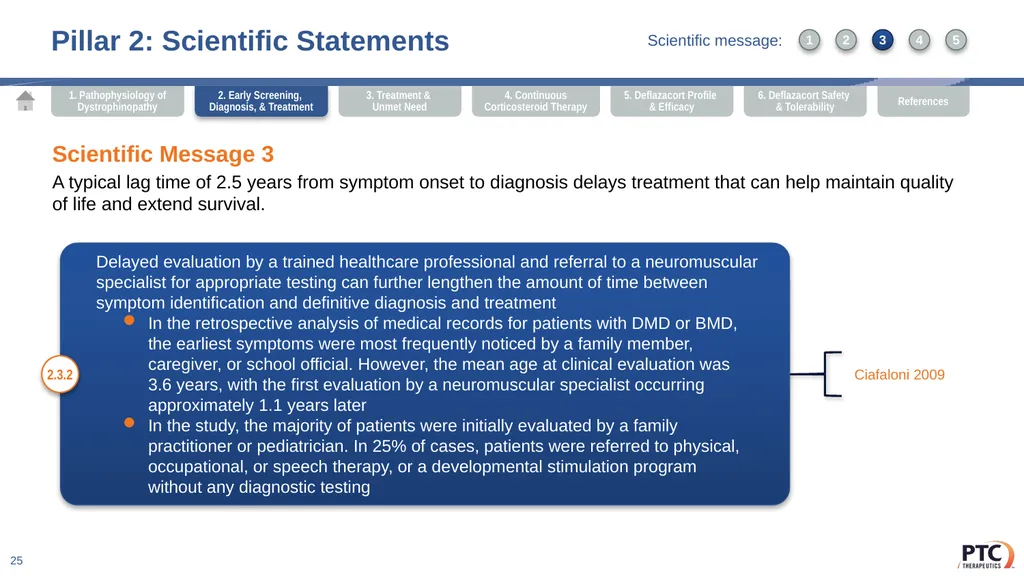
A typical lag time of 2.5 years from symptom onset to diagnosis delays treatment that can help maintain quality of life and extend survival. Delayed evaluation by a trained healthcare professional and referral to a neuromuscular specialist for appropriate testing can further lengthen the amount of time between symptom identification and definitive diagnosis and treatment
In the retrospective analysis of medical records for patients with DMD or BMD, the earliest symptoms were most frequently noticed by a family member, caregiver, or school official. However, the mean age at clinical evaluation was 3.6 years, with the first evaluation by a neuromuscular specialist occurring approximately 1.1 years later
In the study, the majority of patients were initially evaluated by a family practitioner or pediatrician. In 25% of cases, patients were referred to physical, occupational, or speech therapy, or a developmental stimulation program without any diagnostic testing 2.3.2 Ciafaloni 2009 2. Early Screening, Diagnosis, & Treatment 1 3 2 4 5 Scientific message:<br>
A typical lag time of 2.5 years from symptom onset to diagnosis delays treatment that can help maintain quality of life and extend survival. Delayed evaluation by a trained healthcare professional and referral to a neuromuscular specialist for appropriate testing can further lengthen the amount of time between symptom identification and definitive diagnosis and treatment
In the retrospective analysis of medical records for patients with DMD or BMD, the earliest symptoms were most frequently noticed by a family member, caregiver, or school official. However, the mean age at clinical evaluation was 3.6 years, with the first evaluation by a neuromuscular specialist occurring approximately 1.1 years later
In the study, the majority of patients were initially evaluated by a family practitioner or pediatrician. In 25% of cases, patients were referred to physical, occupational, or speech therapy, or a developmental stimulation program without any diagnostic testing 2.3.2 Ciafaloni 2009 2. Early Screening, Diagnosis, & Treatment 1 3 2 4 5 Scientific message:<br>
26
Pillar 2: Scientific Statements 26 Scientific Message 3
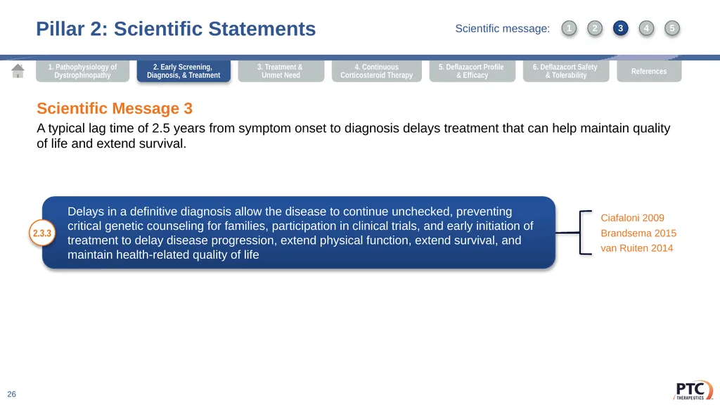
A typical lag time of 2.5 years from symptom onset to diagnosis delays treatment that can help maintain quality of life and extend survival. Delays in a definitive diagnosis allow the disease to continue unchecked, preventing critical genetic counseling for families, participation in clinical trials, and early initiation of treatment to delay disease progression, extend physical function, extend survival, and maintain health-related quality of life 2.3.3 Ciafaloni 2009
Brandsema 2015
van Ruiten 2014 2. Early Screening, Diagnosis, & Treatment 1 3 2 4 5 Scientific message:<br>
A typical lag time of 2.5 years from symptom onset to diagnosis delays treatment that can help maintain quality of life and extend survival. Delays in a definitive diagnosis allow the disease to continue unchecked, preventing critical genetic counseling for families, participation in clinical trials, and early initiation of treatment to delay disease progression, extend physical function, extend survival, and maintain health-related quality of life 2.3.3 Ciafaloni 2009
Brandsema 2015
van Ruiten 2014 2. Early Screening, Diagnosis, & Treatment 1 3 2 4 5 Scientific message:<br>
27
Pillar 2: Scientific Statements 27 Scientific Message 4
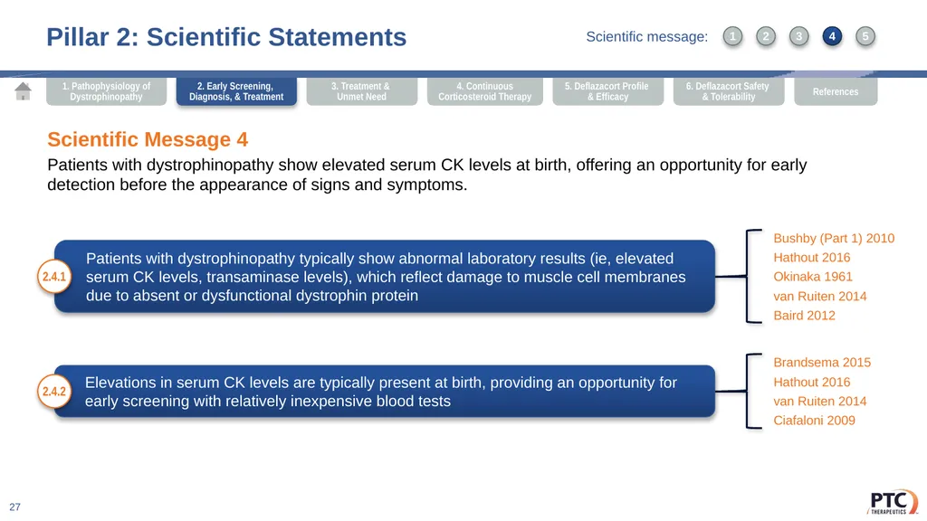
Patients with dystrophinopathy show elevated serum CK levels at birth, offering an opportunity for early detection before the appearance of signs and symptoms. Patients with dystrophinopathy typically show abnormal laboratory results (ie, elevated serum CK levels, transaminase levels), which reflect damage to muscle cell membranes due to absent or dysfunctional dystrophin protein 2.4.1 Bushby (Part 1) 2010
Hathout 2016
Okinaka 1961
van Ruiten 2014
Baird 2012 Elevations in serum CK levels are typically present at birth, providing an opportunity for early screening with relatively inexpensive blood tests 2.4.2 Brandsema 2015
Hathout 2016
van Ruiten 2014
Ciafaloni 2009 2. Early Screening, Diagnosis, & Treatment 1 3 2 4 5 Scientific message:<br>
Patients with dystrophinopathy show elevated serum CK levels at birth, offering an opportunity for early detection before the appearance of signs and symptoms. Patients with dystrophinopathy typically show abnormal laboratory results (ie, elevated serum CK levels, transaminase levels), which reflect damage to muscle cell membranes due to absent or dysfunctional dystrophin protein 2.4.1 Bushby (Part 1) 2010
Hathout 2016
Okinaka 1961
van Ruiten 2014
Baird 2012 Elevations in serum CK levels are typically present at birth, providing an opportunity for early screening with relatively inexpensive blood tests 2.4.2 Brandsema 2015
Hathout 2016
van Ruiten 2014
Ciafaloni 2009 2. Early Screening, Diagnosis, & Treatment 1 3 2 4 5 Scientific message:<br>
28
Serum CK has been used as part of large-scale screening efforts to identify patients
with dystrophinopathies, enabling early diagnosis for patients and appropriate genetic counseling for families
In a program that screened 18,000 newborn males using filter blood spots, 5 affected boys were identified along with 3 likely maternal carriers, allowing early diagnosis and genetic counseling
30,014 samples were screened as part of a Cypriot national pilot program that employed a semiquantitative bioluminescence test, resulting in the identification of 5 cases of BMD or DMD with a low false positive rate (0.10%)
As part of an opt-in screening program in Wales, 343,170 bloodspots were evaluated over a 21-year period. The program resulted in 56 confirmed cases of DMD and 5 cases of BMD, with 13 false-negatives Pillar 2: Scientific Statements 28 Scientific Message 4
Patients with dystrophinopathy show elevated serum CK levels at birth, offering an opportunity for early detection before the appearance of signs and symptoms. 2.4.3 Greenberg 1988
Drousiotou 1998
Moat 2013 2. Early Screening, Diagnosis, & Treatment 1 3 2 4 5 Scientific message:<br>
In a program that screened 18,000 newborn males using filter blood spots, 5 affected boys were identified along with 3 likely maternal carriers, allowing early diagnosis and genetic counseling
30,014 samples were screened as part of a Cypriot national pilot program that employed a semiquantitative bioluminescence test, resulting in the identification of 5 cases of BMD or DMD with a low false positive rate (0.10%)
As part of an opt-in screening program in Wales, 343,170 bloodspots were evaluated over a 21-year period. The program resulted in 56 confirmed cases of DMD and 5 cases of BMD, with 13 false-negatives Pillar 2: Scientific Statements 28 Scientific Message 4
Patients with dystrophinopathy show elevated serum CK levels at birth, offering an opportunity for early detection before the appearance of signs and symptoms. 2.4.3 Greenberg 1988
Drousiotou 1998
Moat 2013 2. Early Screening, Diagnosis, & Treatment 1 3 2 4 5 Scientific message:<br>
29
Pillar 2: Scientific Statements 29 Scientific Message 5
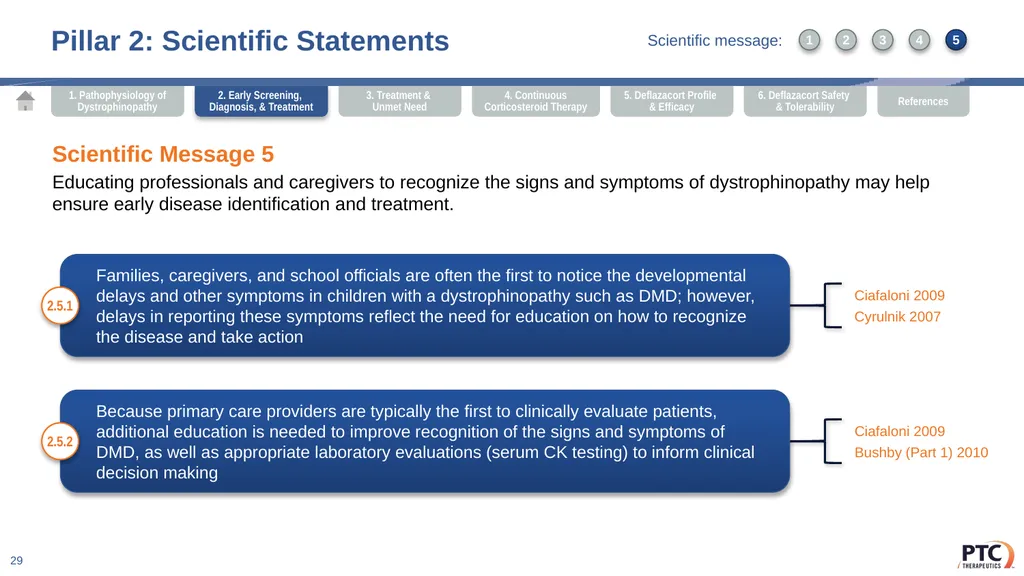
Educating professionals and caregivers to recognize the signs and symptoms of dystrophinopathy may help ensure early disease identification and treatment. Families, caregivers, and school officials are often the first to notice the developmental delays and other symptoms in children with a dystrophinopathy such as DMD; however, delays in reporting these symptoms reflect the need for education on how to recognize the disease and take action 2.5.1 Ciafaloni 2009
Cyrulnik 2007 Because primary care providers are typically the first to clinically evaluate patients, additional education is needed to improve recognition of the signs and symptoms of DMD, as well as appropriate laboratory evaluations (serum CK testing) to inform clinical decision making 2.5.2 Ciafaloni 2009
Bushby (Part 1) 2010 2. Early Screening, Diagnosis, & Treatment 1 3 2 4 5 Scientific message:<br>
Educating professionals and caregivers to recognize the signs and symptoms of dystrophinopathy may help ensure early disease identification and treatment. Families, caregivers, and school officials are often the first to notice the developmental delays and other symptoms in children with a dystrophinopathy such as DMD; however, delays in reporting these symptoms reflect the need for education on how to recognize the disease and take action 2.5.1 Ciafaloni 2009
Cyrulnik 2007 Because primary care providers are typically the first to clinically evaluate patients, additional education is needed to improve recognition of the signs and symptoms of DMD, as well as appropriate laboratory evaluations (serum CK testing) to inform clinical decision making 2.5.2 Ciafaloni 2009
Bushby (Part 1) 2010 2. Early Screening, Diagnosis, & Treatment 1 3 2 4 5 Scientific message:<br>
30
Pillar 2: Scientific Statements 30 Scientific Message 5
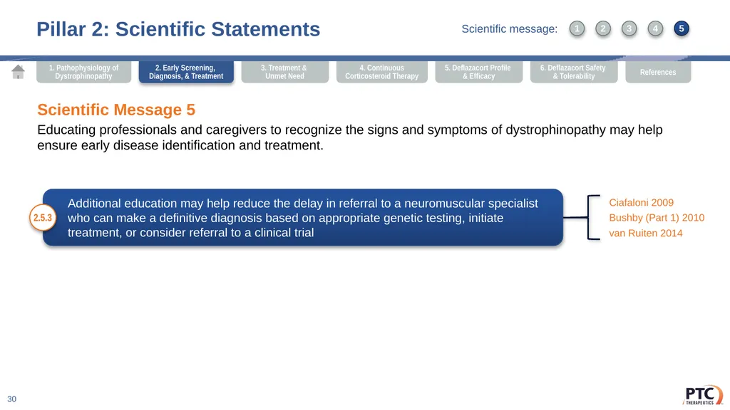
Educating professionals and caregivers to recognize the signs and symptoms of dystrophinopathy may help ensure early disease identification and treatment. Additional education may help reduce the delay in referral to a neuromuscular specialist who can make a definitive diagnosis based on appropriate genetic testing, initiate treatment, or consider referral to a clinical trial 2.5.3 Ciafaloni 2009
Bushby (Part 1) 2010
van Ruiten 2014 2. Early Screening, Diagnosis, & Treatment 1 3 2 4 5 Scientific message:<br>
Educating professionals and caregivers to recognize the signs and symptoms of dystrophinopathy may help ensure early disease identification and treatment. Additional education may help reduce the delay in referral to a neuromuscular specialist who can make a definitive diagnosis based on appropriate genetic testing, initiate treatment, or consider referral to a clinical trial 2.5.3 Ciafaloni 2009
Bushby (Part 1) 2010
van Ruiten 2014 2. Early Screening, Diagnosis, & Treatment 1 3 2 4 5 Scientific message:<br>
31
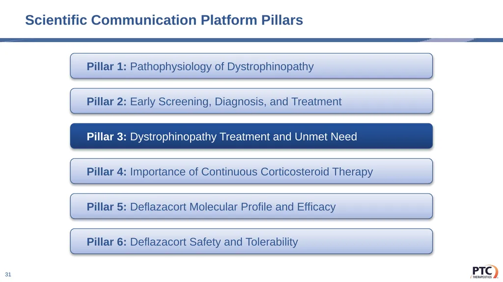
Scientific Communication Platform Pillars 31 Pillar 1: Pathophysiology of Dystrophinopathy Pillar 2: Early Screening, Diagnosis, and Treatment Pillar 3: Dystrophinopathy Treatment and Unmet Need Pillar 4: Importance of Continuous Corticosteroid Therapy Pillar 5: Deflazacort Molecular Profile and Efficacy Pillar 6: Deflazacort Safety and Tolerability<br>
32
Pillar 3: Dystrophinopathy Treatment and Unmet Need 32 Strategic Driver
Corticosteroids are the cornerstone of therapy for patients with dystrophinopathies, helping to delay progression and extend survival while providing a manageable safety profile. Scientific Messages In the absence of a curative therapy, an interdisciplinary approach coordinated among multiple specialists is needed to optimize treatment for each patient.
Patient and caregiver education is critical to navigating the treatment decision-making process in DMD.
Corticosteroid therapy, as the cornerstone of a holistic treatment approach, is the standard of care for patients with DMD.
Early intervention to enhance muscle function, including corticosteroid therapy, may help extend function across muscle types. 3. Treatment &
Unmet Need<br>
Corticosteroids are the cornerstone of therapy for patients with dystrophinopathies, helping to delay progression and extend survival while providing a manageable safety profile. Scientific Messages In the absence of a curative therapy, an interdisciplinary approach coordinated among multiple specialists is needed to optimize treatment for each patient.
Patient and caregiver education is critical to navigating the treatment decision-making process in DMD.
Corticosteroid therapy, as the cornerstone of a holistic treatment approach, is the standard of care for patients with DMD.
Early intervention to enhance muscle function, including corticosteroid therapy, may help extend function across muscle types. 3. Treatment &
Unmet Need<br>
33
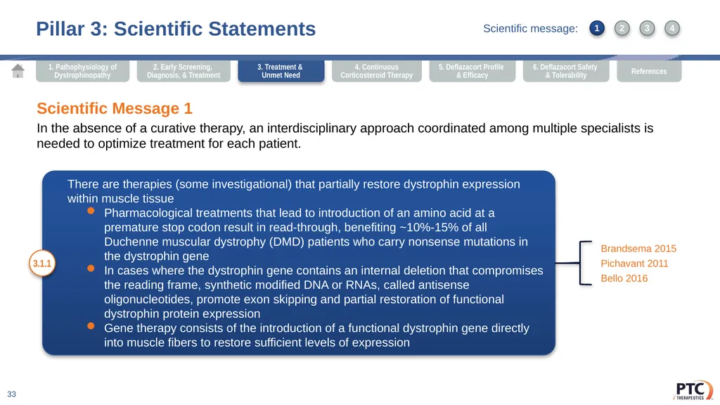
There are therapies (some investigational) that partially restore dystrophin expression within muscle tissue
Pharmacological treatments that lead to introduction of an amino acid at a premature stop codon result in read-through, benefiting ~10%-15% of all Duchenne muscular dystrophy (DMD) patients who carry nonsense mutations in the dystrophin gene
In cases where the dystrophin gene contains an internal deletion that compromises the reading frame, synthetic modified DNA or RNAs, called antisense oligonucleotides, promote exon skipping and partial restoration of functional dystrophin protein expression
Gene therapy consists of the introduction of a functional dystrophin gene directly into muscle fibers to restore sufficient levels of expression Pillar 3: Scientific Statements 33 Scientific Message 1
In the absence of a curative therapy, an interdisciplinary approach coordinated among multiple specialists is needed to optimize treatment for each patient. 3.1.1 Brandsema 2015
Pichavant 2011
Bello 2016 1 3 2 4 3. Treatment &
Unmet Need Scientific message:<br>
Pharmacological treatments that lead to introduction of an amino acid at a premature stop codon result in read-through, benefiting ~10%-15% of all Duchenne muscular dystrophy (DMD) patients who carry nonsense mutations in the dystrophin gene
In cases where the dystrophin gene contains an internal deletion that compromises the reading frame, synthetic modified DNA or RNAs, called antisense oligonucleotides, promote exon skipping and partial restoration of functional dystrophin protein expression
Gene therapy consists of the introduction of a functional dystrophin gene directly into muscle fibers to restore sufficient levels of expression Pillar 3: Scientific Statements 33 Scientific Message 1
In the absence of a curative therapy, an interdisciplinary approach coordinated among multiple specialists is needed to optimize treatment for each patient. 3.1.1 Brandsema 2015
Pichavant 2011
Bello 2016 1 3 2 4 3. Treatment &
Unmet Need Scientific message:<br>
34
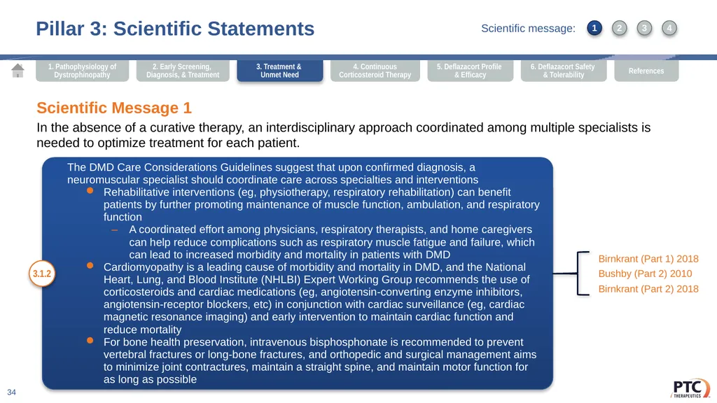
The DMD Care Considerations Guidelines suggest that upon confirmed diagnosis, a neuromuscular specialist should coordinate care across specialties and interventions
Rehabilitative interventions (eg, physiotherapy, respiratory rehabilitation) can benefit patients by further promoting maintenance of muscle function, ambulation, and respiratory function
A coordinated effort among physicians, respiratory therapists, and home caregivers can help reduce complications such as respiratory muscle fatigue and failure, which can lead to increased morbidity and mortality in patients with DMD
Cardiomyopathy is a leading cause of morbidity and mortality in DMD, and the National Heart, Lung, and Blood Institute (NHLBI) Expert Working Group recommends the use of corticosteroids and cardiac medications (eg, angiotensin-converting enzyme inhibitors, angiotensin-receptor blockers, etc) in conjunction with cardiac surveillance (eg, cardiac magnetic resonance imaging) and early intervention to maintain cardiac function and reduce mortality
For bone health preservation, intravenous bisphosphonate is recommended to prevent vertebral fractures or long-bone fractures, and orthopedic and surgical management aims to minimize joint contractures, maintain a straight spine, and maintain motor function for as long as possible Pillar 3: Scientific Statements 34 Scientific Message 1
In the absence of a curative therapy, an interdisciplinary approach coordinated among multiple specialists is needed to optimize treatment for each patient. 3.1.2 Birnkrant (Part 1) 2018
Bushby (Part 2) 2010
Birnkrant (Part 2) 2018 3. Treatment &
Unmet Need 1 3 2 4 Scientific message:<br>
Rehabilitative interventions (eg, physiotherapy, respiratory rehabilitation) can benefit patients by further promoting maintenance of muscle function, ambulation, and respiratory function
A coordinated effort among physicians, respiratory therapists, and home caregivers can help reduce complications such as respiratory muscle fatigue and failure, which can lead to increased morbidity and mortality in patients with DMD
Cardiomyopathy is a leading cause of morbidity and mortality in DMD, and the National Heart, Lung, and Blood Institute (NHLBI) Expert Working Group recommends the use of corticosteroids and cardiac medications (eg, angiotensin-converting enzyme inhibitors, angiotensin-receptor blockers, etc) in conjunction with cardiac surveillance (eg, cardiac magnetic resonance imaging) and early intervention to maintain cardiac function and reduce mortality
For bone health preservation, intravenous bisphosphonate is recommended to prevent vertebral fractures or long-bone fractures, and orthopedic and surgical management aims to minimize joint contractures, maintain a straight spine, and maintain motor function for as long as possible Pillar 3: Scientific Statements 34 Scientific Message 1
In the absence of a curative therapy, an interdisciplinary approach coordinated among multiple specialists is needed to optimize treatment for each patient. 3.1.2 Birnkrant (Part 1) 2018
Bushby (Part 2) 2010
Birnkrant (Part 2) 2018 3. Treatment &
Unmet Need 1 3 2 4 Scientific message:<br>
35
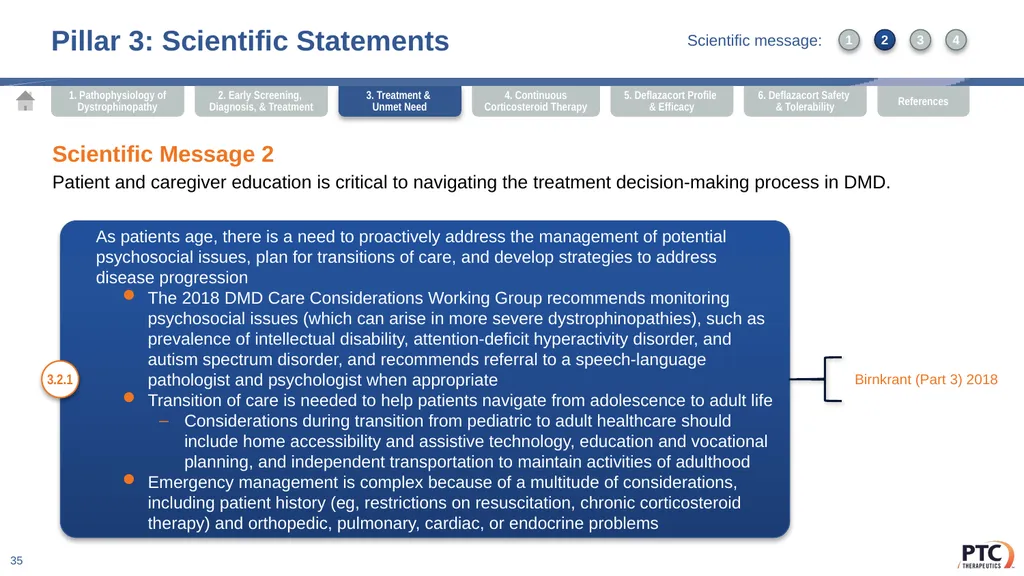
As patients age, there is a need to proactively address the management of potential psychosocial issues, plan for transitions of care, and develop strategies to address disease progression
The 2018 DMD Care Considerations Working Group recommends monitoring psychosocial issues (which can arise in more severe dystrophinopathies), such as prevalence of intellectual disability, attention-deficit hyperactivity disorder, and autism spectrum disorder, and recommends referral to a speech-language pathologist and psychologist when appropriate
Transition of care is needed to help patients navigate from adolescence to adult life
Considerations during transition from pediatric to adult healthcare should include home accessibility and assistive technology, education and vocational planning, and independent transportation to maintain activities of adulthood
Emergency management is complex because of a multitude of considerations, including patient history (eg, restrictions on resuscitation, chronic corticosteroid therapy) and orthopedic, pulmonary, cardiac, or endocrine problems Pillar 3: Scientific Statements 35 Scientific Message 2
Patient and caregiver education is critical to navigating the treatment decision-making process in DMD. 3.2.1 Birnkrant (Part 3) 2018 3. Treatment &
Unmet Need 1 3 2 4 Scientific message:<br>
The 2018 DMD Care Considerations Working Group recommends monitoring psychosocial issues (which can arise in more severe dystrophinopathies), such as prevalence of intellectual disability, attention-deficit hyperactivity disorder, and autism spectrum disorder, and recommends referral to a speech-language pathologist and psychologist when appropriate
Transition of care is needed to help patients navigate from adolescence to adult life
Considerations during transition from pediatric to adult healthcare should include home accessibility and assistive technology, education and vocational planning, and independent transportation to maintain activities of adulthood
Emergency management is complex because of a multitude of considerations, including patient history (eg, restrictions on resuscitation, chronic corticosteroid therapy) and orthopedic, pulmonary, cardiac, or endocrine problems Pillar 3: Scientific Statements 35 Scientific Message 2
Patient and caregiver education is critical to navigating the treatment decision-making process in DMD. 3.2.1 Birnkrant (Part 3) 2018 3. Treatment &
Unmet Need 1 3 2 4 Scientific message:<br>
36
According to recommendations published by the DMD Care Considerations Working Group, patients, caregivers, and providers should engage in yearly discussion of long- term treatment goals and make use of resources that allow for proactive planning for unique health-related issues in DMD Pillar 3: Scientific Statements 36 Scientific Message 2
Patient and caregiver education is critical to navigating the treatment decision-making process in DMD. 3.2.2 Birnkrant (Part 3) 2018 3. Treatment &
Unmet Need 1 3 2 4 Scientific message:<br>
Patient and caregiver education is critical to navigating the treatment decision-making process in DMD. 3.2.2 Birnkrant (Part 3) 2018 3. Treatment &
Unmet Need 1 3 2 4 Scientific message:<br>
37
Corticosteroids are broadly applicable and offer the only method of extending muscle function; the DMD Care Considerations guidelines recommend daily dosing with corticosteroids in patients with DMD
An added outcomes benefit with pharmacological intervention has been noted in patients with DMD when a combination of respiratory, cardiac, orthopedic, and rehabilitative interventions are incorporated into care Pillar 3: Scientific Statements 37 Scientific Message 3
Corticosteroid therapy, as the cornerstone of a holistic treatment approach, is the standard of care for patients with DMD. 3.3.1 Biggar 2006
Bello 2015
Griggs 2016
Narayanan 2017a
McDonald 2018
Wang 2014
Bushby (Part 2) 2010
Birnkrant (Part 2) 2018 3. Treatment &
Unmet Need 1 3 2 4 Scientific message:<br>
An added outcomes benefit with pharmacological intervention has been noted in patients with DMD when a combination of respiratory, cardiac, orthopedic, and rehabilitative interventions are incorporated into care Pillar 3: Scientific Statements 37 Scientific Message 3
Corticosteroid therapy, as the cornerstone of a holistic treatment approach, is the standard of care for patients with DMD. 3.3.1 Biggar 2006
Bello 2015
Griggs 2016
Narayanan 2017a
McDonald 2018
Wang 2014
Bushby (Part 2) 2010
Birnkrant (Part 2) 2018 3. Treatment &
Unmet Need 1 3 2 4 Scientific message:<br>
38
Pillar 3: Scientific Statements 38 Scientific Message 3
Corticosteroid therapy, as the cornerstone of a holistic treatment approach, is the standard of care for patients with DMD. Results from the Cooperative International Neuromuscular Research Group DMD Natural History study demonstrated that a number of clinically meaningful milestones across multiple muscle groups were significantly preserved in boys treated with corticosteroids
Manual muscle test (MMT)-based calculations of global strength showed that in patients <10 years of age treated with corticosteroids, strength declined by 0.14 ± 0.96 MMT unit/year, less than the previously reported decline of 0.4 ± 0.39 MMT unit/year in historical corticosteroid-naïve patients; additionally, pulmonary function tests were relatively preserved in corticosteroid-treated adolescents 3.3.2 Henricson 2013 3. Treatment &
Unmet Need 1 3 2 4 Scientific message:<br>
Corticosteroid therapy, as the cornerstone of a holistic treatment approach, is the standard of care for patients with DMD. Results from the Cooperative International Neuromuscular Research Group DMD Natural History study demonstrated that a number of clinically meaningful milestones across multiple muscle groups were significantly preserved in boys treated with corticosteroids
Manual muscle test (MMT)-based calculations of global strength showed that in patients <10 years of age treated with corticosteroids, strength declined by 0.14 ± 0.96 MMT unit/year, less than the previously reported decline of 0.4 ± 0.39 MMT unit/year in historical corticosteroid-naïve patients; additionally, pulmonary function tests were relatively preserved in corticosteroid-treated adolescents 3.3.2 Henricson 2013 3. Treatment &
Unmet Need 1 3 2 4 Scientific message:<br>
39
Pillar 3: Scientific Statements 39 Scientific Message 3
Corticosteroid therapy, as the cornerstone of a holistic treatment approach, is the standard of care for patients with DMD. Current recommendations published by the DMD Care Considerations Working Group include treatment with corticosteroids in conjunction with physiotherapy as the mainstays of DMD treatment
After an initial discussion with the family, including discussion of side effects and nutritional consultation, corticosteroid therapy should be initiated prior to significant physical decline
Treatment with corticosteroids should continue even after loss of ambulation for the purpose of maintaining upper limb strength, decreasing the risk of scoliosis, and delaying the progressive decline of respiratory and cardiac function
If a patient experiences functional decline while on corticosteroid treatment, a dose increase should be considered, with reassessment in 2-3 months 3.3.3 Birnkrant (Part 1) 2018
Silversides 2003 3. Treatment &
Unmet Need 1 3 2 4 Scientific message:<br>
Corticosteroid therapy, as the cornerstone of a holistic treatment approach, is the standard of care for patients with DMD. Current recommendations published by the DMD Care Considerations Working Group include treatment with corticosteroids in conjunction with physiotherapy as the mainstays of DMD treatment
After an initial discussion with the family, including discussion of side effects and nutritional consultation, corticosteroid therapy should be initiated prior to significant physical decline
Treatment with corticosteroids should continue even after loss of ambulation for the purpose of maintaining upper limb strength, decreasing the risk of scoliosis, and delaying the progressive decline of respiratory and cardiac function
If a patient experiences functional decline while on corticosteroid treatment, a dose increase should be considered, with reassessment in 2-3 months 3.3.3 Birnkrant (Part 1) 2018
Silversides 2003 3. Treatment &
Unmet Need 1 3 2 4 Scientific message:<br>
40
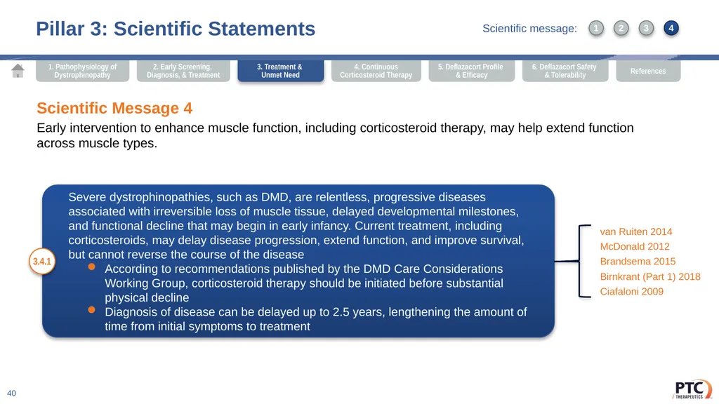
Pillar 3: Scientific Statements 40 Scientific Message 4
Early intervention to enhance muscle function, including corticosteroid therapy, may help extend function across muscle types. Severe dystrophinopathies, such as DMD, are relentless, progressive diseases associated with irreversible loss of muscle tissue, delayed developmental milestones, and functional decline that may begin in early infancy. Current treatment, including corticosteroids, may delay disease progression, extend function, and improve survival, but cannot reverse the course of the disease
According to recommendations published by the DMD Care Considerations Working Group, corticosteroid therapy should be initiated before substantial physical decline
Diagnosis of disease can be delayed up to 2.5 years, lengthening the amount of time from initial symptoms to treatment 3.4.1 van Ruiten 2014
McDonald 2012
Brandsema 2015
Birnkrant (Part 1) 2018
Ciafaloni 2009 3. Treatment &
Unmet Need 1 3 2 4 Scientific message:<br>
Early intervention to enhance muscle function, including corticosteroid therapy, may help extend function across muscle types. Severe dystrophinopathies, such as DMD, are relentless, progressive diseases associated with irreversible loss of muscle tissue, delayed developmental milestones, and functional decline that may begin in early infancy. Current treatment, including corticosteroids, may delay disease progression, extend function, and improve survival, but cannot reverse the course of the disease
According to recommendations published by the DMD Care Considerations Working Group, corticosteroid therapy should be initiated before substantial physical decline
Diagnosis of disease can be delayed up to 2.5 years, lengthening the amount of time from initial symptoms to treatment 3.4.1 van Ruiten 2014
McDonald 2012
Brandsema 2015
Birnkrant (Part 1) 2018
Ciafaloni 2009 3. Treatment &
Unmet Need 1 3 2 4 Scientific message:<br>
41
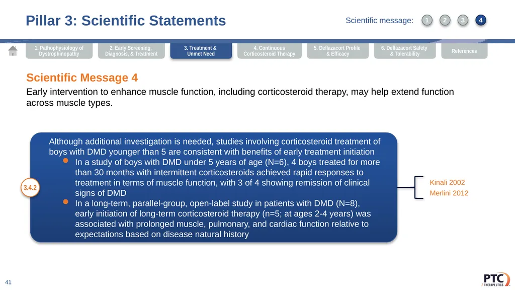
Pillar 3: Scientific Statements 41 Scientific Message 4
Early intervention to enhance muscle function, including corticosteroid therapy, may help extend function across muscle types. Although additional investigation is needed, studies involving corticosteroid treatment of boys with DMD younger than 5 are consistent with benefits of early treatment initiation
In a study of boys with DMD under 5 years of age (N=6), 4 boys treated for more than 30 months with intermittent corticosteroids achieved rapid responses to treatment in terms of muscle function, with 3 of 4 showing remission of clinical signs of DMD
In a long-term, parallel-group, open-label study in patients with DMD (N=8), early initiation of long-term corticosteroid therapy (n=5; at ages 2-4 years) was associated with prolonged muscle, pulmonary, and cardiac function relative to expectations based on disease natural history 3.4.2 Kinali 2002
Merlini 2012 3. Treatment &
Unmet Need 1 3 2 4 Scientific message:<br>
Early intervention to enhance muscle function, including corticosteroid therapy, may help extend function across muscle types. Although additional investigation is needed, studies involving corticosteroid treatment of boys with DMD younger than 5 are consistent with benefits of early treatment initiation
In a study of boys with DMD under 5 years of age (N=6), 4 boys treated for more than 30 months with intermittent corticosteroids achieved rapid responses to treatment in terms of muscle function, with 3 of 4 showing remission of clinical signs of DMD
In a long-term, parallel-group, open-label study in patients with DMD (N=8), early initiation of long-term corticosteroid therapy (n=5; at ages 2-4 years) was associated with prolonged muscle, pulmonary, and cardiac function relative to expectations based on disease natural history 3.4.2 Kinali 2002
Merlini 2012 3. Treatment &
Unmet Need 1 3 2 4 Scientific message:<br>
42
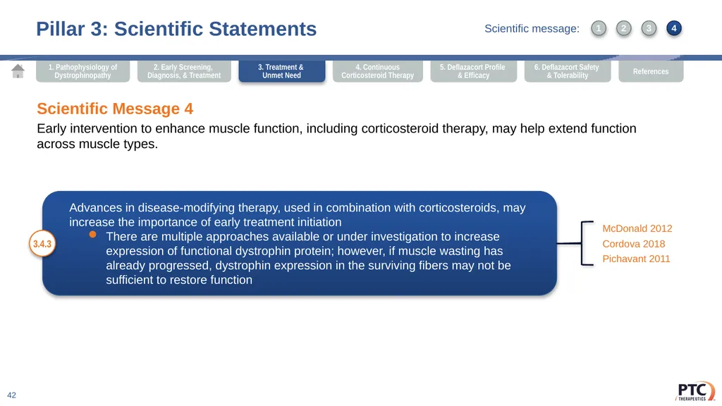
Pillar 3: Scientific Statements 42 Scientific Message 4
Early intervention to enhance muscle function, including corticosteroid therapy, may help extend function across muscle types. Advances in disease-modifying therapy, used in combination with corticosteroids, may increase the importance of early treatment initiation
There are multiple approaches available or under investigation to increase expression of functional dystrophin protein; however, if muscle wasting has already progressed, dystrophin expression in the surviving fibers may not be sufficient to restore function 3.4.3 McDonald 2012
Cordova 2018
Pichavant 2011 3. Treatment &
Unmet Need 1 3 2 4 Scientific message:<br>
Early intervention to enhance muscle function, including corticosteroid therapy, may help extend function across muscle types. Advances in disease-modifying therapy, used in combination with corticosteroids, may increase the importance of early treatment initiation
There are multiple approaches available or under investigation to increase expression of functional dystrophin protein; however, if muscle wasting has already progressed, dystrophin expression in the surviving fibers may not be sufficient to restore function 3.4.3 McDonald 2012
Cordova 2018
Pichavant 2011 3. Treatment &
Unmet Need 1 3 2 4 Scientific message:<br>
43
Scientific Communication Platform Pillars 43 Pillar 1: Pathophysiology of Dystrophinopathy Pillar 2: Early Screening, Diagnosis, and Treatment Pillar 3: Dystrophinopathy Treatment and Unmet Need Pillar 4: Importance of Continuous Corticosteroid Therapy Pillar 5: Deflazacort Molecular Profile and Efficacy Pillar 6: Deflazacort Safety and Tolerability<br>
44
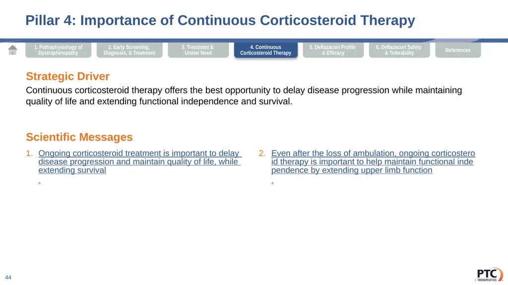
Pillar 4: Importance of Continuous Corticosteroid Therapy 44 Strategic Driver
Continuous corticosteroid therapy offers the best opportunity to delay disease progression while maintaining quality of life and extending functional independence and survival. Scientific Messages Ongoing corticosteroid treatment is important to delay disease progression and maintain quality of life, while extending survival.
Even after the loss of ambulation, ongoing corticosteroid therapy is important to help maintain functional independence by extending upper limb function. 4. Continuous Corticosteroid Therapy<br>
Continuous corticosteroid therapy offers the best opportunity to delay disease progression while maintaining quality of life and extending functional independence and survival. Scientific Messages Ongoing corticosteroid treatment is important to delay disease progression and maintain quality of life, while extending survival.
Even after the loss of ambulation, ongoing corticosteroid therapy is important to help maintain functional independence by extending upper limb function. 4. Continuous Corticosteroid Therapy<br>
45
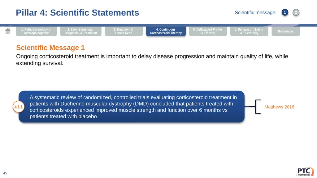
Pillar 4: Scientific Statements 45 Scientific Message 1
Ongoing corticosteroid treatment is important to delay disease progression and maintain quality of life, while extending survival. A systematic review of randomized, controlled trials evaluating corticosteroid treatment in patients with Duchenne muscular dystrophy (DMD) concluded that patients treated with corticosteroids experienced improved muscle strength and function over 6 months vs patients treated with placebo 4.1.1 Matthews 2016 1 2 4. Continuous Corticosteroid Therapy Scientific message:<br>
Ongoing corticosteroid treatment is important to delay disease progression and maintain quality of life, while extending survival. A systematic review of randomized, controlled trials evaluating corticosteroid treatment in patients with Duchenne muscular dystrophy (DMD) concluded that patients treated with corticosteroids experienced improved muscle strength and function over 6 months vs patients treated with placebo 4.1.1 Matthews 2016 1 2 4. Continuous Corticosteroid Therapy Scientific message:<br>
46
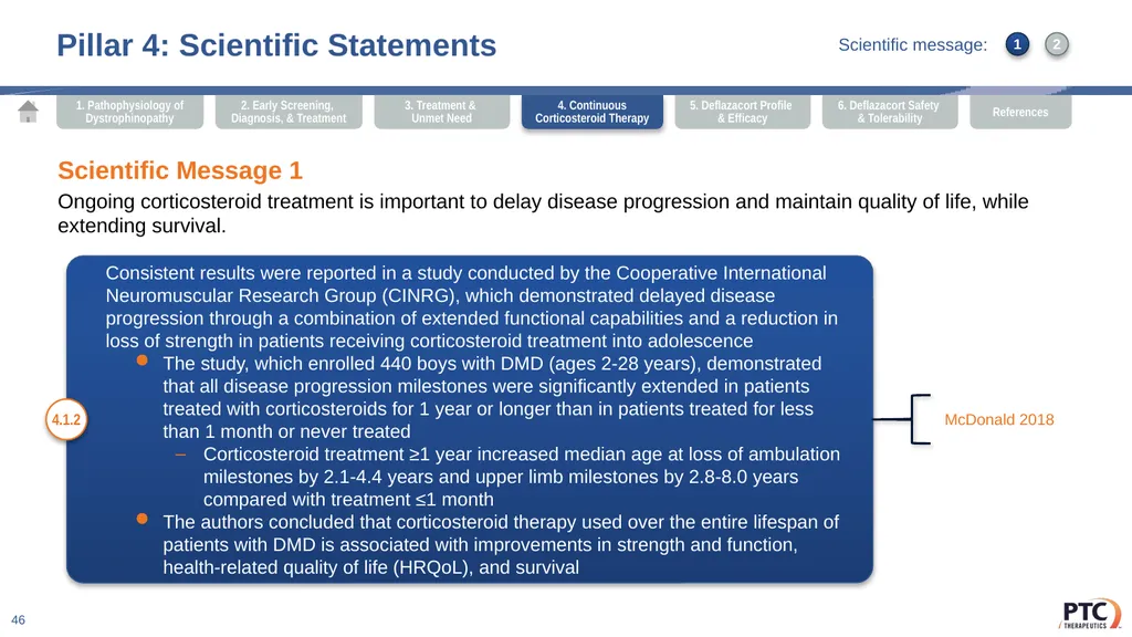
Consistent results were reported in a study conducted by the Cooperative International Neuromuscular Research Group (CINRG), which demonstrated delayed disease progression through a combination of extended functional capabilities and a reduction in loss of strength in patients receiving corticosteroid treatment into adolescence
The study, which enrolled 440 boys with DMD (ages 2-28 years), demonstrated that all disease progression milestones were significantly extended in patients treated with corticosteroids for 1 year or longer than in patients treated for less than 1 month or never treated
Corticosteroid treatment ≥1 year increased median age at loss of ambulation milestones by 2.1-4.4 years and upper limb milestones by 2.8-8.0 years compared with treatment ≤1 month
The authors concluded that corticosteroid therapy used over the entire lifespan of patients with DMD is associated with improvements in strength and function, health-related quality of life (HRQoL), and survival Pillar 4: Scientific Statements 46 Scientific Message 1
Ongoing corticosteroid treatment is important to delay disease progression and maintain quality of life, while extending survival. 4.1.2 McDonald 2018 4. Continuous Corticosteroid Therapy 1 2 Scientific message:<br>
The study, which enrolled 440 boys with DMD (ages 2-28 years), demonstrated that all disease progression milestones were significantly extended in patients treated with corticosteroids for 1 year or longer than in patients treated for less than 1 month or never treated
Corticosteroid treatment ≥1 year increased median age at loss of ambulation milestones by 2.1-4.4 years and upper limb milestones by 2.8-8.0 years compared with treatment ≤1 month
The authors concluded that corticosteroid therapy used over the entire lifespan of patients with DMD is associated with improvements in strength and function, health-related quality of life (HRQoL), and survival Pillar 4: Scientific Statements 46 Scientific Message 1
Ongoing corticosteroid treatment is important to delay disease progression and maintain quality of life, while extending survival. 4.1.2 McDonald 2018 4. Continuous Corticosteroid Therapy 1 2 Scientific message:<br>
47
Clinical evidence suggests a reduced need for ventilation support, scoliosis surgery, and extended survival among boys treated with corticosteroid therapy
In a study of the disease progression of DMD in boys between the ages of 10 and 18 years who received deflazacort (n=40) or no corticosteroid therapy (n=34):
By 18 years of age, nocturnal ventilation was not required in boys treated with deflazacort, whereas 46% of boys not treated required nocturnal ventilation
By 18 years of age, only 10% of boys treated with deflazacort required spinal surgery for scoliosis, whereas 90% of boys not treated had a spinal curvature >20% and required scoliosis surgery
95% of boys treated with deflazacort were alive in the second decade vs 65% alive who were not treated Pillar 4: Scientific Statements 47 Scientific Message 1
Ongoing corticosteroid treatment is important to delay disease progression and maintain quality of life, while extending survival. 4.1.3 McDonald 2018
Biggar 2006 4. Continuous Corticosteroid Therapy 1 2 Scientific message:<br>
In a study of the disease progression of DMD in boys between the ages of 10 and 18 years who received deflazacort (n=40) or no corticosteroid therapy (n=34):
By 18 years of age, nocturnal ventilation was not required in boys treated with deflazacort, whereas 46% of boys not treated required nocturnal ventilation
By 18 years of age, only 10% of boys treated with deflazacort required spinal surgery for scoliosis, whereas 90% of boys not treated had a spinal curvature >20% and required scoliosis surgery
95% of boys treated with deflazacort were alive in the second decade vs 65% alive who were not treated Pillar 4: Scientific Statements 47 Scientific Message 1
Ongoing corticosteroid treatment is important to delay disease progression and maintain quality of life, while extending survival. 4.1.3 McDonald 2018
Biggar 2006 4. Continuous Corticosteroid Therapy 1 2 Scientific message:<br>
48
DMD can place a dramatic burden on patients and their families, ultimately impacting quality of life and functional milestones for patients. Corticosteroid therapy may help maintain HRQoL for patients, while extending muscle strength, physical function, and delaying disease progression
Deflazacort treatment led to significant improvement in HRQoL based on the Sports/Physical Function domain score of the Pediatric Outcomes Data Collection Instrument, which provides surrogate measures of limb function and general feeling Pillar 4: Scientific Statements 48 Scientific Message 1
Ongoing corticosteroid treatment is important to delay disease progression and maintain quality of life, while extending survival. 4.1.4 Landfeldt 2015
Ryder 2017
Shieh 2018
Narayanan 2017a
Matthews 2016 4. Continuous Corticosteroid Therapy 1 2 Scientific message:<br>
Deflazacort treatment led to significant improvement in HRQoL based on the Sports/Physical Function domain score of the Pediatric Outcomes Data Collection Instrument, which provides surrogate measures of limb function and general feeling Pillar 4: Scientific Statements 48 Scientific Message 1
Ongoing corticosteroid treatment is important to delay disease progression and maintain quality of life, while extending survival. 4.1.4 Landfeldt 2015
Ryder 2017
Shieh 2018
Narayanan 2017a
Matthews 2016 4. Continuous Corticosteroid Therapy 1 2 Scientific message:<br>
49
Loss of ambulation is considered a key milestone in the progression of DMD; however, upper limb function can also play an important role in the maintenance of patient independence and quality of life Pillar 4: Scientific Statements 49 Scientific Message 2
Even after the loss of ambulation, ongoing corticosteroid therapy is important to help maintain functional independence by extending upper limb function. 4.2.1 Janssen 2016 Multiple studies have indicated that deflazacort therapy extends upper limb function (as measured by grip and pinch strength, maximum hand-held weight lifted overhead, and Brooke upper extremity functional rating scale), thereby allowing patients to maintain independence for activities such as feeding oneself, dressing, and self-care 4.2.2 McDonald 2018
Balaban 2005 4. Continuous Corticosteroid Therapy 1 2 Scientific message:<br>
Even after the loss of ambulation, ongoing corticosteroid therapy is important to help maintain functional independence by extending upper limb function. 4.2.1 Janssen 2016 Multiple studies have indicated that deflazacort therapy extends upper limb function (as measured by grip and pinch strength, maximum hand-held weight lifted overhead, and Brooke upper extremity functional rating scale), thereby allowing patients to maintain independence for activities such as feeding oneself, dressing, and self-care 4.2.2 McDonald 2018
Balaban 2005 4. Continuous Corticosteroid Therapy 1 2 Scientific message:<br>
50
Current recommendations published by the DMD Care Considerations Working Group indicate that treatment with corticosteroids should continue even after loss of ambulation for the purpose of maintaining upper limb strength, decreasing the risk of scoliosis, and delaying the progressive decline of respiratory and cardiac function Pillar 4: Scientific Statements 50 Scientific Message 2
Even after the loss of ambulation, ongoing corticosteroid therapy is important to help maintain functional independence by extending upper limb function. 4.2.3 Birnkrant (Part 1) 2018
Silversides 2003
McDonald 2018 4. Continuous Corticosteroid Therapy 1 2 Scientific message:<br>
Even after the loss of ambulation, ongoing corticosteroid therapy is important to help maintain functional independence by extending upper limb function. 4.2.3 Birnkrant (Part 1) 2018
Silversides 2003
McDonald 2018 4. Continuous Corticosteroid Therapy 1 2 Scientific message:<br>
51
Scientific Communication Platform Pillars 51 Pillar 1: Pathophysiology of Dystrophinopathy Pillar 2: Early Screening, Diagnosis, and Treatment Pillar 3: Dystrophinopathy Treatment and Unmet Need Pillar 4: Importance of Continuous Corticosteroid Therapy Pillar 5: Deflazacort Molecular Profile and Efficacy Pillar 6: Deflazacort Safety and Tolerability<br>
52
Pillar 5: Deflazacort Molecular Profile and Efficacy 52 Strategic Driver
Corticosteroids are a standard of care that delays progression, helps maintain health-related quality of life (HRQoL), and may extend survival. Deflazacort is the first and only US Food and Drug Administration (FDA)-approved corticosteroid for the treatment of DMD. Scientific Messages Deflazacort is the first and only corticosteroid that is FDA approved for the treatment of all DMD patients ≥5 years of age and that can be used throughout life.
Deflazacort treatment led to significant improvements in muscle strength and motor function vs placebo.
Deflazacort treatment significantly improved muscle strength, pulmonary function, and was more effective at extending motor function vs prednisone or prednisolone.
Deflazacort treatment delayed disease progression across key DMD milestones by approximately 1-3 years.
Deflazacort was associated with numerical delays in the need for nocturnal ventilation or scoliosis surgery, and numerically extended survival for patients with DMD.
Deflazacort was associated with better maintenance of HRQoL for patients with DMD than prednisone or prednisolone. 5. Deflazacort Profile & Efficacy<br>
Corticosteroids are a standard of care that delays progression, helps maintain health-related quality of life (HRQoL), and may extend survival. Deflazacort is the first and only US Food and Drug Administration (FDA)-approved corticosteroid for the treatment of DMD. Scientific Messages Deflazacort is the first and only corticosteroid that is FDA approved for the treatment of all DMD patients ≥5 years of age and that can be used throughout life.
Deflazacort treatment led to significant improvements in muscle strength and motor function vs placebo.
Deflazacort treatment significantly improved muscle strength, pulmonary function, and was more effective at extending motor function vs prednisone or prednisolone.
Deflazacort treatment delayed disease progression across key DMD milestones by approximately 1-3 years.
Deflazacort was associated with numerical delays in the need for nocturnal ventilation or scoliosis surgery, and numerically extended survival for patients with DMD.
Deflazacort was associated with better maintenance of HRQoL for patients with DMD than prednisone or prednisolone. 5. Deflazacort Profile & Efficacy<br>
53
Deflazacort (Emflaza®) was approved by the FDA on February 9, 2017, for the treatment of patients 5 years and older with DMD. It is the first approved treatment that could be used in a wide range of patients with the disease. The approved dose of deflazacort is
0.9 mg/kg/d Pillar 5: Scientific Statements 53 Scientific Message 1
Deflazacort is the first and only corticosteroid that is FDA approved for the treatment of all DMD patients ≥5 years of age and that can be used throughout life. 5.1.1 Emflaza PI 2017
US FDA Press Release 2017 1 3 2 4 6 5 5. Deflazacort Profile & Efficacy Scientific message:<br>
Deflazacort is the first and only corticosteroid that is FDA approved for the treatment of all DMD patients ≥5 years of age and that can be used throughout life. 5.1.1 Emflaza PI 2017
US FDA Press Release 2017 1 3 2 4 6 5 5. Deflazacort Profile & Efficacy Scientific message:<br>
54
The efficacy and safety of deflazacort were evaluated in a pivotal, multicenter,
phase 3 study of deflazacort (at doses of 0.9 mg/kg/d or 1.2 mg/kg/d) and prednisone (0.75 mg/kg/d) vs placebo, which enrolled 196 boys with DMD
Boys treated with deflazacort demonstrated significant improvement in the primary endpoint of muscle strength on the Medical Research Council (MRC) index compared with placebo (0.9 mg/kg/d [0.25 vs 20.1; P=.017]; 1.2 mg/kg/d [0.36 vs 20.1; P=.0003])
At 12 weeks, boys treated with deflazacort demonstrated significant improvements in tests of motor function at doses of 0.9 mg/kg/d and 1.2 mg/kg/d, which included:
Time from supine to standing vs placebo: deflazacort 0.9 mg/kg/d (P=.0018), deflazacort 1.2 mg/kg/d (P=.0002)
Time to climb 4 stairs vs placebo (P=.0001 for all active treatments)
Time to run or walk 30 feet vs placebo (P=.0001 for all active treatments) Pillar 5: Scientific Statements 54 Scientific Message 2
Deflazacort treatment led to significant improvements in muscle strength and motor function vs placebo. 5.2.1 Griggs 2016 5. Deflazacort Profile & Efficacy 1 3 2 4 6 5 Scientific message:<br>
Boys treated with deflazacort demonstrated significant improvement in the primary endpoint of muscle strength on the Medical Research Council (MRC) index compared with placebo (0.9 mg/kg/d [0.25 vs 20.1; P=.017]; 1.2 mg/kg/d [0.36 vs 20.1; P=.0003])
At 12 weeks, boys treated with deflazacort demonstrated significant improvements in tests of motor function at doses of 0.9 mg/kg/d and 1.2 mg/kg/d, which included:
Time from supine to standing vs placebo: deflazacort 0.9 mg/kg/d (P=.0018), deflazacort 1.2 mg/kg/d (P=.0002)
Time to climb 4 stairs vs placebo (P=.0001 for all active treatments)
Time to run or walk 30 feet vs placebo (P=.0001 for all active treatments) Pillar 5: Scientific Statements 54 Scientific Message 2
Deflazacort treatment led to significant improvements in muscle strength and motor function vs placebo. 5.2.1 Griggs 2016 5. Deflazacort Profile & Efficacy 1 3 2 4 6 5 Scientific message:<br>
55
In a supporting randomized, double-blind, controlled trial conducted in 28 boys with DMD, treatment with deflazacort at 2 mg/kg on an alternate-day schedule (vs placebo) stabilized muscle strength, as measured by change in MRC index, through 24 months vs placebo
Compared with patients receiving placebo, patients treated with deflazacort also performed significantly better at 24 months in other functional parameters, including grade of gait (P<.025), grade of rising from a chair (P<.025), and Gower grade and time (P<.05) Pillar 5: Scientific Statements 55 Scientific Message 2
Deflazacort treatment led to significant improvements in muscle strength and motor function vs placebo. 5.2.2 Angelini 1994 5. Deflazacort Profile & Efficacy 1 3 2 4 6 5 Scientific message:<br>
Compared with patients receiving placebo, patients treated with deflazacort also performed significantly better at 24 months in other functional parameters, including grade of gait (P<.025), grade of rising from a chair (P<.025), and Gower grade and time (P<.05) Pillar 5: Scientific Statements 55 Scientific Message 2
Deflazacort treatment led to significant improvements in muscle strength and motor function vs placebo. 5.2.2 Angelini 1994 5. Deflazacort Profile & Efficacy 1 3 2 4 6 5 Scientific message:<br>
56
In secondary analyses of the pivotal phase 3 study, the efficacy and safety of deflazacort were evaluated vs prednisone from week 12 to week 52, after patients in the placebo group were randomly allocated to either deflazacort (0.9 mg/kg/d or 1.2 mg/kg/d) or prednisone (0.75 mg/kg/d)
Patients treated with deflazacort demonstrated continued improvement in muscle strength from weeks 12 to 52, while patients treated with prednisone experienced worsening over time. Patients treated with deflazacort (0.9 mg/kg/d) demonstrated significant improvement in muscle strength vs those treated with prednisone at week 52, as measured by change in MRC index from week 12 to 52 (0.17 vs -0.12; P=.0044)
Patients treated with deflazacort at either dose demonstrated greater numerical improvement in time from supine to stand, time to climb 4 stairs, and time to run or walk 30 feet compared with those on prednisone. From baseline to week 52, patients in the deflazacort 0.9 mg/kg/d (P=.0461) and deflazacort 1.2 mg/kg/d (P=.0012) groups had significant improvement in the time to climb 4 stairs compared with those taking prednisone
Deflazacort (0.9 mg/kg/d) was associated with improvement in maximum voluntary ventilation from baseline, which was numerically greater than that observed with prednisone. Assessment of patients’ pulmonary function demonstrated significantly greater benefit with deflazacort (1.2 mg/kg/d) vs prednisone in terms of forced vital capacity Pillar 5: Scientific Statements 56 Scientific Message 3
Deflazacort treatment significantly improved muscle strength, pulmonary function, and was more effective at extending motor function vs prednisone or prednisolone. 5.3.1 Griggs 2016
Data on file 2015 5. Deflazacort Profile & Efficacy 1 3 2 4 6 5 Scientific message:<br>
Patients treated with deflazacort demonstrated continued improvement in muscle strength from weeks 12 to 52, while patients treated with prednisone experienced worsening over time. Patients treated with deflazacort (0.9 mg/kg/d) demonstrated significant improvement in muscle strength vs those treated with prednisone at week 52, as measured by change in MRC index from week 12 to 52 (0.17 vs -0.12; P=.0044)
Patients treated with deflazacort at either dose demonstrated greater numerical improvement in time from supine to stand, time to climb 4 stairs, and time to run or walk 30 feet compared with those on prednisone. From baseline to week 52, patients in the deflazacort 0.9 mg/kg/d (P=.0461) and deflazacort 1.2 mg/kg/d (P=.0012) groups had significant improvement in the time to climb 4 stairs compared with those taking prednisone
Deflazacort (0.9 mg/kg/d) was associated with improvement in maximum voluntary ventilation from baseline, which was numerically greater than that observed with prednisone. Assessment of patients’ pulmonary function demonstrated significantly greater benefit with deflazacort (1.2 mg/kg/d) vs prednisone in terms of forced vital capacity Pillar 5: Scientific Statements 56 Scientific Message 3
Deflazacort treatment significantly improved muscle strength, pulmonary function, and was more effective at extending motor function vs prednisone or prednisolone. 5.3.1 Griggs 2016
Data on file 2015 5. Deflazacort Profile & Efficacy 1 3 2 4 6 5 Scientific message:<br>
57
The efficacy and safety of deflazacort (n=53) vs prednisone/prednisolone (n=61) were evaluated in a post hoc analysis of the placebo arm of the Ataluren Confirmatory Trial in patients with nonsense mutation DMD (ACT DMD)
The ACT DMD trial examined the safety and efficacy of ataluren, an orally bioavailable nonsense mutation read-through agent. Patients in both treatment arms were required to be on corticosteroid therapy for more than 6 months at study entry and to maintain a stable dose throughout the study, allowing for adjustment due to weight change
At week 48, deflazacort treatment was associated with significantly greater walking ability and physical function as measured by change from baseline in the 6-minute walk distance scores vs prednisone (-39.01 vs -70.59; difference: 31.6; 95% CI, 0.22, 62.94)
At week 48, deflazacort treatment was associated with significantly greater benefit in physical functioning, as captured by the change from baseline in the 4-stair climb test vs prednisone (3.79 sec vs 6.67 sec; difference: -2.88; 95% CI, -5.27, -0.48)
A numerical advantage for deflazacort vs prednisone was observed in terms of other timed function tests, including 4-stair descend, rise from supine position, 10-meter walk/run, as well as the North Star Ambulatory Assessment, which evaluates 17 DMD-relevant functional abilities Pillar 5: Scientific Statements 57 Scientific Message 3
Deflazacort treatment significantly improved muscle strength, pulmonary function, and was more effective at extending motor function vs prednisone or prednisolone. 5.3.2 Shieh 2018
McDonald 2017 5. Deflazacort Profile & Efficacy 1 3 2 4 6 5 Scientific message:<br>
The ACT DMD trial examined the safety and efficacy of ataluren, an orally bioavailable nonsense mutation read-through agent. Patients in both treatment arms were required to be on corticosteroid therapy for more than 6 months at study entry and to maintain a stable dose throughout the study, allowing for adjustment due to weight change
At week 48, deflazacort treatment was associated with significantly greater walking ability and physical function as measured by change from baseline in the 6-minute walk distance scores vs prednisone (-39.01 vs -70.59; difference: 31.6; 95% CI, 0.22, 62.94)
At week 48, deflazacort treatment was associated with significantly greater benefit in physical functioning, as captured by the change from baseline in the 4-stair climb test vs prednisone (3.79 sec vs 6.67 sec; difference: -2.88; 95% CI, -5.27, -0.48)
A numerical advantage for deflazacort vs prednisone was observed in terms of other timed function tests, including 4-stair descend, rise from supine position, 10-meter walk/run, as well as the North Star Ambulatory Assessment, which evaluates 17 DMD-relevant functional abilities Pillar 5: Scientific Statements 57 Scientific Message 3
Deflazacort treatment significantly improved muscle strength, pulmonary function, and was more effective at extending motor function vs prednisone or prednisolone. 5.3.2 Shieh 2018
McDonald 2017 5. Deflazacort Profile & Efficacy 1 3 2 4 6 5 Scientific message:<br>
58
In a prospective, multicenter, cohort study to evaluate the long-term effects of corticosteroid use on 440 patients with DMD 2-28 years of age:
A set of 8 clinically meaningful milestones of disease progression throughout the lifespan were established, and all were predictive of future trajectories of functional decline
Deflazacort treatment, vs prednisone or prednisolone, significantly delayed the loss of 3 key milestones and was numerically superior in all milestones associated with future progression and quality of life
Median age at loss of ability to stand from supine was delayed by approximately 2.1 years (P=.0114)
Median age at loss of ambulation was delayed by approximately 2.7 years (P=.0102)
Median age at loss of hand-to-mouth function with retained hand function was delayed by approximately 2.7 years (Brooke score=5; P=.0110) Pillar 5: Scientific Statements 58 Scientific Message 4
Deflazacort treatment delayed disease progression across key DMD milestones by approximately 1-3 years. 5.4.1 McDonald 2018
Janssen 2016 5. Deflazacort Profile & Efficacy 1 3 2 4 6 5 Scientific message:<br>
A set of 8 clinically meaningful milestones of disease progression throughout the lifespan were established, and all were predictive of future trajectories of functional decline
Deflazacort treatment, vs prednisone or prednisolone, significantly delayed the loss of 3 key milestones and was numerically superior in all milestones associated with future progression and quality of life
Median age at loss of ability to stand from supine was delayed by approximately 2.1 years (P=.0114)
Median age at loss of ambulation was delayed by approximately 2.7 years (P=.0102)
Median age at loss of hand-to-mouth function with retained hand function was delayed by approximately 2.7 years (Brooke score=5; P=.0110) Pillar 5: Scientific Statements 58 Scientific Message 4
Deflazacort treatment delayed disease progression across key DMD milestones by approximately 1-3 years. 5.4.1 McDonald 2018
Janssen 2016 5. Deflazacort Profile & Efficacy 1 3 2 4 6 5 Scientific message:<br>
59
In an observational study of 340 patients with DMD to characterize the natural history of the disease:
Patients treated with corticosteroids for ≥1 year (while remaining ambulatory) experienced an approximately 3-year delay in loss of ambulation relative to patients who were untreated, or who were treated for less than a year
Treatment with deflazacort (n=80) delayed the loss of ambulation by 2.7 years vs daily prednisone (n=94) (log-rank P=.0001) Pillar 5: Scientific Statements 59 Scientific Message 4
Deflazacort treatment delayed disease progression across key DMD milestones by approximately 1-3 years. 5.4.2 Bello 2015 In a similar analysis based on data gathered from the Duchenne Connect Registry between 2007 and 2011:
Deflazacort significantly extended wheelchair-free survival by 1 year compared with prednisone (P<.0013) 5.4.3 Wang 2014 5. Deflazacort Profile & Efficacy 1 3 2 4 6 5 Scientific message:<br>
Patients treated with corticosteroids for ≥1 year (while remaining ambulatory) experienced an approximately 3-year delay in loss of ambulation relative to patients who were untreated, or who were treated for less than a year
Treatment with deflazacort (n=80) delayed the loss of ambulation by 2.7 years vs daily prednisone (n=94) (log-rank P=.0001) Pillar 5: Scientific Statements 59 Scientific Message 4
Deflazacort treatment delayed disease progression across key DMD milestones by approximately 1-3 years. 5.4.2 Bello 2015 In a similar analysis based on data gathered from the Duchenne Connect Registry between 2007 and 2011:
Deflazacort significantly extended wheelchair-free survival by 1 year compared with prednisone (P<.0013) 5.4.3 Wang 2014 5. Deflazacort Profile & Efficacy 1 3 2 4 6 5 Scientific message:<br>
60
In a study of the clinical course of DMD in boys between the ages of 10 and 18 years who received deflazacort (n=40) or no corticosteroid therapy (n=34):
By 18 years of age, nocturnal ventilation was not required in patients treated with corticosteroids, whereas 46% of boys not treated required nocturnal ventilation
By 18 years of age, only 10% of boys treated with deflazacort required spinal surgery for scoliosis, whereas 90% of boys not treated had a spinal curvature >20% and required scoliosis surgery
95% of boys treated with deflazacort were alive in the second decade vs 65% alive who were not treated Pillar 5: Scientific Statements 60 Scientific Message 5
Deflazacort was associated with numerical delays in the need for nocturnal ventilation or scoliosis surgery, and numerically extended survival for patients with DMD. 5.5.1 Biggar 2006 5. Deflazacort Profile & Efficacy 1 3 2 4 6 5 Scientific message:<br>
By 18 years of age, nocturnal ventilation was not required in patients treated with corticosteroids, whereas 46% of boys not treated required nocturnal ventilation
By 18 years of age, only 10% of boys treated with deflazacort required spinal surgery for scoliosis, whereas 90% of boys not treated had a spinal curvature >20% and required scoliosis surgery
95% of boys treated with deflazacort were alive in the second decade vs 65% alive who were not treated Pillar 5: Scientific Statements 60 Scientific Message 5
Deflazacort was associated with numerical delays in the need for nocturnal ventilation or scoliosis surgery, and numerically extended survival for patients with DMD. 5.5.1 Biggar 2006 5. Deflazacort Profile & Efficacy 1 3 2 4 6 5 Scientific message:<br>
61
In a post hoc analysis of the placebo arm from the ACT DMD trial, treatment with deflazacort was associated with better maintenance of HRQoL vs prednisone/prednisolone as measured by change from baseline in the Pediatric Outcomes Data Collection Instrument (PODCI) domains of Sports and Physical Functioning (-4.80 vs -10.76; P=.03; difference: 5.96; 95% CI, 0.65, 11.28)
Treatment difference for the Transfers/Basic Mobility domain of the PODCI also favored deflazacort, although this was not statistically significant (-7.53 vs -9.20; P=.55; difference: 1.67; 95% CI, -3.87, 7.21) Pillar 5: Scientific Statements 61 Scientific Message 6
Deflazacort was associated with better maintenance of HRQoL for patients with DMD than prednisone or prednisolone. 5.6.1 Shieh 2018
Narayanan 2017a 5. Deflazacort Profile & Efficacy 1 3 2 4 6 5 Scientific message:<br>
Treatment difference for the Transfers/Basic Mobility domain of the PODCI also favored deflazacort, although this was not statistically significant (-7.53 vs -9.20; P=.55; difference: 1.67; 95% CI, -3.87, 7.21) Pillar 5: Scientific Statements 61 Scientific Message 6
Deflazacort was associated with better maintenance of HRQoL for patients with DMD than prednisone or prednisolone. 5.6.1 Shieh 2018
Narayanan 2017a 5. Deflazacort Profile & Efficacy 1 3 2 4 6 5 Scientific message:<br>
62
Scientific Communication Platform Pillars 62 Pillar 1: Pathophysiology of Dystrophinopathy Pillar 2: Early Screening, Diagnosis, and Treatment Pillar 3: Dystrophinopathy Treatment and Unmet Need Pillar 4: Importance of Continuous Corticosteroid Therapy Pillar 5: Deflazacort Molecular Profile and Efficacy Pillar 6: Deflazacort Safety and Tolerability<br>
63
Pillar 6: Deflazacort Safety and Tolerability 63 Strategic Driver
Deflazacort allows patients with DMD the opportunity to maintain a higher percentage of recommended daily dose vs prednisone, while experiencing numerically fewer adverse events (AEs) and significantly less weight gain. Scientific Messages Deflazacort has a unique molecular profile with potential benefits in terms of bone sparing, as well as favorable lipid and glucose metabolism profiles.
Deflazacort treatment was associated with numerically fewer AEs, serious AEs, and AEs leading to discontinuation relative to prednisone.
Weight gain, which may impact ambulation because of increased mechanical load, was significantly lower in patients treated with deflazacort vs prednisone.
Deflazacort treatment resulted in biomechanical changes when compared with prednisone over 52 weeks of treatment. These biomechanical changes may be related to functional benefits.
More patients were able to tolerate treatment with a higher percentage of recommended daily dose of deflazacort than with prednisone or prednisolone. 6. Deflazacort Safety & Tolerability<br>
Deflazacort allows patients with DMD the opportunity to maintain a higher percentage of recommended daily dose vs prednisone, while experiencing numerically fewer adverse events (AEs) and significantly less weight gain. Scientific Messages Deflazacort has a unique molecular profile with potential benefits in terms of bone sparing, as well as favorable lipid and glucose metabolism profiles.
Deflazacort treatment was associated with numerically fewer AEs, serious AEs, and AEs leading to discontinuation relative to prednisone.
Weight gain, which may impact ambulation because of increased mechanical load, was significantly lower in patients treated with deflazacort vs prednisone.
Deflazacort treatment resulted in biomechanical changes when compared with prednisone over 52 weeks of treatment. These biomechanical changes may be related to functional benefits.
More patients were able to tolerate treatment with a higher percentage of recommended daily dose of deflazacort than with prednisone or prednisolone. 6. Deflazacort Safety & Tolerability<br>
64
Pillar 6: Scientific Statements 64 Synthetic corticosteroids, including deflazacort, were developed to improve safety and tolerability while optimizing anti-inflammatory potency
Deflazacort was synthesized in 1969 as an oxazoline derivative of prednisolone and demonstrates potent anti-inflammatory activity, limited sodium-retaining activity, limited impact on carbohydrate metabolism, and limited activation of the mineralocorticoid receptor 6.1.1 Parente 2017
Markham 1995
Nathansohn 1969 Scientific Message 1
Deflazacort has a unique molecular profile with potential benefits in terms of bone sparing, as well as favorable lipid and glucose metabolism profiles. Deflazacort is less lipophilic than prednisolone, which may lead to reduced accumulation in body fat and reduced penetration of the blood−brain barrier 6.1.2 Parente 2017
Luzzani 1981 1 3 2 4 5 6. Deflazacort Safety & Tolerability Scientific message:<br>
Deflazacort was synthesized in 1969 as an oxazoline derivative of prednisolone and demonstrates potent anti-inflammatory activity, limited sodium-retaining activity, limited impact on carbohydrate metabolism, and limited activation of the mineralocorticoid receptor 6.1.1 Parente 2017
Markham 1995
Nathansohn 1969 Scientific Message 1
Deflazacort has a unique molecular profile with potential benefits in terms of bone sparing, as well as favorable lipid and glucose metabolism profiles. Deflazacort is less lipophilic than prednisolone, which may lead to reduced accumulation in body fat and reduced penetration of the blood−brain barrier 6.1.2 Parente 2017
Luzzani 1981 1 3 2 4 5 6. Deflazacort Safety & Tolerability Scientific message:<br>
65
Pillar 6: Scientific Statements 65 Scientific Message 1
Deflazacort has a unique molecular profile with potential benefits in terms of bone sparing, as well as favorable lipid and glucose metabolism profiles. Studies across a range of conditions or in healthy volunteers have established the favorable profile of deflazacort in terms of lipid, glucose, and phosphocalcium metabolism/bone sparing
In a study of 27 prepubertal patients with kidney transplantation, fat body mass and serum leptin increased significantly, along with total cholesterol and low-density lipoprotein cholesterol in patients treated with methylprednisone. In contrast, high density-lipoprotein cholesterol increased, and apolipoprotein B decreased significantly in patients treated with deflazacort
Patients treated with methylprednisone lost 50% more bone than did patients treated with deflazacort, and bone mineral content decreased only in methylprednisone-treated patients
In a study in healthy volunteers of the effects of long-term exposure to deflazacort or betamethasone, fasting plasma glucose and insulin concentrations were increased significantly by prednisone and betamethasone, whereas deflazacort increased only fasting plasma insulin. After oral glucose, increases in blood glucose and insulin were significantly higher with betamethasone, and to a lesser extent, with prednisone compared with deflazacort
In a study of 10 patients with conditions requiring corticosteroid therapy (either deflazacort or prednisone), elevated urinary calcium, phosphate, hydroxyproline, and nephrogenous cyclic adenosine monophosphate were observed during treatment with prednisone, in addition to an increase in the exchangeable calcium pool. In contrast, deflazacort treatment had minimal, and in some cases, no impact on these indices 6.1.3 Parente 2017
Markham 1995
Ferraris 2000
Bruno 1992
Gennari 1984
Pagano 1989 6. Deflazacort Safety & Tolerability 1 3 2 4 5 Scientific message:<br>
Deflazacort has a unique molecular profile with potential benefits in terms of bone sparing, as well as favorable lipid and glucose metabolism profiles. Studies across a range of conditions or in healthy volunteers have established the favorable profile of deflazacort in terms of lipid, glucose, and phosphocalcium metabolism/bone sparing
In a study of 27 prepubertal patients with kidney transplantation, fat body mass and serum leptin increased significantly, along with total cholesterol and low-density lipoprotein cholesterol in patients treated with methylprednisone. In contrast, high density-lipoprotein cholesterol increased, and apolipoprotein B decreased significantly in patients treated with deflazacort
Patients treated with methylprednisone lost 50% more bone than did patients treated with deflazacort, and bone mineral content decreased only in methylprednisone-treated patients
In a study in healthy volunteers of the effects of long-term exposure to deflazacort or betamethasone, fasting plasma glucose and insulin concentrations were increased significantly by prednisone and betamethasone, whereas deflazacort increased only fasting plasma insulin. After oral glucose, increases in blood glucose and insulin were significantly higher with betamethasone, and to a lesser extent, with prednisone compared with deflazacort
In a study of 10 patients with conditions requiring corticosteroid therapy (either deflazacort or prednisone), elevated urinary calcium, phosphate, hydroxyproline, and nephrogenous cyclic adenosine monophosphate were observed during treatment with prednisone, in addition to an increase in the exchangeable calcium pool. In contrast, deflazacort treatment had minimal, and in some cases, no impact on these indices 6.1.3 Parente 2017
Markham 1995
Ferraris 2000
Bruno 1992
Gennari 1984
Pagano 1989 6. Deflazacort Safety & Tolerability 1 3 2 4 5 Scientific message:<br>
66
Pillar 6: Scientific Statements 66 Scientific Message 2
Deflazacort treatment was associated with numerically fewer AEs, serious AEs, and AEs leading to discontinuation relative to prednisone. In a pivotal, multicenter phase 3 study of deflazacort (at doses of 0.9 mg/kg/d or 1.2 mg/kg/d) and prednisone (0.75 mg/kg/d) vs placebo that enrolled 196 boys with DMD:
The percentage of patients with ≥1 treatment-emergent adverse event (TEAE) was 85.3%, 86.2%, 92.1%, and 76.0% in the 2 deflazacort treatment arms, the prednisone treatment arm, and the placebo arm, respectively
The 5 most commonly reported TEAEs were Cushingoid appearance, erythema (redness of the skin), hirsutism (excessive hair growth), headache, and weight gain, which were numerically more frequent with prednisone than with deflazacort 6.2.1 Griggs 2016 The apparent safety profile from a supporting study of deflazacort vs placebo and a post hoc analysis of the Ataluren Confirmatory Trial in patients with nonsense mutation DMD (ACT DMD) placebo arm were consistent with the data from the pivotal study 6.2.2 Angelini 1994
Narayanan 2017b
Shieh 2018 6. Deflazacort Safety & Tolerability 1 3 2 4 5 Scientific message:<br>
Deflazacort treatment was associated with numerically fewer AEs, serious AEs, and AEs leading to discontinuation relative to prednisone. In a pivotal, multicenter phase 3 study of deflazacort (at doses of 0.9 mg/kg/d or 1.2 mg/kg/d) and prednisone (0.75 mg/kg/d) vs placebo that enrolled 196 boys with DMD:
The percentage of patients with ≥1 treatment-emergent adverse event (TEAE) was 85.3%, 86.2%, 92.1%, and 76.0% in the 2 deflazacort treatment arms, the prednisone treatment arm, and the placebo arm, respectively
The 5 most commonly reported TEAEs were Cushingoid appearance, erythema (redness of the skin), hirsutism (excessive hair growth), headache, and weight gain, which were numerically more frequent with prednisone than with deflazacort 6.2.1 Griggs 2016 The apparent safety profile from a supporting study of deflazacort vs placebo and a post hoc analysis of the Ataluren Confirmatory Trial in patients with nonsense mutation DMD (ACT DMD) placebo arm were consistent with the data from the pivotal study 6.2.2 Angelini 1994
Narayanan 2017b
Shieh 2018 6. Deflazacort Safety & Tolerability 1 3 2 4 5 Scientific message:<br>
67
Pillar 6: Scientific Statements 67 Scientific Message 3
Weight gain, which may impact ambulation because of increased mechanical load, was significantly lower in patients treated with deflazacort vs prednisone. In the pivotal phase 3 study, there was no significant difference between placebo and deflazacort in terms of patient weight gain at 12 weeks (0.9 mg/kg/d; least-squares [LS] mean change 1.72 kg [95% CI, 0.51, 2.93]; 1.2 mg/kg/d LS mean change 1.71 kg [95% CI, 0.47, 2.94], placebo LS mean change 1.23 kg [95% CI, 0.00, 2.46]); however, patients treated with prednisone gained significantly more weight than patients treated with placebo (3.23 kg vs 1.23 kg; P=.0459)
Patients treated with deflazacort at 0.9 mg/kg/d (5.05 kg vs 8.45 kg; P<.0001) and 1.2 mg/kg/d (5.60 kg vs 8.45 kg; P<.0001) experienced significantly less weight gain than patients treated with prednisone when comparing change from baseline to week 52 6.3.2 Griggs 2016 In patients with DMD, weight gain may have implications for gait and ambulation because of increased mechanical load on impaired muscles 6.3.1 Griggs 2016
Wahlgren 2014
Cunniff 2016 6. Deflazacort Safety & Tolerability 1 3 2 4 5 Scientific message:<br>
Weight gain, which may impact ambulation because of increased mechanical load, was significantly lower in patients treated with deflazacort vs prednisone. In the pivotal phase 3 study, there was no significant difference between placebo and deflazacort in terms of patient weight gain at 12 weeks (0.9 mg/kg/d; least-squares [LS] mean change 1.72 kg [95% CI, 0.51, 2.93]; 1.2 mg/kg/d LS mean change 1.71 kg [95% CI, 0.47, 2.94], placebo LS mean change 1.23 kg [95% CI, 0.00, 2.46]); however, patients treated with prednisone gained significantly more weight than patients treated with placebo (3.23 kg vs 1.23 kg; P=.0459)
Patients treated with deflazacort at 0.9 mg/kg/d (5.05 kg vs 8.45 kg; P<.0001) and 1.2 mg/kg/d (5.60 kg vs 8.45 kg; P<.0001) experienced significantly less weight gain than patients treated with prednisone when comparing change from baseline to week 52 6.3.2 Griggs 2016 In patients with DMD, weight gain may have implications for gait and ambulation because of increased mechanical load on impaired muscles 6.3.1 Griggs 2016
Wahlgren 2014
Cunniff 2016 6. Deflazacort Safety & Tolerability 1 3 2 4 5 Scientific message:<br>
68
Pillar 6: Scientific Statements 68 Scientific Message 3
Weight gain, which may impact ambulation because of increased mechanical load, was significantly lower in patients treated with deflazacort vs prednisone. A more recent post hoc analysis of the ACT DMD placebo arm appears to confirm that patients receiving deflazacort had a smaller increase in weight than patients receiving prednisone/prednisolone at week 48 (mean change 3.9 kg [95% CI, 3.2, 4.6] vs 4.6 kg [95% CI, 3.8, 5.4]) 6.3.3 Narayanan 2017a
Shieh 2018 6. Deflazacort Safety & Tolerability 1 3 2 4 5 Scientific message:<br>
Weight gain, which may impact ambulation because of increased mechanical load, was significantly lower in patients treated with deflazacort vs prednisone. A more recent post hoc analysis of the ACT DMD placebo arm appears to confirm that patients receiving deflazacort had a smaller increase in weight than patients receiving prednisone/prednisolone at week 48 (mean change 3.9 kg [95% CI, 3.2, 4.6] vs 4.6 kg [95% CI, 3.8, 5.4]) 6.3.3 Narayanan 2017a
Shieh 2018 6. Deflazacort Safety & Tolerability 1 3 2 4 5 Scientific message:<br>
69
Pillar 6: Scientific Statements 69 Scientific Message 4
Deflazacort treatment resulted in biomechanical changes when compared with prednisone over 52 weeks of treatment. These biomechanical changes may be related to functional benefits. In the pivotal study, treatment with deflazacort was associated with a significantly greater decrease in forearm length percentile than treatment with prednisone, when measured from baseline to 52 weeks (0.9 mg/kg/d LS mean -8.05; P=.0011; 1.2 mg/kg/d LS mean -4.96; P=.0491; prednisone 0.75 mg/kg/d LS mean 0.85) 6.4.1 Griggs 2016 There were no differences between the active treatments and placebo in terms of changes in height percentile at 12 weeks
At week 52, deflazacort 1.2 mg/kg/d had significantly greater decreases in height percentile from baseline to 52 weeks vs prednisone (deflazacort 1.2 mg/kg/d LS mean -17.04; P=.0015; prednisone 0.75 mg/kg/d LS mean -7.04)
It is possible that the resulting shorter stature provides a biomechanical advantage that may assist in prolonging ambulation 6.4.2 Griggs 2016
Bodor 2013 6. Deflazacort Safety & Tolerability 1 3 2 4 5 Scientific message:<br>
Deflazacort treatment resulted in biomechanical changes when compared with prednisone over 52 weeks of treatment. These biomechanical changes may be related to functional benefits. In the pivotal study, treatment with deflazacort was associated with a significantly greater decrease in forearm length percentile than treatment with prednisone, when measured from baseline to 52 weeks (0.9 mg/kg/d LS mean -8.05; P=.0011; 1.2 mg/kg/d LS mean -4.96; P=.0491; prednisone 0.75 mg/kg/d LS mean 0.85) 6.4.1 Griggs 2016 There were no differences between the active treatments and placebo in terms of changes in height percentile at 12 weeks
At week 52, deflazacort 1.2 mg/kg/d had significantly greater decreases in height percentile from baseline to 52 weeks vs prednisone (deflazacort 1.2 mg/kg/d LS mean -17.04; P=.0015; prednisone 0.75 mg/kg/d LS mean -7.04)
It is possible that the resulting shorter stature provides a biomechanical advantage that may assist in prolonging ambulation 6.4.2 Griggs 2016
Bodor 2013 6. Deflazacort Safety & Tolerability 1 3 2 4 5 Scientific message:<br>
70
Pillar 6: Scientific Statements 70 Scientific Message 5
More patients were able to tolerate treatment with a higher percentage of recommended daily dose of deflazacort than with prednisone or prednisolone. In an observational study of 340 patients to characterize the natural history of DMD, the average dose was lower for daily prednisone (0.56 mg/kg/d, 75% of recommended) than daily deflazacort (0.75 mg/kg/d, 83% of recommended; P<.001) 6.5.1 Bello 2015 From the post hoc analysis of the ACT DMD trial, among patients on a daily dosing regimen, the mean dose was higher for deflazacort (0.695 mg/kg/d, recommended 0.9 mg/kg/d; 77% of recommended) than for prednisone/prednisolone (0.515 mg/kg/d, recommended 0.75 mg/kg/d; 69% of recommended) 6.5.2 Shieh 2018 6. Deflazacort Safety & Tolerability 1 3 2 4 5 Scientific message:<br>
More patients were able to tolerate treatment with a higher percentage of recommended daily dose of deflazacort than with prednisone or prednisolone. In an observational study of 340 patients to characterize the natural history of DMD, the average dose was lower for daily prednisone (0.56 mg/kg/d, 75% of recommended) than daily deflazacort (0.75 mg/kg/d, 83% of recommended; P<.001) 6.5.1 Bello 2015 From the post hoc analysis of the ACT DMD trial, among patients on a daily dosing regimen, the mean dose was higher for deflazacort (0.695 mg/kg/d, recommended 0.9 mg/kg/d; 77% of recommended) than for prednisone/prednisolone (0.515 mg/kg/d, recommended 0.75 mg/kg/d; 69% of recommended) 6.5.2 Shieh 2018 6. Deflazacort Safety & Tolerability 1 3 2 4 5 Scientific message:<br>
71
References Aartsma-Rus A, Straub V, Hemmings R, et al. Development of exon skipping therapies for Duchenne muscular dystrophy: a critical review and a perspective on the outstanding issues. Nucleic Acid Ther. 2017;27(5):251-259.
Akima H, Lott D, Senesac C, et al. Relationships of thigh muscle contractile and non-contractile tissue with function, strength, and age in boys with Duchenne muscular dystrophy. Neuromuscul Disord. 2012;22(1):16-25.
Angelini C, Pegoraro E, Turella E, Intino MT, Pini A, Costa C. Deflazacort in Duchenne dystrophy: study of long-term effect. Muscle Nerve. 1994;17(4):386-391.
Baird MF, Graham SM, Baker JS, Bickerstaff GF. Creatine-kinase- and exercise-related muscle damage implications for muscle performance and recovery. J Nutr Metab. 2012;2012:960363.
Balaban B, Matthews DJ, Clayton GH, et al. Corticosteroid treatment and functional improvement in Duchenne muscular dystrophy: long-term effect. Am J Phys Med Rehabil. 2005;84(11):843-850.
Barnard AM, Willcocks RJ, Finanger EL, et al. Skeletal muscle magnetic resonance biomarkers correlate with function and sentinel events in Duchenne muscular dystrophy. PLoS One. 2018;13(3);e0194283.
Bello L, Gordish-Dressman H, Morgenroth LP, et al. Prednisone/prednisolone and deflazacort regimens in the CINRG Duchenne Natural History Study. Neurology. 2015;85(12):1048-1055.
Bello L, Pegoraro E. Genetic diagnosis as a tool for personalized treatment of Duchenne muscular dystrophy. Acta Myol. 2016;35(3):122-127.
Biggar WD, Harris VA, Eliasoph L, Alman B. Long-term benefits of deflazacort treatment for boys with Duchenne muscular dystrophy in their second decade. Neuromuscul Disord. 2006;16(4):249-255.
Birnkrant DJ, Bushby K, Bann CM, et al. Diagnosis and management of Duchenne muscular dystrophy, part 1: diagnosis, and neuromuscular, rehabilitation, endocrine, and gastrointestinal and nutritional management. Lancet Neurol. 2018;17(3):251-267.
Birnkrant DJ, Bushby K, Bann CM, et al. Diagnosis and management of Duchenne muscular dystrophy, part 2: respiratory, cardiac, bone health, and orthopedic management. Lancet Neurol. 2018;17(4):347-361.
Birnkrant DJ, Bushby K, Bann CM, et al. Diagnosis and management of Duchenne muscular dystrophy, part 3: primary care, emergency management, psychosocial care, and transitions of care across the lifespan. Lancet Neurol. 2018;17(5):445-455.
Bladen CL, Salgado D, Foncuberta ME, et al. The TREAT-NMD DMD Global Database: analysis of more than 7,000 Duchenne muscular dystrophy mutations. Hum Mutat. 2015;36(4):395-402.
Bodor M, McDonald CM. Why short stature is beneficial in Duchenne muscular dystrophy. Muscle Nerve. 2013;48(3):336-342.
Brandsema JF, Darras BT. Dystrophinopathies. Semin Neurol. 2015;35(4):369-384.
Bruno A, Pagano G, Benzi L, et al. Change in glucose metabolism after long-term treatment with deflazacort and betamethasone. Eur J Clin Pharmacol. 1992;43(1):47-50. 71 References<br>
Akima H, Lott D, Senesac C, et al. Relationships of thigh muscle contractile and non-contractile tissue with function, strength, and age in boys with Duchenne muscular dystrophy. Neuromuscul Disord. 2012;22(1):16-25.
Angelini C, Pegoraro E, Turella E, Intino MT, Pini A, Costa C. Deflazacort in Duchenne dystrophy: study of long-term effect. Muscle Nerve. 1994;17(4):386-391.
Baird MF, Graham SM, Baker JS, Bickerstaff GF. Creatine-kinase- and exercise-related muscle damage implications for muscle performance and recovery. J Nutr Metab. 2012;2012:960363.
Balaban B, Matthews DJ, Clayton GH, et al. Corticosteroid treatment and functional improvement in Duchenne muscular dystrophy: long-term effect. Am J Phys Med Rehabil. 2005;84(11):843-850.
Barnard AM, Willcocks RJ, Finanger EL, et al. Skeletal muscle magnetic resonance biomarkers correlate with function and sentinel events in Duchenne muscular dystrophy. PLoS One. 2018;13(3);e0194283.
Bello L, Gordish-Dressman H, Morgenroth LP, et al. Prednisone/prednisolone and deflazacort regimens in the CINRG Duchenne Natural History Study. Neurology. 2015;85(12):1048-1055.
Bello L, Pegoraro E. Genetic diagnosis as a tool for personalized treatment of Duchenne muscular dystrophy. Acta Myol. 2016;35(3):122-127.
Biggar WD, Harris VA, Eliasoph L, Alman B. Long-term benefits of deflazacort treatment for boys with Duchenne muscular dystrophy in their second decade. Neuromuscul Disord. 2006;16(4):249-255.
Birnkrant DJ, Bushby K, Bann CM, et al. Diagnosis and management of Duchenne muscular dystrophy, part 1: diagnosis, and neuromuscular, rehabilitation, endocrine, and gastrointestinal and nutritional management. Lancet Neurol. 2018;17(3):251-267.
Birnkrant DJ, Bushby K, Bann CM, et al. Diagnosis and management of Duchenne muscular dystrophy, part 2: respiratory, cardiac, bone health, and orthopedic management. Lancet Neurol. 2018;17(4):347-361.
Birnkrant DJ, Bushby K, Bann CM, et al. Diagnosis and management of Duchenne muscular dystrophy, part 3: primary care, emergency management, psychosocial care, and transitions of care across the lifespan. Lancet Neurol. 2018;17(5):445-455.
Bladen CL, Salgado D, Foncuberta ME, et al. The TREAT-NMD DMD Global Database: analysis of more than 7,000 Duchenne muscular dystrophy mutations. Hum Mutat. 2015;36(4):395-402.
Bodor M, McDonald CM. Why short stature is beneficial in Duchenne muscular dystrophy. Muscle Nerve. 2013;48(3):336-342.
Brandsema JF, Darras BT. Dystrophinopathies. Semin Neurol. 2015;35(4):369-384.
Bruno A, Pagano G, Benzi L, et al. Change in glucose metabolism after long-term treatment with deflazacort and betamethasone. Eur J Clin Pharmacol. 1992;43(1):47-50. 71 References<br>
72
References Bushby KM, Thambyayah M, Gardner-Medwin D. Prevalence and incidence of Becker muscular dystrophy. Lancet. 1991;337(8748):1022-1024.
Bushby K, Finkel R, Birnkrant DJ, et al. Diagnosis and management of Duchenne muscular dystrophy, part 1: diagnosis, and pharmacological and psychosocial management. Lancet Neurol. 2010;9(1):77-93.
Bushby K, Finkel R, Birnkrant DJ, et al. Diagnosis and management of Duchenne muscular dystrophy, part 2: implementation of multidisciplinary care. Lancet. 2010;9(2):177-189.
Ciafaloni E, Fox DJ, Pandya S, et al. Delayed diagnosis in Duchenne muscular dystrophy: data from the Muscular Dystrophy Surveillance, Tracking, and Research Network (MD STARnet). J Pediatr. 2009;155(3):380-385.
Cordova G, Negroni E, Cabello-Verrugio C, et al. Combined therapies for Duchenne muscular dystrophy to optimize treatment efficacy. Front Genet. 2018;9:114.
Cunniff T, Wanaski S, Dubow J, Meyer J. Potential mechanisms for prolonged loss of ambulation with deflazacort in Duchenne muscular dystrophy - tolerability profile and effects on growth. Neurology. 2016;86(16 suppl):P5.059.
Cyrulnik SE, Fee RJ, De Vivo DC, et al. Delayed developmental language milestones in children with Duchenne’s muscular dystrophy. J Pediatr. 2007;150(5):474-478.
Darras BT, Miller DR, Urion DK. Dystrophinopathies. In: Adam MP, Ardinger HH, Pagon RA, et al, eds. GeneReviews. Updated April 26, 2018. Seattle, WA: University of Washington, Seattle. http://www.ncbi.nlm.nih.gov/books/NBK1119/. Accessed May 4, 2018.
Data on file. MP-104-NM-001 Clinical Study Report. Marathon Pharmaceuticals; 2015.
Drousiotou A, Ioannou P, Georgiou T, et al. Neonatal screening for Duchenne muscular dystrophy: a novel semiquantitative application of the bioluminescence test for creatine kinase in a pilot national program in Cyprus. Genet Test. 1998;2(1):55-60.
Emflaza® (deflazacort) [US prescribing information]. South Plainfield, NJ: PTC Therapeutics; 2017.
Ervasti JM. Dystrophin, its interactions with other proteins, and implications for muscular dystrophy. Biochim Biophys Acta. 2007;1772(2):108-117.
Falzarano MS, Scotton C, Passarelli C, et al. Duchenne muscular dystrophy: from diagnosis to therapy. Molecules. 2015;20(10):18168-18184.
Ferlini A, Neri M, Gualandi F. The medical genetics of dystrophinopathies: molecular genetic diagnosis and its impact on clinical practice. Neuromuscul Disord. 2013;23(1):4-14.
Ferraris JR, Pasqualini T, Legal S, Sorroche, et al; The Deflazacort Study Group. Effect of deflazacort versus methylprednisone on growth, body composition, lipid profile, and bone mass after renal transplantation. Pediatr Nephrol. 2000;14:682-688.
Gao Q, McNally EM. The dystrophin complex: structure, function and implications for therapy. Compr Physiol. 2015;5(3):1223-1239. 72 References<br>
Bushby K, Finkel R, Birnkrant DJ, et al. Diagnosis and management of Duchenne muscular dystrophy, part 1: diagnosis, and pharmacological and psychosocial management. Lancet Neurol. 2010;9(1):77-93.
Bushby K, Finkel R, Birnkrant DJ, et al. Diagnosis and management of Duchenne muscular dystrophy, part 2: implementation of multidisciplinary care. Lancet. 2010;9(2):177-189.
Ciafaloni E, Fox DJ, Pandya S, et al. Delayed diagnosis in Duchenne muscular dystrophy: data from the Muscular Dystrophy Surveillance, Tracking, and Research Network (MD STARnet). J Pediatr. 2009;155(3):380-385.
Cordova G, Negroni E, Cabello-Verrugio C, et al. Combined therapies for Duchenne muscular dystrophy to optimize treatment efficacy. Front Genet. 2018;9:114.
Cunniff T, Wanaski S, Dubow J, Meyer J. Potential mechanisms for prolonged loss of ambulation with deflazacort in Duchenne muscular dystrophy - tolerability profile and effects on growth. Neurology. 2016;86(16 suppl):P5.059.
Cyrulnik SE, Fee RJ, De Vivo DC, et al. Delayed developmental language milestones in children with Duchenne’s muscular dystrophy. J Pediatr. 2007;150(5):474-478.
Darras BT, Miller DR, Urion DK. Dystrophinopathies. In: Adam MP, Ardinger HH, Pagon RA, et al, eds. GeneReviews. Updated April 26, 2018. Seattle, WA: University of Washington, Seattle. http://www.ncbi.nlm.nih.gov/books/NBK1119/. Accessed May 4, 2018.
Data on file. MP-104-NM-001 Clinical Study Report. Marathon Pharmaceuticals; 2015.
Drousiotou A, Ioannou P, Georgiou T, et al. Neonatal screening for Duchenne muscular dystrophy: a novel semiquantitative application of the bioluminescence test for creatine kinase in a pilot national program in Cyprus. Genet Test. 1998;2(1):55-60.
Emflaza® (deflazacort) [US prescribing information]. South Plainfield, NJ: PTC Therapeutics; 2017.
Ervasti JM. Dystrophin, its interactions with other proteins, and implications for muscular dystrophy. Biochim Biophys Acta. 2007;1772(2):108-117.
Falzarano MS, Scotton C, Passarelli C, et al. Duchenne muscular dystrophy: from diagnosis to therapy. Molecules. 2015;20(10):18168-18184.
Ferlini A, Neri M, Gualandi F. The medical genetics of dystrophinopathies: molecular genetic diagnosis and its impact on clinical practice. Neuromuscul Disord. 2013;23(1):4-14.
Ferraris JR, Pasqualini T, Legal S, Sorroche, et al; The Deflazacort Study Group. Effect of deflazacort versus methylprednisone on growth, body composition, lipid profile, and bone mass after renal transplantation. Pediatr Nephrol. 2000;14:682-688.
Gao Q, McNally EM. The dystrophin complex: structure, function and implications for therapy. Compr Physiol. 2015;5(3):1223-1239. 72 References<br>
73
References Gaur L, Hanna A, Bandettini WP, et al. Upper arm and cardiac magnetic resonance imaging in Duchenne muscular dystrophy. Ann Clin Transl Neurol. 2016;3(12):948-955.
Gayraud J, Ramonatxo M, Rivier F, Humberclaude V, Petrof B, Matecki S. Ventilatory parameters and maximal respiratory pressure changes with age in Duchenne muscular dystrophy patients. Pediatr Pulmonol. 2010;45(6):552-559.
Gennari C, Imbimbo B, Montagnani M, Bernini M, Nardi P, Avioli LV. Effects of prednisone and deflazacort on mineral metabolism and parathyroid hormone activity in humans. Calcif Tissue Int. 1984;36(3):245-252.
Goemans N, Buyse G. Current treatment and management of dystrophinopathies. Curr Treat Options Neurol. 2014;16(5):287.
Greenberg CR, Rohringer M, Jacobs HK, et al. Gene studies in newborn males with Duchenne muscular dystrophy detected by neonatal screening. Lancet. 1988;2(8608):425-427.
Griggs RC, Miller JP, Greenberg CR, et al. Efficacy and safety of deflazacort vs prednisone and placebo for Duchenne muscular dystrophy. Neurology. 2016;87:2123-2131.
Hathout Y, Seol H, Han MHJ, et al. Clinical utility of serum biomarkers in Duchenne muscular dystrophy. Clin Proteomics. 2016;13:9.
Henricson EK, Abresch RT, Cnaan A, et al. The Cooperative International Neuromuscular Research Group Duchenne Natural History Study: glucocorticoid treatment preserves clinically meaningful functional milestones and reduces rate of disease progression as measured by manual muscle testing and other commonly used clinical trial outcome measures. Muscle Nerve. 2013;48(1):55-67.
Holtzer C, Meaney FJ, Andrews J, et al. Disparities in the diagnostic process of Duchenne and Becker muscular dystrophy. Genet Med. 2011;13(11):942-947.
Humbertclaude V, Hamroun D, Bezzou K, et al. Motor and respiratory heterogeneity in Duchenne patients: implication for clinical trials. Eur J Paediatr Neurol. 2012;16(2):149-160.
Janssen MM, Hendriks JC, Geurts AC, de Groot IJ. Variables associated with upper extremity function in patients with Duchenne muscular dystrophy. J Neurol. 2016;263(9):1810-1818.
Kim HK, Laor T, Horn PS, et al. T2 mapping in Duchenne muscular dystrophy: distribution of disease activity and correlation with clinical assessments. Radiology. 2010;255(3):899-908.
Kinali M, Mercuri E, Main M, Muntoni F, Dubowitz V. An effective, low-dosage, intermittent schedule of prednisolone in the long-term treatment of early cases of Duchenne dystrophy. Neuromuscul Disord. 2002;12(suppl 1):S169-S174.
Landfeldt E, Lindgren P, Bell CF, et al. Health-related quality of life in patients with Duchenne muscular dystrophy: a multinational, cross-sectional study. Dev Med Child Neurol. 2016;58(5):508-515.
Li W, Zheng Y, Zhang W, et al. Progression and variation of fatty infiltration of the thigh muscles in Duchenne muscular dystrophy, a muscle magnetic resonance imaging study. Neuromuscul Disord. 2015;25(5):375-380.
Lieber RT, Ward SR. Cellular mechanisms of tissue fibrosis, 4: structural and functional consequences of skeletal muscle fibrosis. Am J Physiol Cell Physiol. 2013;305(3):C241-C252. 73 References<br>
Gayraud J, Ramonatxo M, Rivier F, Humberclaude V, Petrof B, Matecki S. Ventilatory parameters and maximal respiratory pressure changes with age in Duchenne muscular dystrophy patients. Pediatr Pulmonol. 2010;45(6):552-559.
Gennari C, Imbimbo B, Montagnani M, Bernini M, Nardi P, Avioli LV. Effects of prednisone and deflazacort on mineral metabolism and parathyroid hormone activity in humans. Calcif Tissue Int. 1984;36(3):245-252.
Goemans N, Buyse G. Current treatment and management of dystrophinopathies. Curr Treat Options Neurol. 2014;16(5):287.
Greenberg CR, Rohringer M, Jacobs HK, et al. Gene studies in newborn males with Duchenne muscular dystrophy detected by neonatal screening. Lancet. 1988;2(8608):425-427.
Griggs RC, Miller JP, Greenberg CR, et al. Efficacy and safety of deflazacort vs prednisone and placebo for Duchenne muscular dystrophy. Neurology. 2016;87:2123-2131.
Hathout Y, Seol H, Han MHJ, et al. Clinical utility of serum biomarkers in Duchenne muscular dystrophy. Clin Proteomics. 2016;13:9.
Henricson EK, Abresch RT, Cnaan A, et al. The Cooperative International Neuromuscular Research Group Duchenne Natural History Study: glucocorticoid treatment preserves clinically meaningful functional milestones and reduces rate of disease progression as measured by manual muscle testing and other commonly used clinical trial outcome measures. Muscle Nerve. 2013;48(1):55-67.
Holtzer C, Meaney FJ, Andrews J, et al. Disparities in the diagnostic process of Duchenne and Becker muscular dystrophy. Genet Med. 2011;13(11):942-947.
Humbertclaude V, Hamroun D, Bezzou K, et al. Motor and respiratory heterogeneity in Duchenne patients: implication for clinical trials. Eur J Paediatr Neurol. 2012;16(2):149-160.
Janssen MM, Hendriks JC, Geurts AC, de Groot IJ. Variables associated with upper extremity function in patients with Duchenne muscular dystrophy. J Neurol. 2016;263(9):1810-1818.
Kim HK, Laor T, Horn PS, et al. T2 mapping in Duchenne muscular dystrophy: distribution of disease activity and correlation with clinical assessments. Radiology. 2010;255(3):899-908.
Kinali M, Mercuri E, Main M, Muntoni F, Dubowitz V. An effective, low-dosage, intermittent schedule of prednisolone in the long-term treatment of early cases of Duchenne dystrophy. Neuromuscul Disord. 2002;12(suppl 1):S169-S174.
Landfeldt E, Lindgren P, Bell CF, et al. Health-related quality of life in patients with Duchenne muscular dystrophy: a multinational, cross-sectional study. Dev Med Child Neurol. 2016;58(5):508-515.
Li W, Zheng Y, Zhang W, et al. Progression and variation of fatty infiltration of the thigh muscles in Duchenne muscular dystrophy, a muscle magnetic resonance imaging study. Neuromuscul Disord. 2015;25(5):375-380.
Lieber RT, Ward SR. Cellular mechanisms of tissue fibrosis, 4: structural and functional consequences of skeletal muscle fibrosis. Am J Physiol Cell Physiol. 2013;305(3):C241-C252. 73 References<br>
74
References Luzzani F, Glasser A. Differential binding in vitro to glucocorticoid receptors of deflazacort and prednisolone. Eur J Pharmacol. 1981;76:427-430.
Marden FA, Connolly AM, Siegel MJ, Rubin DA. Compositional analysis of muscle in boys with Duchenne muscular dystrophy using MR imaging. Skeletal Radiol. 2005;34(3):140-148.
Markham A, Bryson HM. Deflazacort. A review of its pharmacological properties and therapeutic efficacy. Drugs. 1995;50(2):317-333.
Matthews E, Brassington R, Kuntzer T, Jichi F, Manzur AY. Corticosteroids for the treatment of Duchenne muscular dystrophy. Cochrane Database Syst Rev. 2016;(5):CD003725.
McDonald CM, Han JJ, Mah JK, et al. Corticosteroids and Duchenne muscular dystrophy: does earlier treatment really matter? Muscle Nerve. 2012;45(6):777-779.
McDonald CM, Henricson EK, Abresch RT, et al. The Cooperative International Neuromuscular Research Group Duchenne Natural History Study−a longitudinal investigation in the era of glucocorticoid therapy: design of protocol and the methods used. Muscle Nerve. 2013;48(1):32-54.
McDonald CM, Campbell C, Torricelli RE, et al. Ataluren in patients with nonsense mutation Duchenne muscular dystrophy (ACT DMD): a multicentre, randomised, double-blind, placebo-controlled, phase 3 trial. Lancet. 2017;390(10101):1489-1498.
McDonald CM, Henricson HK, Abresch RT, et al; the CINRG Investigators. Long-term effects of glucocorticoids on function, quality of life, and survival in patients with Duchenne muscular dystrophy: a prospective cohort study. Lancet. 2018;391(10119):451-461.
Mercuri E, Coratti G, Messina S, et al. Revised North Star Ambulatory Assessment for young boys with Duchenne muscular dystrophy. PLoS One. 2016;11(8):e0160195.
Merlini L, Gennari M, Malaspina E, et al. Early corticosteroid treatment in 4 Duchenne muscular dystrophy patients: 14-year follow-up. Muscle Nerve. 2012;45(6):796-802.
Moat SJ, Bradley DM, Salmon R, et al. Newborn bloodspot screening for Duchenne muscular dystrophy: 21 years experience in Wales (UK). Eur J Hum Genet. 2013;21(10):1049-1053.
Narayanan S, Jin F, Souza M, et al. Comparison of deflazacort and prednisone/prednisolone in Duchenne muscular dystrophy: results from the post-hoc analysis of placebo arm of phase-3 ACT DMD. Poster presented at: Academy of Managed Care Pharmacy Nexus Meeting; October 16-19, 2017a; Dallas, TX.
Narayanan S, Jin F, Souza M, et al. Comparison of deflazacort and prednisone/prednisolone in Duchenne muscular dystrophy: results from the post-hoc analysis of placebo arm of phase-3 ACT DMD. Poster presented at: ISPOR 22nd Annual International Conference; May 23, 2017b; Boston, MA.
Nathansohn G, Pasqualucci CR, Radaelli P, Schiatti P, Selva D, Winters G. Steroids possessing nitrogen atoms. V. Some pharmacological and chemico-physical aspects of a new class of active corticoids: [17alpha,16alpha-d]-oxazolino-steroids. Steroids. 1969;13(3):365-381. 74 References<br>
Marden FA, Connolly AM, Siegel MJ, Rubin DA. Compositional analysis of muscle in boys with Duchenne muscular dystrophy using MR imaging. Skeletal Radiol. 2005;34(3):140-148.
Markham A, Bryson HM. Deflazacort. A review of its pharmacological properties and therapeutic efficacy. Drugs. 1995;50(2):317-333.
Matthews E, Brassington R, Kuntzer T, Jichi F, Manzur AY. Corticosteroids for the treatment of Duchenne muscular dystrophy. Cochrane Database Syst Rev. 2016;(5):CD003725.
McDonald CM, Han JJ, Mah JK, et al. Corticosteroids and Duchenne muscular dystrophy: does earlier treatment really matter? Muscle Nerve. 2012;45(6):777-779.
McDonald CM, Henricson EK, Abresch RT, et al. The Cooperative International Neuromuscular Research Group Duchenne Natural History Study−a longitudinal investigation in the era of glucocorticoid therapy: design of protocol and the methods used. Muscle Nerve. 2013;48(1):32-54.
McDonald CM, Campbell C, Torricelli RE, et al. Ataluren in patients with nonsense mutation Duchenne muscular dystrophy (ACT DMD): a multicentre, randomised, double-blind, placebo-controlled, phase 3 trial. Lancet. 2017;390(10101):1489-1498.
McDonald CM, Henricson HK, Abresch RT, et al; the CINRG Investigators. Long-term effects of glucocorticoids on function, quality of life, and survival in patients with Duchenne muscular dystrophy: a prospective cohort study. Lancet. 2018;391(10119):451-461.
Mercuri E, Coratti G, Messina S, et al. Revised North Star Ambulatory Assessment for young boys with Duchenne muscular dystrophy. PLoS One. 2016;11(8):e0160195.
Merlini L, Gennari M, Malaspina E, et al. Early corticosteroid treatment in 4 Duchenne muscular dystrophy patients: 14-year follow-up. Muscle Nerve. 2012;45(6):796-802.
Moat SJ, Bradley DM, Salmon R, et al. Newborn bloodspot screening for Duchenne muscular dystrophy: 21 years experience in Wales (UK). Eur J Hum Genet. 2013;21(10):1049-1053.
Narayanan S, Jin F, Souza M, et al. Comparison of deflazacort and prednisone/prednisolone in Duchenne muscular dystrophy: results from the post-hoc analysis of placebo arm of phase-3 ACT DMD. Poster presented at: Academy of Managed Care Pharmacy Nexus Meeting; October 16-19, 2017a; Dallas, TX.
Narayanan S, Jin F, Souza M, et al. Comparison of deflazacort and prednisone/prednisolone in Duchenne muscular dystrophy: results from the post-hoc analysis of placebo arm of phase-3 ACT DMD. Poster presented at: ISPOR 22nd Annual International Conference; May 23, 2017b; Boston, MA.
Nathansohn G, Pasqualucci CR, Radaelli P, Schiatti P, Selva D, Winters G. Steroids possessing nitrogen atoms. V. Some pharmacological and chemico-physical aspects of a new class of active corticoids: [17alpha,16alpha-d]-oxazolino-steroids. Steroids. 1969;13(3):365-381. 74 References<br>
75
References Okinaka S, Kumagai H, Ebashi S, et al. Serum creatine phosphokinase. Activity in progressive muscular dystrophy and neuromuscular diseases. Arch Neurol. 1961;4:520-525.
Pagano G, Bruno A, Cavallo-Perin P, Cesco L, Imbimbo B. Glucose intolerance after short-term administration of corticosteroids in healthy subjects. Prednisone, deflazacort, and betamethasone. Arch Intern Med. 1989;149(5):1093-1101.
Parente L. Deflazacort: therapeutic index, relative potency and equivalent doses versus other corticosteroids. BMC Pharmacol Toxicol. 2017;18(1):1.
Passamano L, Taglia A, Palladino A, et al. Improvement of survival in Duchenne muscular dystrophy: retrospective analysis of 835 patients. Acta Myol. 2012;31(2):121-125.
Pichavant C, Aartsma-Rus A, Clemens PR, et al. Current status of pharmaceutical and genetic therapeutic approaches to treat DMD. Mol Ther. 2011;19(5):830-840.
Ryder S, Leadley RM, Armstrong N, et al. The burden, epidemiology, costs and treatment for Duchenne muscular dystrophy: an evidence review. Orphanet J Rare Dis. 2017;12(1):79.
Shieh PB, Mcintosh J, Jin F, et al. Deflazacort vs. prednisone/prednisolone for maintaining motor function and delaying loss of ambulation: a post hoc analysis from the ACT DMD trial. Muscle Nerve. July 20, 2018 [Epub ahead of print].
Silversides CK, Webb GD, Harris VA, Biggar DW. Effects of deflazacort on left ventricular function in patients with Duchenne muscular dystrophy. Am J Cardiol. 2003;91(6):769-772.
US Food and Drug Administration. FDA approves drug to treat Duchenne muscular dystrophy [press release]. February 9, 2017. https://www.fda.gov/NewsEvents/Newsroom/PressAnnouncements/ucm540945.htm. Accessed April 19, 2018.
van Ruiten HJ, Straub V, Bushby K, Guglieri M. Improving recognition of Duchenne muscular dystrophy: a retrospective case note review. Arch Dis Child. 2014;99(12):1074-1077.
Verhaert D, Richards K, Rafael-Fortney JA, Raman SV. Cardiac involvement in patients with muscular dystrophies: magnetic resonance imaging phenotype and genotypic considerations. Circ Cardiovasc Imaging. 2011;4(1):67-76.
Villalta SA, Rosenberg AS, Bluestone JA. The immune system in Duchenne muscular dystrophy: friend or foe. Rare Dis. 2015;3(1):e1010966.
Wahlgren L, Kroksmark AK. Gait velocity in boys with Duchenne muscular dystrophy - impact of overweight, muscle strength and range of motion on gait velocity. Neuromuscul Disord. 2014;24(9-10):861.
Wang RT, Silverstein Fadlon CA, Ulm JW, et al. Online self-report data for Duchenne muscular dystrophy confirms natural history and can be used to assess for therapeutic benefits. PLoS Curr. 2014;6. doi:10.1371/currents.md.e1e8f2be7c949f9ffe81ec6fca1cce6a. 75 References<br>
Pagano G, Bruno A, Cavallo-Perin P, Cesco L, Imbimbo B. Glucose intolerance after short-term administration of corticosteroids in healthy subjects. Prednisone, deflazacort, and betamethasone. Arch Intern Med. 1989;149(5):1093-1101.
Parente L. Deflazacort: therapeutic index, relative potency and equivalent doses versus other corticosteroids. BMC Pharmacol Toxicol. 2017;18(1):1.
Passamano L, Taglia A, Palladino A, et al. Improvement of survival in Duchenne muscular dystrophy: retrospective analysis of 835 patients. Acta Myol. 2012;31(2):121-125.
Pichavant C, Aartsma-Rus A, Clemens PR, et al. Current status of pharmaceutical and genetic therapeutic approaches to treat DMD. Mol Ther. 2011;19(5):830-840.
Ryder S, Leadley RM, Armstrong N, et al. The burden, epidemiology, costs and treatment for Duchenne muscular dystrophy: an evidence review. Orphanet J Rare Dis. 2017;12(1):79.
Shieh PB, Mcintosh J, Jin F, et al. Deflazacort vs. prednisone/prednisolone for maintaining motor function and delaying loss of ambulation: a post hoc analysis from the ACT DMD trial. Muscle Nerve. July 20, 2018 [Epub ahead of print].
Silversides CK, Webb GD, Harris VA, Biggar DW. Effects of deflazacort on left ventricular function in patients with Duchenne muscular dystrophy. Am J Cardiol. 2003;91(6):769-772.
US Food and Drug Administration. FDA approves drug to treat Duchenne muscular dystrophy [press release]. February 9, 2017. https://www.fda.gov/NewsEvents/Newsroom/PressAnnouncements/ucm540945.htm. Accessed April 19, 2018.
van Ruiten HJ, Straub V, Bushby K, Guglieri M. Improving recognition of Duchenne muscular dystrophy: a retrospective case note review. Arch Dis Child. 2014;99(12):1074-1077.
Verhaert D, Richards K, Rafael-Fortney JA, Raman SV. Cardiac involvement in patients with muscular dystrophies: magnetic resonance imaging phenotype and genotypic considerations. Circ Cardiovasc Imaging. 2011;4(1):67-76.
Villalta SA, Rosenberg AS, Bluestone JA. The immune system in Duchenne muscular dystrophy: friend or foe. Rare Dis. 2015;3(1):e1010966.
Wahlgren L, Kroksmark AK. Gait velocity in boys with Duchenne muscular dystrophy - impact of overweight, muscle strength and range of motion on gait velocity. Neuromuscul Disord. 2014;24(9-10):861.
Wang RT, Silverstein Fadlon CA, Ulm JW, et al. Online self-report data for Duchenne muscular dystrophy confirms natural history and can be used to assess for therapeutic benefits. PLoS Curr. 2014;6. doi:10.1371/currents.md.e1e8f2be7c949f9ffe81ec6fca1cce6a. 75 References<br>