Gastroitestinal tracts( GIT ) part 1))practical anatomy By Dr. Marwa Alhamedy Dr. Safa Hasoon Digestive organs collectively make up the gastrointestinal (GI) tract, also called the digestive tract or alimentary canal The GI tract organs is
"Gastroitestinal tracts( GIT ) part 1))practical" is the property of its rightful owner. Permission is granted to
download and print the materials on this website for personal, non-commercial use only, and to display it
on your personal computer provided you do not modify the materials and that you retain all copyright
notices contained in the materials. By downloading content from our website, you accept the terms of this
agreement.
Presentation Transcript
01
Gastroitestinal tracts( GIT ) part 1))practical anatomy By
Dr. Marwa Alhamedy
Dr. Safa Hasoon<br>
02
Digestive organs collectively make up the
gastrointestinal (GI) tract, also called the digestive tract or alimentary canal
❖The GI tract organs is continuous tube about 30 feet
(9–10 meters) from mouth to anus.<br>
03
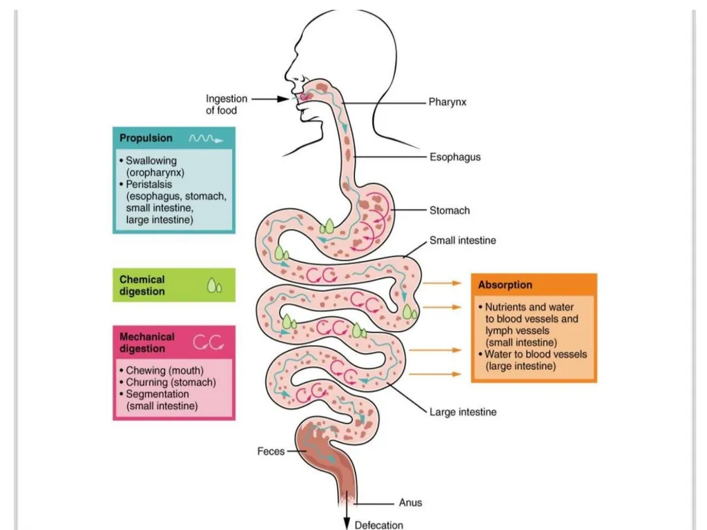
Functions of the Digestive System ❖Ingestion: the entry of food into the alimentary canal through the mouth.
❖Propulsion: the movement of food through the digestive tract either voluntarily (swallowing) or involuntarily (peristalsis).
❖Mechanical Digestion Physical breakdown of food in preparation for chemical digestion and includes chewing, mixing, and churning
❖Digestion: Enzymes breakdown complex food molecules into simpler building blocks (nutrients)
❖Absorption: Passage of nutrients into the bloodstream. ❖Defecation: undigested materials are removed from the body as faeces.<br>
04
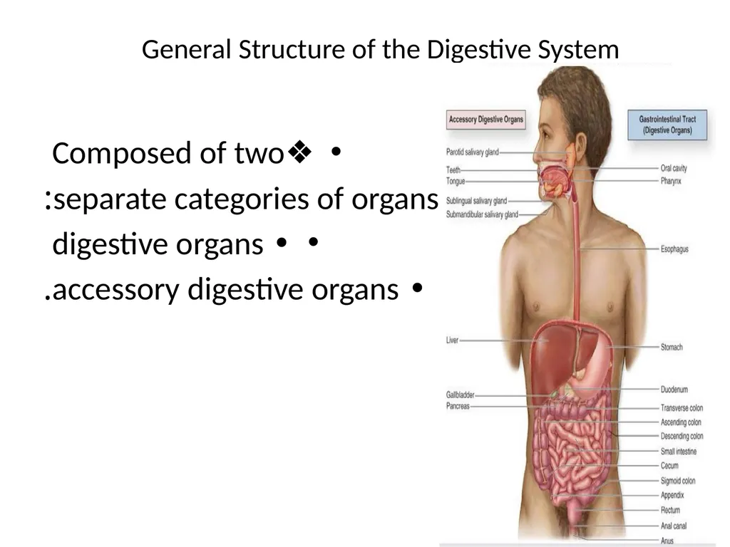
General Structure of the Digestive System ❖Composed of two
separate categories of organs:
• digestive organs
• accessory digestive organs.<br>
05
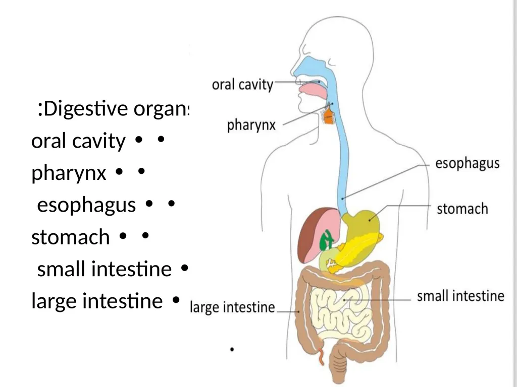
Digestive organs include:
• oral cavity
• pharynx
• esophagus
• stomach
• small intestine
• large intestine<br>
06
Accessory digestive organs: Assist the GI tract in the digestion of food, Include:
• Teeth
• Tongue
• Salivary glands
• Liver
• Gallbladder
• Pancreas<br>
07
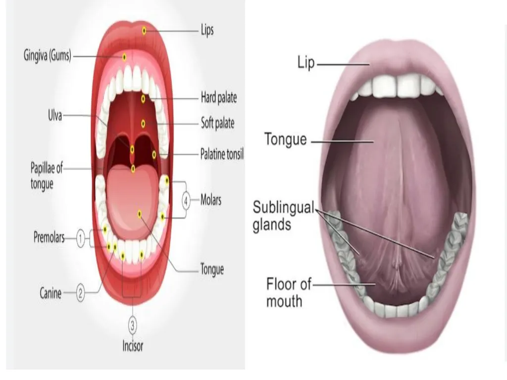
Oral Cavity (mouth) ❖Entrance to the GI tract
❖Bounded anteriorly by the teeth and lips ❖Bounded posteriorly by the oropharynx ❖Superior boundary (roof) is formed by the hard and soft palates.
*it is the initial site of digestion:
• mechanical digestion (via mastication)
• chemical digestion (via enzymes in saliva).<br>
08
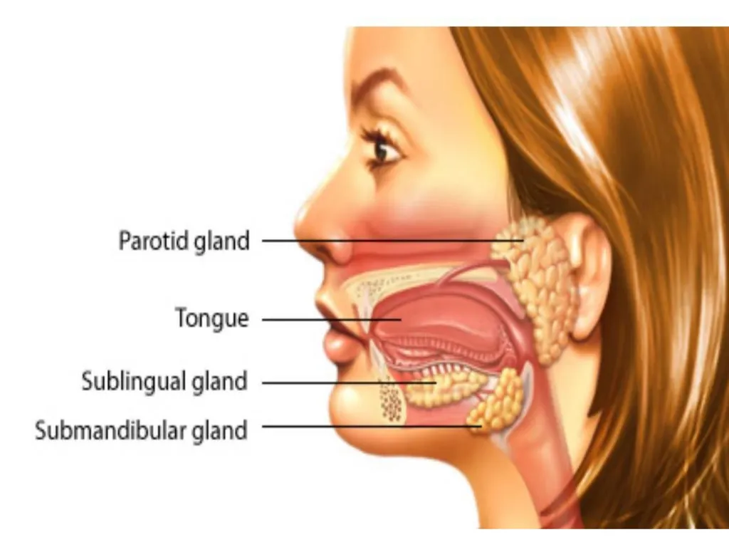
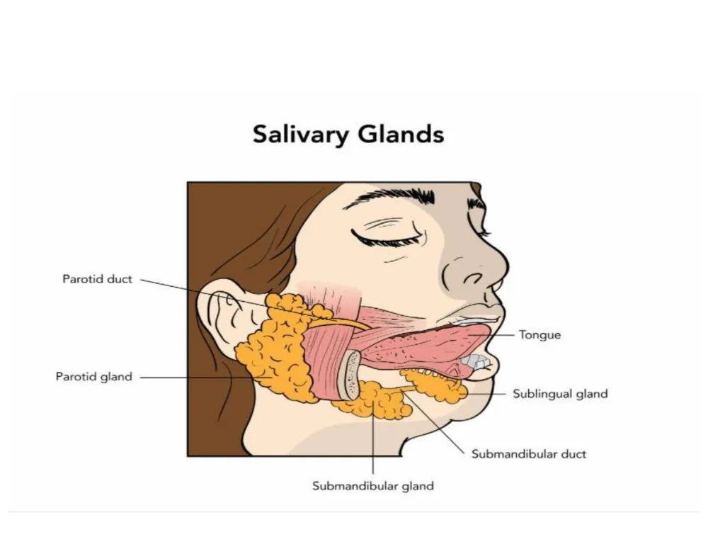
salivary glands The major salivary glands are:
* Parotid : lies anterior to the ear between the masseter muscle and skin. Parotid duct opens into the vestibule next to second upper molar. *Submandibular : lies along the medial aspect of the mandibular body , Its ducts open at the base of the lingual frenulum.
* Sublingual : lies anterior to the submandibular gland under the tongue.It opens via 10-12 ducts into the floor of the mouth.<br>
09
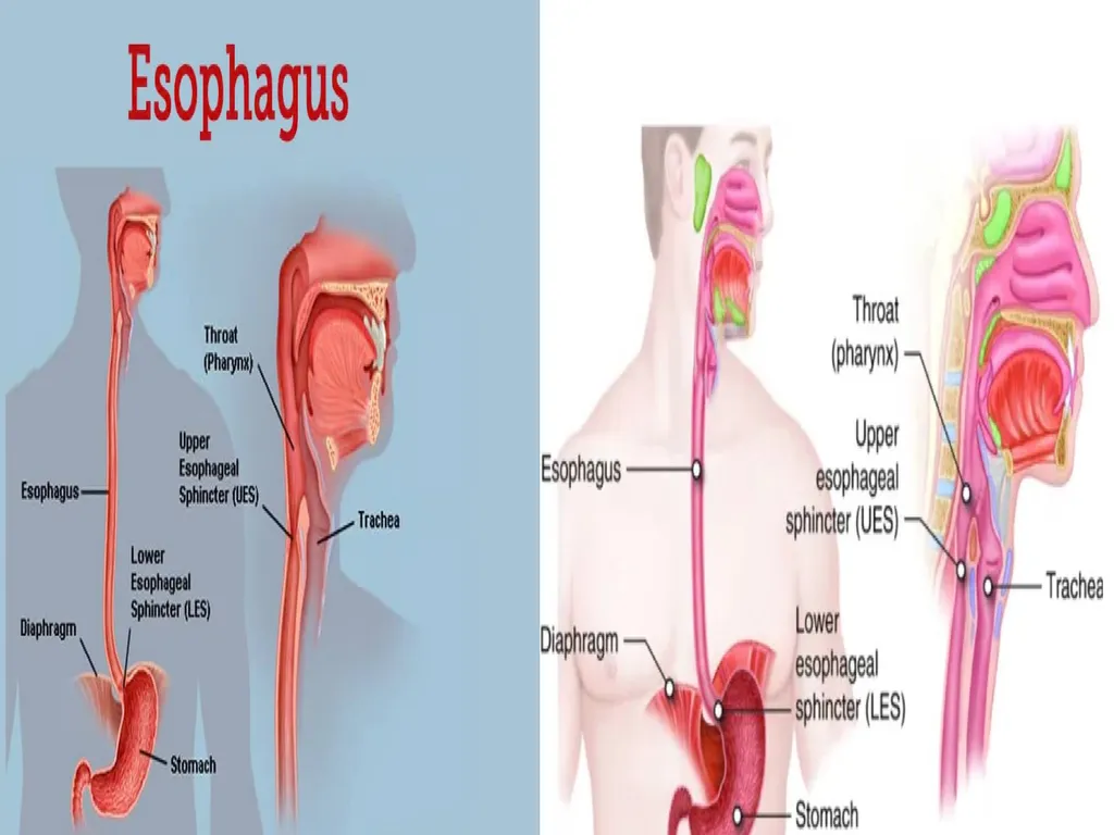
Esophagus *Tubular passageway
* About 25 cm in adult
* Connects Pharynx to stomach
Superior esophageal sphincter:
- between pharynx and esophagus
- Skeletal muscle
Inferior esophageal sphincter
- between esophagus and stomach
- smooth muscle
- Also called cardiac sphincter<br>
10
Stomach ❖J-shaped elastic sac which is the widest part of the digestive system
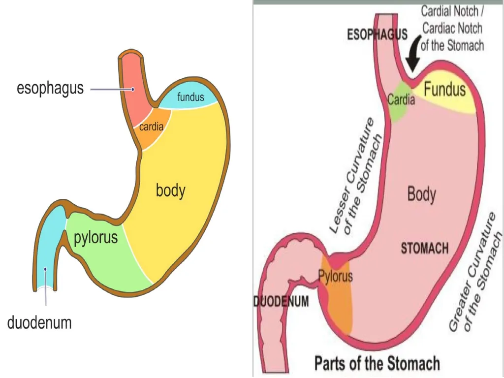
❖The stomach is divided into 5 regions:
*Cardia
* Fundus
* Body
* Pylorus
* Pyloric sphincter<br>
11
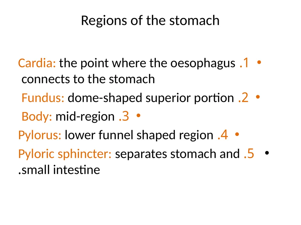
Regions of the stomach 1. Cardia: the point where the oesophagus connects to the stomach
2. Fundus: dome-shaped superior portion
3. Body: mid-region
4. Pylorus: lower funnel shaped region
5. Pyloric sphincter: separates stomach and small intestine.<br>
12
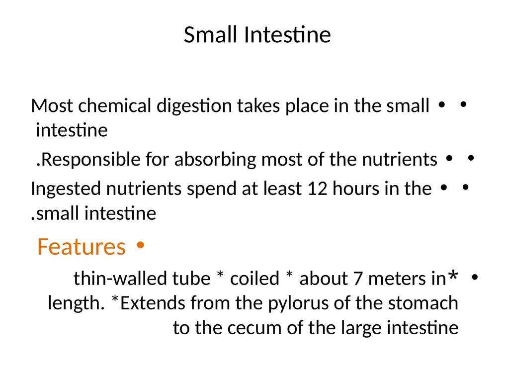
Small Intestine • Most chemical digestion takes place in the small intestine
• Responsible for absorbing most of the nutrients.
• Ingested nutrients spend at least 12 hours in the small intestine.
Features
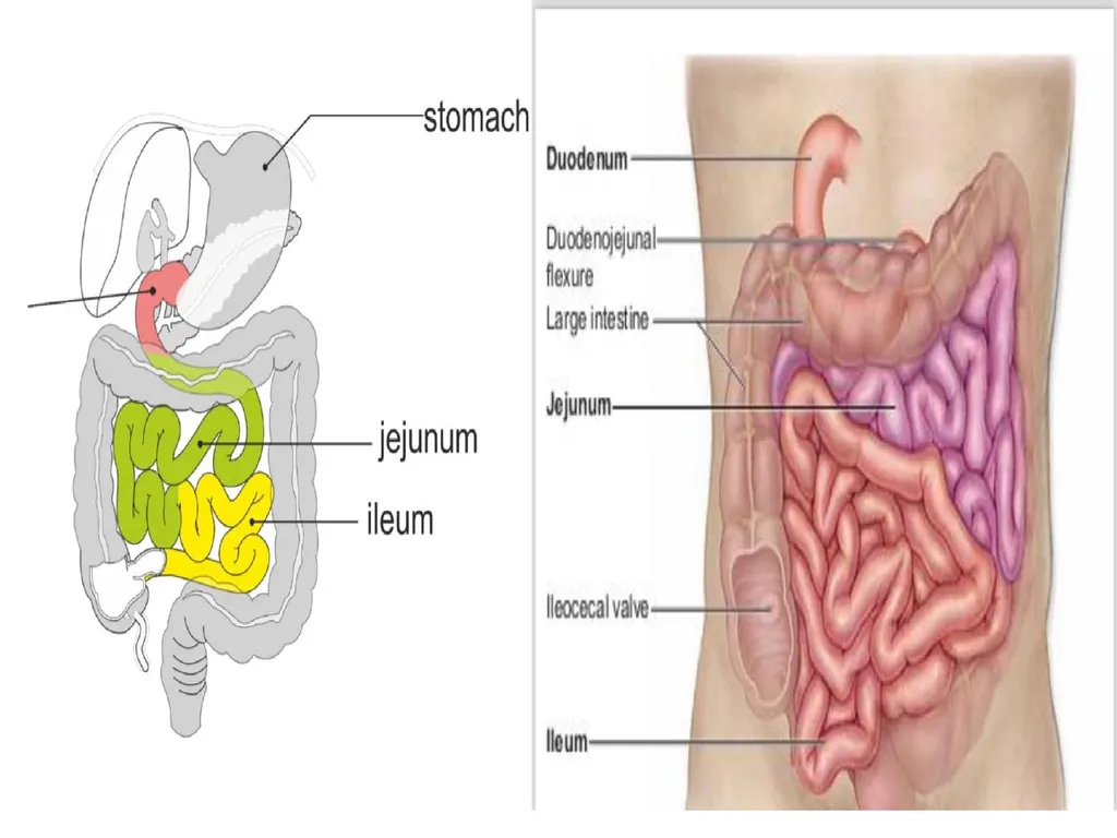
*thin-walled tube * coiled * about 7 meters in length. *Extends from the pylorus of the stomach to the cecum of the large intestine<br>