
Growth and Development Post-natal Isra Dilshad







Description: Growth and Development Post-natal Isra Dilshad Email: isra.dilshadtiu.edu.iq 3rd grade 1st semester 7th week 13-17 Oct Outline Postnatal craniofacial growth Cranial vault Cranial base Maxillary complex Mandible Objectives At the end of
Related Topics
Download Presentation
"Growth and Development Post-natal Isra Dilshad" is the property of its rightful owner. Permission is granted to download and print the materials on this website for personal, non-commercial use only, and to display it on your personal computer provided you do not modify the materials and that you retain all copyright notices contained in the materials. By downloading content from our website, you accept the terms of this agreement.
Presentation Transcript
slide1. Growth and Development Post-natal Isra Dilshad
Email: isra.dilshad@tiu.edu.iq
3rd grade – 1st semester
7th week
13-17 Oct<br>
slide2. Outline Postnatal craniofacial growth
Cranial vault
Cranial base
Maxillary complex
Mandible<br>
slide3. Objectives At the end of this lecture, you will be able to;
Explain the processes of post-natal craniofacial growth. 13 October 2024 3<br>
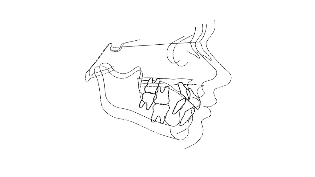
slide4. Postnatal craniofacial growth Growth studies clearly show that overall, as the face enlarges, it grows forwards and downwards away from the cranial base.
The calvarium, the cranial base, the maxilla, and the mandible all grow at different rates and at different times and are under the influence of different genetic and environmental factors.
A harmonious facial form relies on a harmonious pattern of facial development.
Variations in facial development can lead to variations in facial form and jaw relationships.<br>
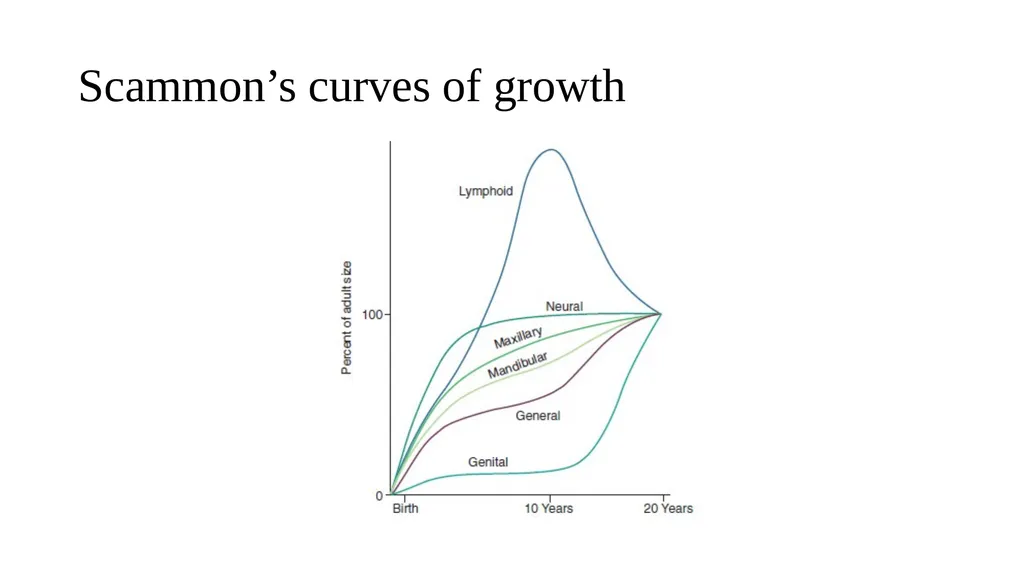
slide6. Scammon’s curves of growth<br>
slide7. Postnatal craniofacial growth Facial growth in the first few years of life is determined principally by growth of the brain (neural growth).
The calvarium, eyes, and surrounding bony orbits grow rapidly, slowing by age 7 when most growth is complete
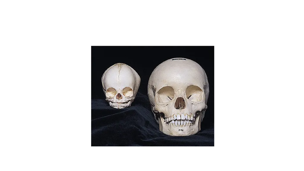
In children, the face makes up a smaller proportion of the skull compared to adolescents or adults.<br>
slide9. Postnatal craniofacial growth The rest of the face grows as the child increases in height, slowing down around puberty.
The pubertal growth spurt, a period of rapid growth, occurs around ages
10–12 in girls
12–15 in boys
The maxilla follows a pattern of growth closer to neural growth and declines to adult levels around the age of 12.
The mandible follows a pattern of growth more closely related to the rest of the body (somatic growth).
Somatic growth increases significantly during puberty and continues until growth declines to adult levels around the age of 16 in girls and 18–20 in boys.<br>
slide10. Small growth spurts have been reported for males in their early twenties.
Facial growth never completely stops, but reduces to adult levels, with subtle long-term changes throughout life.<br>
slide11. Craniofacial complex divided to four areas that grow rather differently: The cranial vault (Calvarium), the bones that cover the upper and outer surface of the brain.
The cranial base, the bony floor under the brain, which also is the dividing line between the cranium and the face.
The nasomaxillary complex, made up of the nose, maxilla, and associated small bones.
The mandible<br>
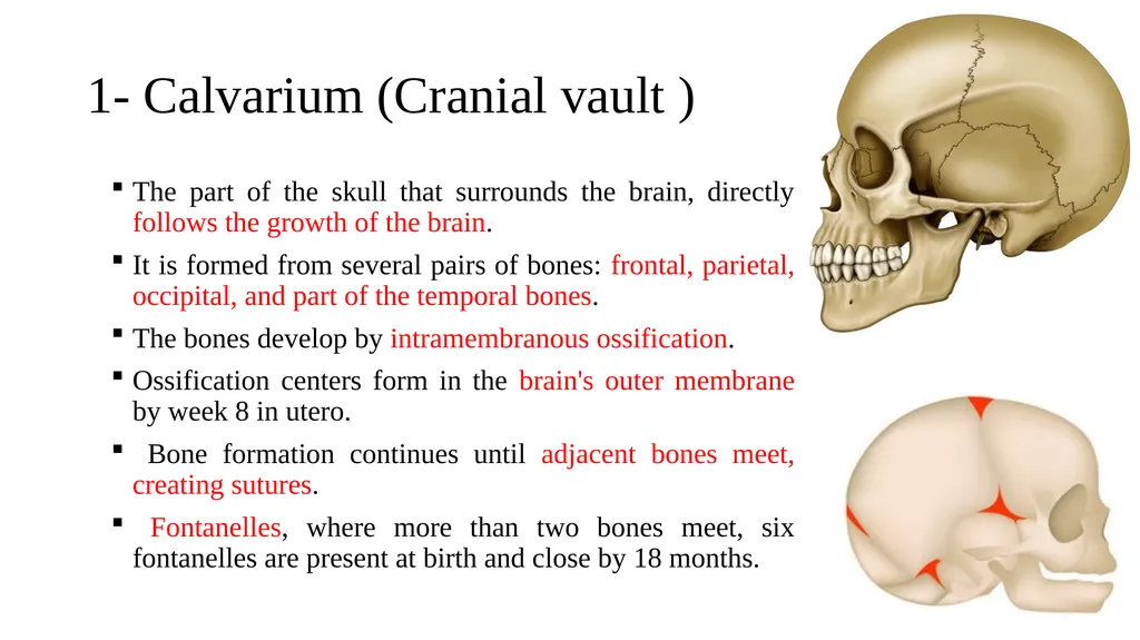
slide12. 1- Calvarium (Cranial vault ) The part of the skull that surrounds the brain, directly follows the growth of the brain.
It is formed from several pairs of bones: frontal, parietal, occipital, and part of the temporal bones.
The bones develop by intramembranous ossification.
Ossification centers form in the brain's outer membrane by week 8 in utero.
Bone formation continues until adjacent bones meet, creating sutures.
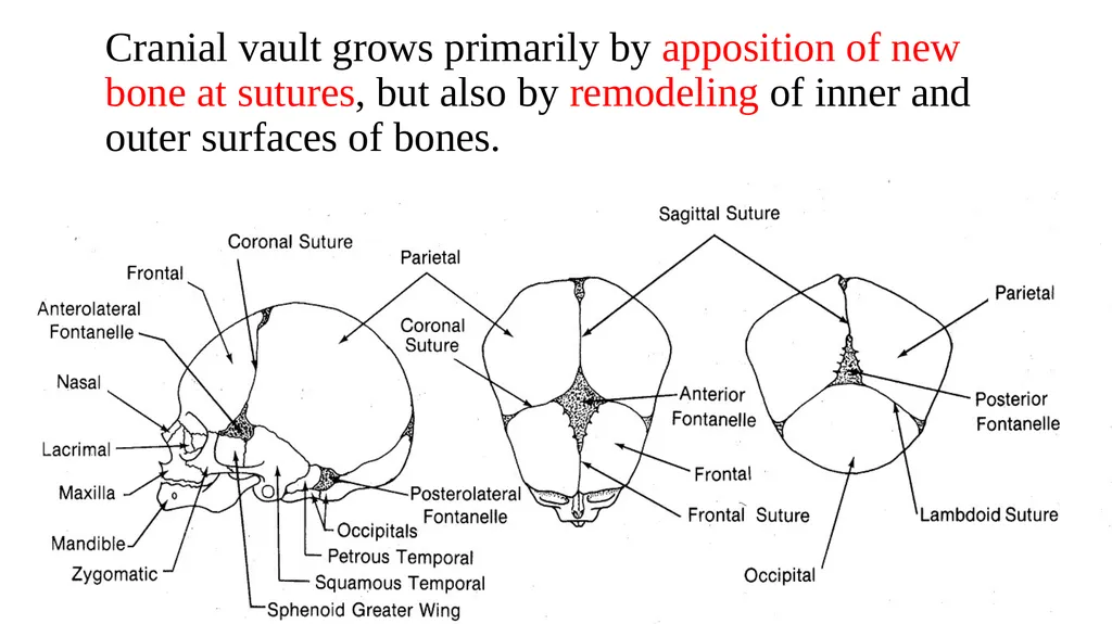
Fontanelles, where more than two bones meet, six fontanelles are present at birth and close by 18 months.<br>
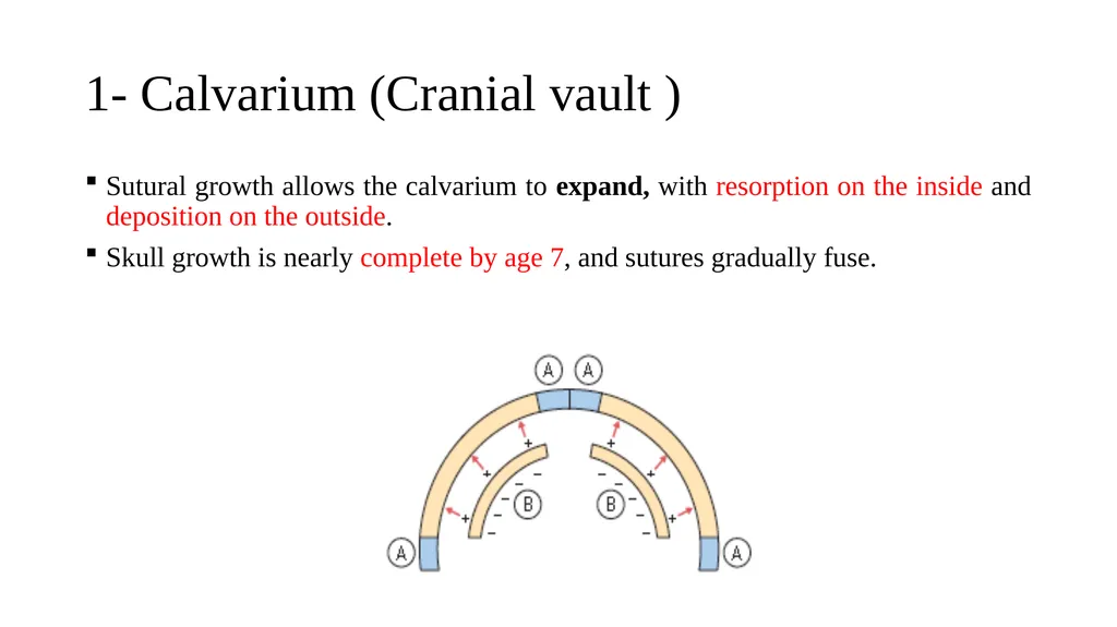
slide13. 1- Calvarium (Cranial vault ) Sutural growth allows the calvarium to expand, with resorption on the inside and deposition on the outside.
Skull growth is nearly complete by age 7, and sutures gradually fuse.<br>
slide14. Cranial vault grows primarily by apposition of new bone at sutures, but also by remodeling of inner and outer surfaces of bones.<br>
slide15. 2- Cranial base The cranial base develops by endochondral ossification, with cartilage forming in three regions of the cranial base around week 6 in utero and ossification centers appearing by 3–5 months.
Brain growth drives most cranial base growth, with half of postnatal growth completed by age 3.
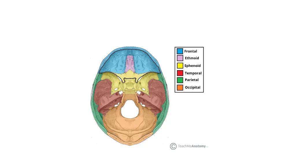
A number of bones contribute to the cranial base, including the frontal, ethmoid, sphenoid, temporal and occipital.
Remodeling and sutural infilling occur as the brain enlarges.
There are also primary cartilaginous growth sites in the cranial base, the synchondroses.<br>
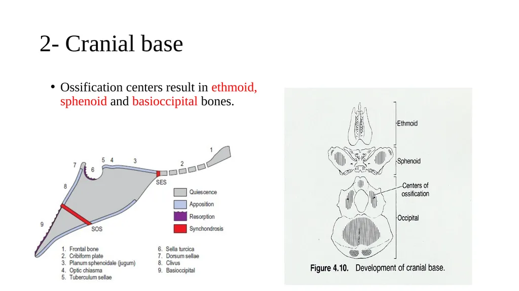
slide17. 2- Cranial base Ossification centers result in ethmoid, sphenoid and basioccipital bones.<br>
slide18. Synchondroses of the cranial base Cartilaginous joints between the bones of the cranial base
Immovable joints
Most important synchondrosis: spheno-ethmoidal, inter-sphenoidal, and spheno-occipital.<br>
slide19. 2- Cranial base The spheno-occipital synchondrosis significantly contributes to cranial base growth until ages 13–15 in females and 15–17 in males, fusing around 20 years.
The other two synchondroses in the anterior cranial base fuse by age 7, making this region useful for analyzing facial changes via cephalograms during growth and orthodontics.
Growth at the spheno-occipital synchondrosis, located between the anterior cranial base and the TMJ, lengthens the cranial base, moving the TMJs and lower jaw further back from the maxilla.
The shape, or angle, of the cranial base also affects the jaw relationship.<br>
slide20. Spheno-occipital synchondrosis<br>
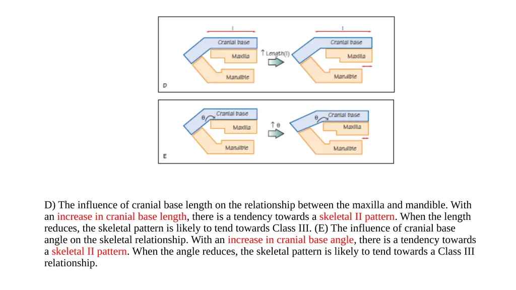
slide21. D) The influence of cranial base length on the relationship between the maxilla and mandible. With an increase in cranial base length, there is a tendency towards a skeletal II pattern. When the length reduces, the skeletal pattern is likely to tend towards Class III. (E) The influence of cranial base angle on the skeletal relationship. With an increase in cranial base angle, there is a tendency towards a skeletal II pattern. When the angle reduces, the skeletal pattern is likely to tend towards a Class III relationship.<br>
slide22. 10 min Break<br>
slide23. 3- Nasomaxillary complex Middle third of the facial skeleton is a complex structure including the maxilla, palatal, zygomatic, ethmoid*, vomer, and nasal bones.
These separate bones join with each other and the anterior cranial base at sutures.
Growth of the nasomaxillary complex is clearly an important factor in the position of the upper teeth relative to the lower teeth and the final maxillary position and facial appearance.
Until the age 7, growth occurs by downward and forward displacement and remodeling.<br>
slide24. Growth of nasomaxillary complex can be attributed to the following mechanisms: A- Displacement (Primary Vs. Secondary):
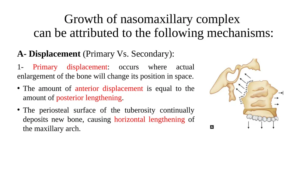
1- Primary displacement: occurs where actual enlargement of the bone will change its position in space.
The amount of anterior displacement is equal to the amount of posterior lengthening.
The periosteal surface of the tuberosity continually deposits new bone, causing horizontal lengthening of the maxillary arch.<br>
slide25. Growth of nasomaxillary complex can be attributed to the following mechanisms: A- Displacement (Primary Vs. Secondary):
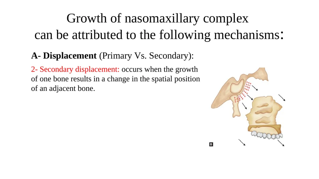
2- Secondary displacement: occurs when the growth of one bone results in a change in the spatial position of an adjacent bone.<br>
slide26. Growth of nasomaxillary complex can be attributed to the following mechanisms: B- Growth at sutures:
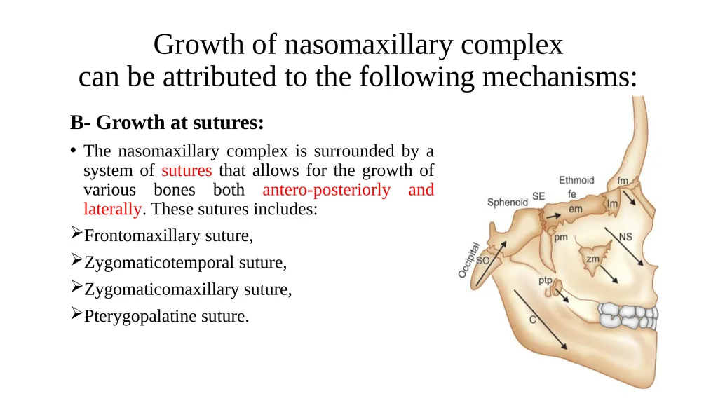
The nasomaxillary complex is surrounded by a system of sutures that allows for the growth of various bones both antero-posteriorly and laterally. These sutures includes:
Frontomaxillary suture,
Zygomaticotemporal suture,
Zygomaticomaxillary suture,
Pterygopalatine suture.<br>
slide27. Tension from the downward and forward displacement of the maxilla stimulates sutural bone growth.
New bone forms on either side of the suture in response to this displacement.
As the maxilla moves forward and downward, osteogenic sutural membranes generate new bone, increasing the maxilla's size while maintaining bone-to-bone contact.<br>
slide28. Growth of nasomaxillary complex can be attributed to the following mechanisms: C- Surface Remodeling
All bony surfaces undergo selective remodeling through deposition and resorption, along with endosteal and periosteal surfaces of bone. This remodeling increases size and alters the bone's shape and functional relationships.<br>
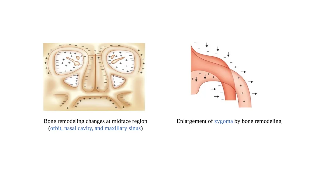
slide29. Bone remodeling changes at midface region
(orbit, nasal cavity, and maxillary sinus) Enlargement of zygoma by bone remodeling<br>
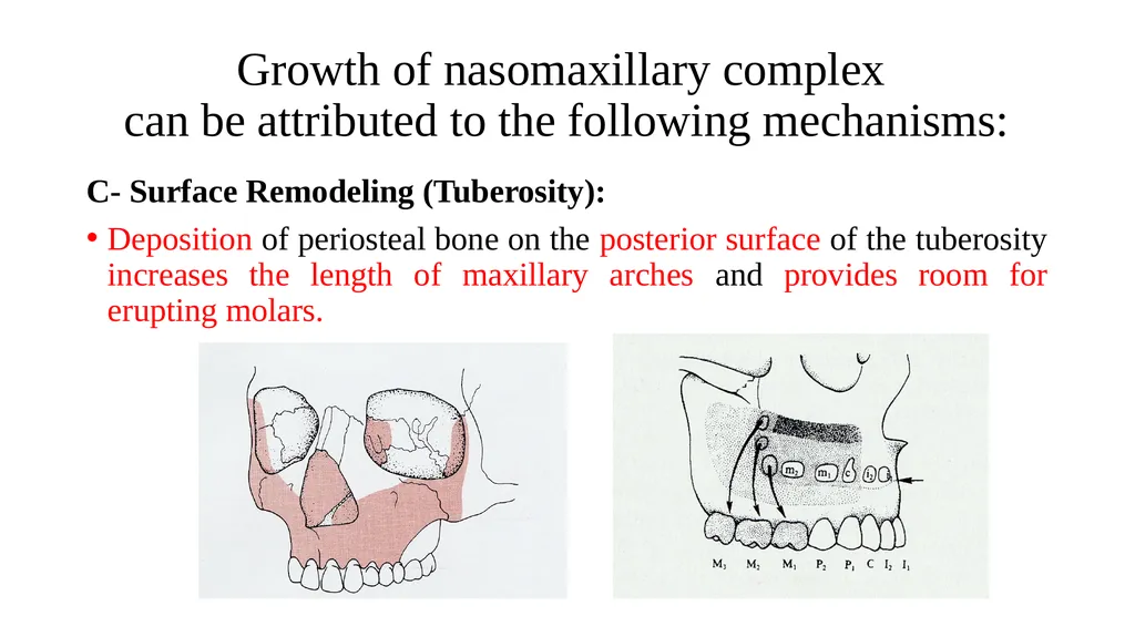
slide30. Growth of nasomaxillary complex can be attributed to the following mechanisms: C- Surface Remodeling (Tuberosity):
Deposition of periosteal bone on the posterior surface of the tuberosity increases the length of maxillary arches and provides room for erupting molars.<br>
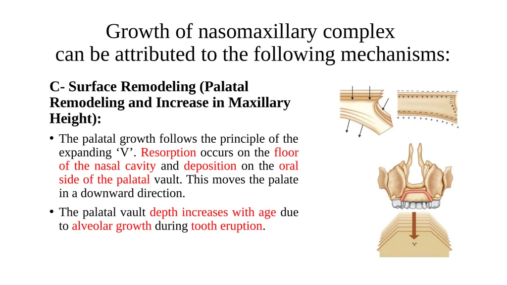
slide31. Growth of nasomaxillary complex can be attributed to the following mechanisms: C- Surface Remodeling (Palatal Remodeling and Increase in Maxillary Height):
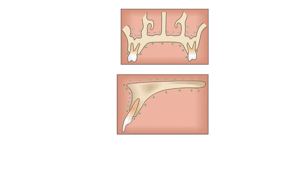
The palatal growth follows the principle of the expanding ‘V’. Resorption occurs on the floor of the nasal cavity and deposition on the oral side of the palatal vault. This moves the palate in a downward direction.
The palatal vault depth increases with age due to alveolar growth during tooth eruption.<br>
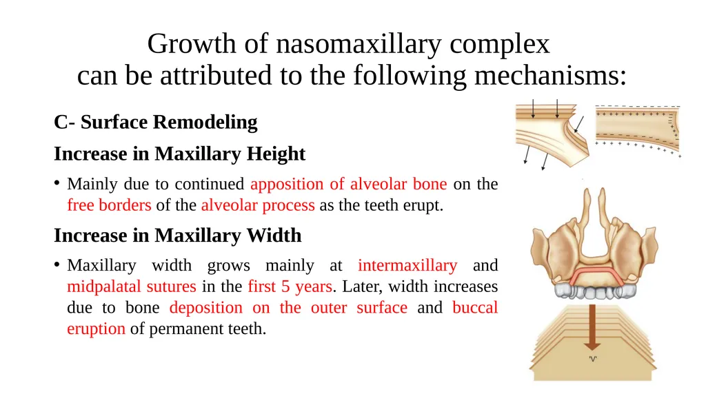
slide33. Growth of nasomaxillary complex can be attributed to the following mechanisms: C- Surface Remodeling
Increase in Maxillary Height
Mainly due to continued apposition of alveolar bone on the free borders of the alveolar process as the teeth erupt.
Increase in Maxillary Width
Maxillary width grows mainly at intermaxillary and midpalatal sutures in the first 5 years. Later, width increases due to bone deposition on the outer surface and buccal eruption of permanent teeth.<br>
slide34. 3- Maxillary complex (conclusion) As growth of the brain and calvarium slows so does maxillary growth.
Forward displacement of the maxillary complex creates space for backward growth, lengthening the dental arch in the tuberosity area for molar eruption.
Downward growth occurs through drift of the palate and vertical development of alveolar processes with tooth eruption.
Lateral growth occurs by displacement of the two halves of the maxilla and infill at the mid-palatal suture.
Complex patterns of surface remodeling maintain and develop the shape of the bones of the midfacial skeleton.<br>
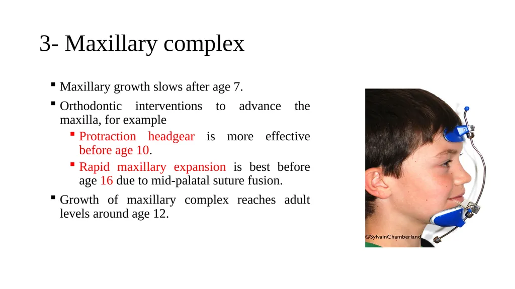
slide35. 3- Maxillary complex Maxillary growth slows after age 7.
Orthodontic interventions to advance the maxilla, for example
Protraction headgear is more effective before age 10.
Rapid maxillary expansion is best before age 16 due to mid-palatal suture fusion.
Growth of maxillary complex reaches adult levels around age 12.<br>
slide36. 4- Mandible The mandible shows the most postnatal growth among facial bones, growing downward and forward, with growth spurts during puberty.
It can be divided into functional and developmental subunits, including the body, alveolar process, condyles, ramus, lingual tuberosity with the chin, and angular and coronoid processes.<br>
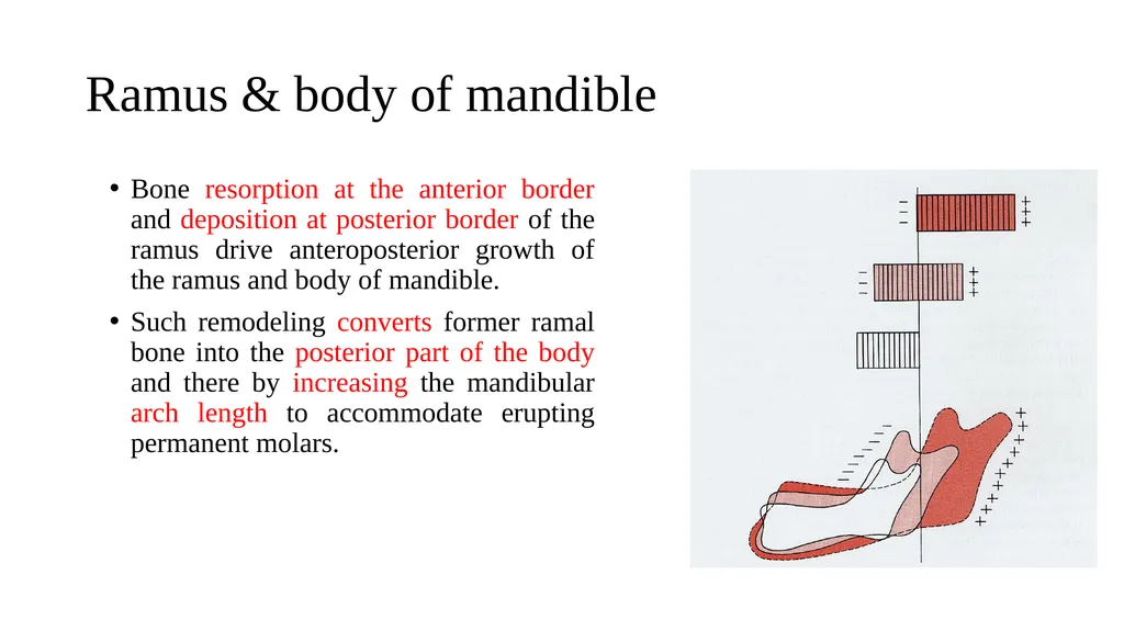
slide37. Ramus & body of mandible Bone resorption at the anterior border and deposition at posterior border of the ramus drive anteroposterior growth of the ramus and body of mandible.
Such remodeling converts former ramal bone into the posterior part of the body and there by increasing the mandibular arch length to accommodate erupting permanent molars.<br>
slide38. Mandibular Condyle The condyle shows minimum growth at birth. It is an anatomic part of special interest because it is a major site of growth of mandible, having considerable clinical significance.
Growth of the condylar cartilage would increase the length and height of the mandible.<br>
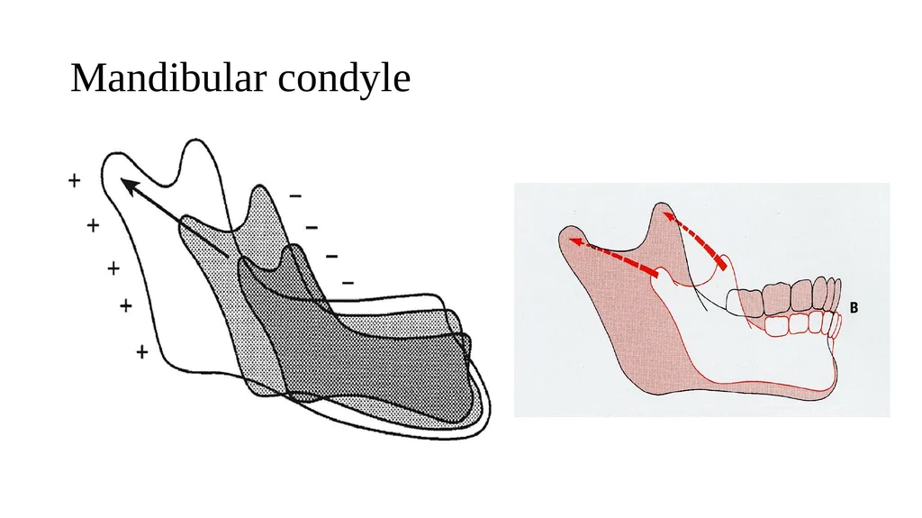
slide39. The role of condylar cartilage in mandibular growth has been a subject of controversy. There are two major schools of thought about the role of condyle: 1. Weimann and Sicker considered condyle as the major growth center of mandible with an intrinsic genetic potential. Others thought that the condylar cartilage was analogous to an epiphyseal cartilage. It was believed that condyle grows towards cranial base by deposition of bone at condylar cartilage. As the condyle pushes against the cranial base, the entire mandible gets displaced in a forward and downward direction.<br>
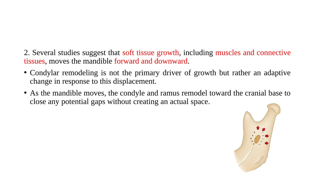
slide40. 2. Several studies suggest that soft tissue growth, including muscles and connective tissues, moves the mandible forward and downward.
Condylar remodeling is not the primary driver of growth but rather an adaptive change in response to this displacement.
As the mandible moves, the condyle and ramus remodel toward the cranial base to close any potential gaps without creating an actual space.<br>
slide41. Mandibular condyle<br>
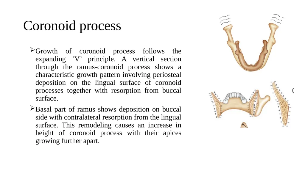
slide42. Coronoid process Growth of coronoid process follows the expanding ‘V’ principle. A vertical section through the ramus-coronoid process shows a characteristic growth pattern involving periosteal deposition on the lingual surface of coronoid processes together with resorption from buccal surface.
Basal part of ramus shows deposition on buccal side with contralateral resorption from the lingual surface. This remodeling causes an increase in height of coronoid process with their apices growing further apart.<br>
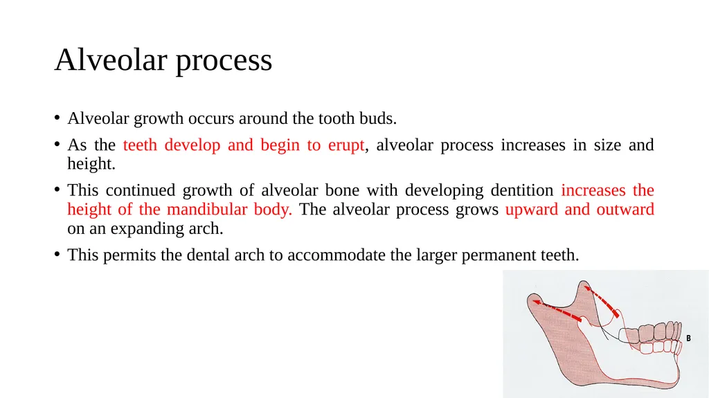
slide43. Alveolar process Alveolar growth occurs around the tooth buds.
As the teeth develop and begin to erupt, alveolar process increases in size and height.
This continued growth of alveolar bone with developing dentition increases the height of the mandibular body. The alveolar process grows upward and outward on an expanding arch.
This permits the dental arch to accommodate the larger permanent teeth.<br>
slide44. Chin The chin is not well developed at birth but grow significantly during puberty.
It is influenced by sexual and genetic factors.
Chin becomes prominent at puberty especially in male, selective bone remodeling enhances prominence, with bone resorption above creating a concavity and apposition occurring at the inferior aspect
Angle of mandible
Selective bone remodeling at the angle of mandible, causes flaring of the angle as age advances.<br>
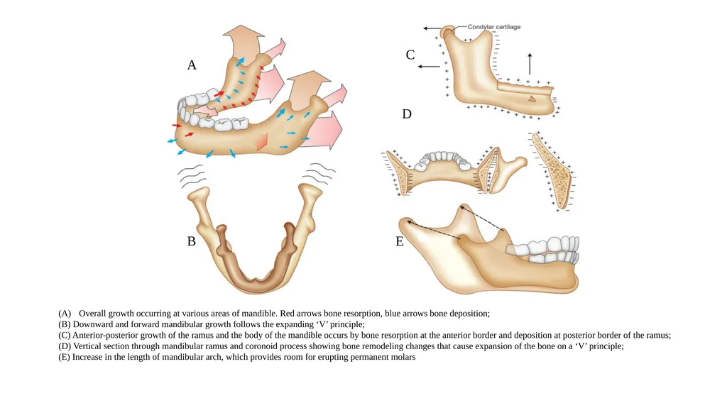
slide45. Overall growth occurring at various areas of mandible. Red arrows bone resorption, blue arrows bone deposition;
(B) Downward and forward mandibular growth follows the expanding ‘V’ principle;
(C) Anterior-posterior growth of the ramus and the body of the mandible occurs by bone resorption at the anterior border and deposition at posterior border of the ramus;
(D) Vertical section through mandibular ramus and coronoid process showing bone remodeling changes that cause expansion of the bone on a ‘V’ principle;
(E) Increase in the length of mandibular arch, which provides room for erupting permanent molars A B C E D<br>
slide46. 4- Mandible As with the maxilla, complex patterns of surface remodeling maintain and develop the shape of the mandible.
The mandible grows steadily at 2–3 mm per year in length of the body of mandible until puberty, when the growth rate doubles.
Growth slows to adult levels by age
17 in girls
19 in boys<br>
Email: isra.dilshad@tiu.edu.iq
3rd grade – 1st semester
7th week
13-17 Oct<br>
slide2. Outline Postnatal craniofacial growth
Cranial vault
Cranial base
Maxillary complex
Mandible<br>
slide3. Objectives At the end of this lecture, you will be able to;
Explain the processes of post-natal craniofacial growth. 13 October 2024 3<br>
slide4. Postnatal craniofacial growth Growth studies clearly show that overall, as the face enlarges, it grows forwards and downwards away from the cranial base.
The calvarium, the cranial base, the maxilla, and the mandible all grow at different rates and at different times and are under the influence of different genetic and environmental factors.
A harmonious facial form relies on a harmonious pattern of facial development.
Variations in facial development can lead to variations in facial form and jaw relationships.<br>
slide6. Scammon’s curves of growth<br>
slide7. Postnatal craniofacial growth Facial growth in the first few years of life is determined principally by growth of the brain (neural growth).
The calvarium, eyes, and surrounding bony orbits grow rapidly, slowing by age 7 when most growth is complete
In children, the face makes up a smaller proportion of the skull compared to adolescents or adults.<br>
slide9. Postnatal craniofacial growth The rest of the face grows as the child increases in height, slowing down around puberty.
The pubertal growth spurt, a period of rapid growth, occurs around ages
10–12 in girls
12–15 in boys
The maxilla follows a pattern of growth closer to neural growth and declines to adult levels around the age of 12.
The mandible follows a pattern of growth more closely related to the rest of the body (somatic growth).
Somatic growth increases significantly during puberty and continues until growth declines to adult levels around the age of 16 in girls and 18–20 in boys.<br>
slide10. Small growth spurts have been reported for males in their early twenties.
Facial growth never completely stops, but reduces to adult levels, with subtle long-term changes throughout life.<br>
slide11. Craniofacial complex divided to four areas that grow rather differently: The cranial vault (Calvarium), the bones that cover the upper and outer surface of the brain.
The cranial base, the bony floor under the brain, which also is the dividing line between the cranium and the face.
The nasomaxillary complex, made up of the nose, maxilla, and associated small bones.
The mandible<br>
slide12. 1- Calvarium (Cranial vault ) The part of the skull that surrounds the brain, directly follows the growth of the brain.
It is formed from several pairs of bones: frontal, parietal, occipital, and part of the temporal bones.
The bones develop by intramembranous ossification.
Ossification centers form in the brain's outer membrane by week 8 in utero.
Bone formation continues until adjacent bones meet, creating sutures.
Fontanelles, where more than two bones meet, six fontanelles are present at birth and close by 18 months.<br>
slide13. 1- Calvarium (Cranial vault ) Sutural growth allows the calvarium to expand, with resorption on the inside and deposition on the outside.
Skull growth is nearly complete by age 7, and sutures gradually fuse.<br>
slide14. Cranial vault grows primarily by apposition of new bone at sutures, but also by remodeling of inner and outer surfaces of bones.<br>
slide15. 2- Cranial base The cranial base develops by endochondral ossification, with cartilage forming in three regions of the cranial base around week 6 in utero and ossification centers appearing by 3–5 months.
Brain growth drives most cranial base growth, with half of postnatal growth completed by age 3.
A number of bones contribute to the cranial base, including the frontal, ethmoid, sphenoid, temporal and occipital.
Remodeling and sutural infilling occur as the brain enlarges.
There are also primary cartilaginous growth sites in the cranial base, the synchondroses.<br>
slide17. 2- Cranial base Ossification centers result in ethmoid, sphenoid and basioccipital bones.<br>
slide18. Synchondroses of the cranial base Cartilaginous joints between the bones of the cranial base
Immovable joints
Most important synchondrosis: spheno-ethmoidal, inter-sphenoidal, and spheno-occipital.<br>
slide19. 2- Cranial base The spheno-occipital synchondrosis significantly contributes to cranial base growth until ages 13–15 in females and 15–17 in males, fusing around 20 years.
The other two synchondroses in the anterior cranial base fuse by age 7, making this region useful for analyzing facial changes via cephalograms during growth and orthodontics.
Growth at the spheno-occipital synchondrosis, located between the anterior cranial base and the TMJ, lengthens the cranial base, moving the TMJs and lower jaw further back from the maxilla.
The shape, or angle, of the cranial base also affects the jaw relationship.<br>
slide20. Spheno-occipital synchondrosis<br>
slide21. D) The influence of cranial base length on the relationship between the maxilla and mandible. With an increase in cranial base length, there is a tendency towards a skeletal II pattern. When the length reduces, the skeletal pattern is likely to tend towards Class III. (E) The influence of cranial base angle on the skeletal relationship. With an increase in cranial base angle, there is a tendency towards a skeletal II pattern. When the angle reduces, the skeletal pattern is likely to tend towards a Class III relationship.<br>
slide22. 10 min Break<br>
slide23. 3- Nasomaxillary complex Middle third of the facial skeleton is a complex structure including the maxilla, palatal, zygomatic, ethmoid*, vomer, and nasal bones.
These separate bones join with each other and the anterior cranial base at sutures.
Growth of the nasomaxillary complex is clearly an important factor in the position of the upper teeth relative to the lower teeth and the final maxillary position and facial appearance.
Until the age 7, growth occurs by downward and forward displacement and remodeling.<br>
slide24. Growth of nasomaxillary complex can be attributed to the following mechanisms: A- Displacement (Primary Vs. Secondary):
1- Primary displacement: occurs where actual enlargement of the bone will change its position in space.
The amount of anterior displacement is equal to the amount of posterior lengthening.
The periosteal surface of the tuberosity continually deposits new bone, causing horizontal lengthening of the maxillary arch.<br>
slide25. Growth of nasomaxillary complex can be attributed to the following mechanisms: A- Displacement (Primary Vs. Secondary):
2- Secondary displacement: occurs when the growth of one bone results in a change in the spatial position of an adjacent bone.<br>
slide26. Growth of nasomaxillary complex can be attributed to the following mechanisms: B- Growth at sutures:
The nasomaxillary complex is surrounded by a system of sutures that allows for the growth of various bones both antero-posteriorly and laterally. These sutures includes:
Frontomaxillary suture,
Zygomaticotemporal suture,
Zygomaticomaxillary suture,
Pterygopalatine suture.<br>
slide27. Tension from the downward and forward displacement of the maxilla stimulates sutural bone growth.
New bone forms on either side of the suture in response to this displacement.
As the maxilla moves forward and downward, osteogenic sutural membranes generate new bone, increasing the maxilla's size while maintaining bone-to-bone contact.<br>
slide28. Growth of nasomaxillary complex can be attributed to the following mechanisms: C- Surface Remodeling
All bony surfaces undergo selective remodeling through deposition and resorption, along with endosteal and periosteal surfaces of bone. This remodeling increases size and alters the bone's shape and functional relationships.<br>
slide29. Bone remodeling changes at midface region
(orbit, nasal cavity, and maxillary sinus) Enlargement of zygoma by bone remodeling<br>
slide30. Growth of nasomaxillary complex can be attributed to the following mechanisms: C- Surface Remodeling (Tuberosity):
Deposition of periosteal bone on the posterior surface of the tuberosity increases the length of maxillary arches and provides room for erupting molars.<br>
slide31. Growth of nasomaxillary complex can be attributed to the following mechanisms: C- Surface Remodeling (Palatal Remodeling and Increase in Maxillary Height):
The palatal growth follows the principle of the expanding ‘V’. Resorption occurs on the floor of the nasal cavity and deposition on the oral side of the palatal vault. This moves the palate in a downward direction.
The palatal vault depth increases with age due to alveolar growth during tooth eruption.<br>
slide33. Growth of nasomaxillary complex can be attributed to the following mechanisms: C- Surface Remodeling
Increase in Maxillary Height
Mainly due to continued apposition of alveolar bone on the free borders of the alveolar process as the teeth erupt.
Increase in Maxillary Width
Maxillary width grows mainly at intermaxillary and midpalatal sutures in the first 5 years. Later, width increases due to bone deposition on the outer surface and buccal eruption of permanent teeth.<br>
slide34. 3- Maxillary complex (conclusion) As growth of the brain and calvarium slows so does maxillary growth.
Forward displacement of the maxillary complex creates space for backward growth, lengthening the dental arch in the tuberosity area for molar eruption.
Downward growth occurs through drift of the palate and vertical development of alveolar processes with tooth eruption.
Lateral growth occurs by displacement of the two halves of the maxilla and infill at the mid-palatal suture.
Complex patterns of surface remodeling maintain and develop the shape of the bones of the midfacial skeleton.<br>
slide35. 3- Maxillary complex Maxillary growth slows after age 7.
Orthodontic interventions to advance the maxilla, for example
Protraction headgear is more effective before age 10.
Rapid maxillary expansion is best before age 16 due to mid-palatal suture fusion.
Growth of maxillary complex reaches adult levels around age 12.<br>
slide36. 4- Mandible The mandible shows the most postnatal growth among facial bones, growing downward and forward, with growth spurts during puberty.
It can be divided into functional and developmental subunits, including the body, alveolar process, condyles, ramus, lingual tuberosity with the chin, and angular and coronoid processes.<br>
slide37. Ramus & body of mandible Bone resorption at the anterior border and deposition at posterior border of the ramus drive anteroposterior growth of the ramus and body of mandible.
Such remodeling converts former ramal bone into the posterior part of the body and there by increasing the mandibular arch length to accommodate erupting permanent molars.<br>
slide38. Mandibular Condyle The condyle shows minimum growth at birth. It is an anatomic part of special interest because it is a major site of growth of mandible, having considerable clinical significance.
Growth of the condylar cartilage would increase the length and height of the mandible.<br>
slide39. The role of condylar cartilage in mandibular growth has been a subject of controversy. There are two major schools of thought about the role of condyle: 1. Weimann and Sicker considered condyle as the major growth center of mandible with an intrinsic genetic potential. Others thought that the condylar cartilage was analogous to an epiphyseal cartilage. It was believed that condyle grows towards cranial base by deposition of bone at condylar cartilage. As the condyle pushes against the cranial base, the entire mandible gets displaced in a forward and downward direction.<br>
slide40. 2. Several studies suggest that soft tissue growth, including muscles and connective tissues, moves the mandible forward and downward.
Condylar remodeling is not the primary driver of growth but rather an adaptive change in response to this displacement.
As the mandible moves, the condyle and ramus remodel toward the cranial base to close any potential gaps without creating an actual space.<br>
slide41. Mandibular condyle<br>
slide42. Coronoid process Growth of coronoid process follows the expanding ‘V’ principle. A vertical section through the ramus-coronoid process shows a characteristic growth pattern involving periosteal deposition on the lingual surface of coronoid processes together with resorption from buccal surface.
Basal part of ramus shows deposition on buccal side with contralateral resorption from the lingual surface. This remodeling causes an increase in height of coronoid process with their apices growing further apart.<br>
slide43. Alveolar process Alveolar growth occurs around the tooth buds.
As the teeth develop and begin to erupt, alveolar process increases in size and height.
This continued growth of alveolar bone with developing dentition increases the height of the mandibular body. The alveolar process grows upward and outward on an expanding arch.
This permits the dental arch to accommodate the larger permanent teeth.<br>
slide44. Chin The chin is not well developed at birth but grow significantly during puberty.
It is influenced by sexual and genetic factors.
Chin becomes prominent at puberty especially in male, selective bone remodeling enhances prominence, with bone resorption above creating a concavity and apposition occurring at the inferior aspect
Angle of mandible
Selective bone remodeling at the angle of mandible, causes flaring of the angle as age advances.<br>
slide45. Overall growth occurring at various areas of mandible. Red arrows bone resorption, blue arrows bone deposition;
(B) Downward and forward mandibular growth follows the expanding ‘V’ principle;
(C) Anterior-posterior growth of the ramus and the body of the mandible occurs by bone resorption at the anterior border and deposition at posterior border of the ramus;
(D) Vertical section through mandibular ramus and coronoid process showing bone remodeling changes that cause expansion of the bone on a ‘V’ principle;
(E) Increase in the length of mandibular arch, which provides room for erupting permanent molars A B C E D<br>
slide46. 4- Mandible As with the maxilla, complex patterns of surface remodeling maintain and develop the shape of the mandible.
The mandible grows steadily at 2–3 mm per year in length of the body of mandible until puberty, when the growth rate doubles.
Growth slows to adult levels by age
17 in girls
19 in boys<br>