
Gynecological endoscopy Laparoscopy and

Description: Gynecological endoscopy Laparoscopy and hysteroscopy Dr Amna Fadhil Objectives: At the end of this lecture the 5th year students should be able to: Describe the advantages of laparoscopy over laparotomy . Recognize its contraindications and
Related Topics
Download Presentation
"Gynecological endoscopy Laparoscopy and" is the property of its rightful owner. Permission is granted to download and print the materials on this website for personal, non-commercial use only, and to display it on your personal computer provided you do not modify the materials and that you retain all copyright notices contained in the materials. By downloading content from our website, you accept the terms of this agreement.
Presentation Transcript
slide1. Gynecological endoscopy
Laparoscopy and hysteroscopy Dr Amna Fadhil<br>
slide2. Objectives: At the end of this lecture the 5th year students should be able to:
Describe the advantages of laparoscopy over laparotomy .
Recognize its contraindications and possible complications<br>
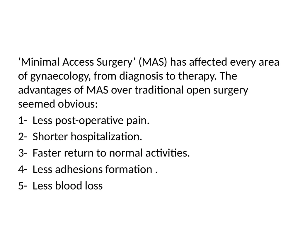
slide3. ‘Minimal Access Surgery’ (MAS) has affected every area of gynaecology, from diagnosis to therapy. The advantages of MAS over traditional open surgery seemed obvious:
1- Less post-operative pain.
2- Shorter hospitalization.
3- Faster return to normal activities.
4- Less adhesions formation .
5- Less blood loss<br>
slide4. Laparoscopy
Laparoscopy allows visualization of the peritoneal cavity. This involves insertion of a needle called a Veress needle into a suitable puncture point in the umbilicus. This allows insufflation of the peritoneal cavity with carbon dioxide so that a larger instrument such as trocar and cannula can be inserted. The procedure usually performed as an inpatient procedure under general anesthesia.<br>
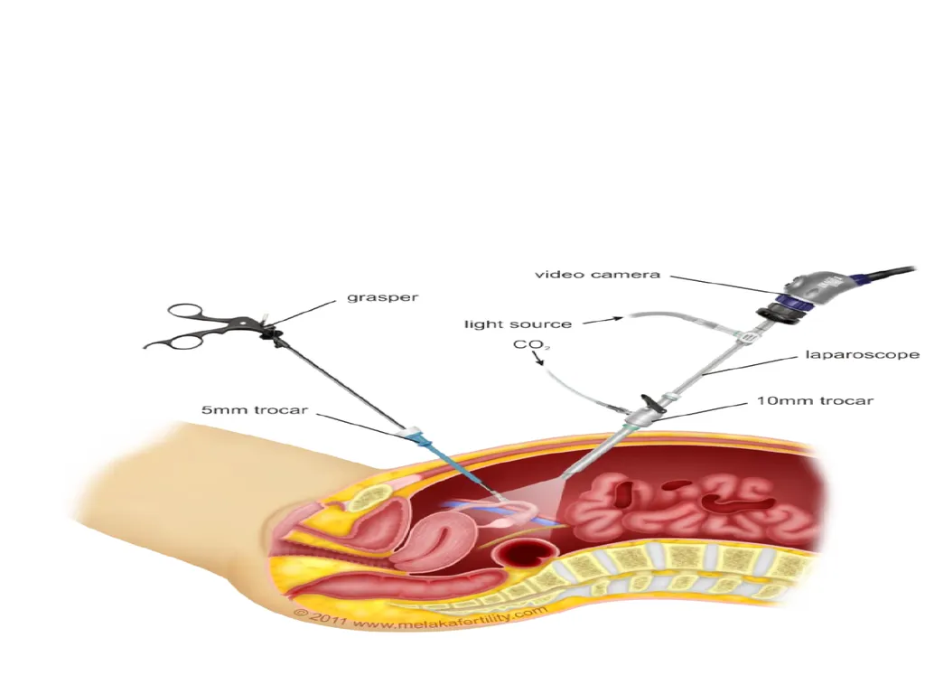
slide5. Instruments used: veress needle, trocar and cannula, laparoscope with the camera and light source attached to it, other various instruments like grasper, bipolar and monopolar cautery and scissors.
The majority of instruments used for:
diagnostic laparoscopy are 5 mm in diameter
operative laparoscopy are 10 mm in diameter<br>
slide7. A safe entry technique for laparoscopy:
The patient should be lying flat
Ensure the bladder is empty
check the abdomen for masses
Make the primary incision at the base of the umbilicus
Insert the veress needle through the base of the umbilicus, sensing a double click
Insert 2-3 ml of saline through the veress needle; it should run in freely
Aspirate back; nothing should be aspirated
Fill with co2 to 25mmhg about 2-3 L
Insert the primary trocar<br>
slide8. Indications of laparoscopy:
1. Diagnostic indications:
a. pelvic pain (acute and chronic)
b. ectopic pregnancy
c. pelvic inflammatory disease
d. endometriosis
e. infertility
f. others like pelvic mass and staging of ovarian malignancy<br>
slide9. Therapeutic indications: various surgical procedures can be performed by laparoscopy:
a. sterilization
b. aspiration of an ovarian cyst, ovarian biopsy and drilling.
c. salpingectomy and salpingostomy for ectopic pregnancy
d. treatment of endometriosis with laser or cautery
e. myomectomy
f. others like laparoscopic assisted vaginal hysterectomy, prolapse and incontinence procedures.<br>
slide10. Contraindications:
Conditions that increase the risk of complications such as:
Mechanical or paralytic bowel obstruction, generalized peritonitis, Diaphragmatic hernia, Major intraperitoneal haemorrhage, severe cardiorespiratory disease, Massive obesity, inflammatory bowel disease, large abdominal mass and multiple abdominal incisions.<br>
slide11. Complications of laparoscopy: 1. When the abdomen is being instrumented injury to the inferior epigastric vessels or intraperitoneal vessels and organs like bladder and bowel injury may occur (emptying the bladder prior to surgery can reduce bladder injury).
2. Anaesthetic complications
3. Surgical emphysema due to leakage of co2 into subcutaneous tissue
4. Infection
5. Venous thromboembolism and port site hernia
6. Incisional hernia has been reported<br>
slide12. Hysteroscopy
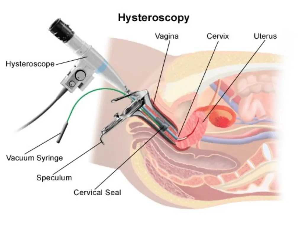
Hysteroscopy involves passing a small-diameter telescope, either flexible or rigid, through the cervix to directly inspect the uterine cavity.
Types of hysteroscope:
A flexible hysteroscope may be used in the outpatient setting, with carbon dioxide as a filling medium.
Rigid instruments employ circulating fluids and therefore can be used to visualize the uterine cavity even if the woman is bleeding.
The procedure can be performed under local, regional or general anaesthesia.<br>
slide14. Indications of hysteroscopy:
1. Diagnostic hysteroscopy: with target biopsy
a. Abnormal menstruation, intermenstrual bleeding, postcoital and postmenopausal bleeding.
b. Abnormal pelvic ultrasound findings (e.g. endometrial polyps, submucous fibroids)
c. Subfertility
d. Recurrent miscarriage
e. Asherman’s syndrome
f. Congenital uterine anomaly
g. Lost intrauterine contraceptive device (IUCD)<br>
slide15. . Operative hysteroscopy:
Hysteroscopic surgery has a number of well-defined indications and is the treatment of choice for:
1. polypectomy.
2. myomectomy for intracavitary or submucous fibroids
3. adhesiolysis of intrauterine adhesions
4. metroplasty (division/resection of uterine septum).
5. endometrial ablation (destruction of the endometrium).
6. hysteroscopic sterilization<br>
slide16. Contraindications of hysteroscopy:
1. Pelvic infection
2. Pregnancy
3. Cervical cancer
4. Heavy uterine bleeding<br>
slide17. Complications of hysteroscopy:
Diagnostic hysteroscopy is a safe procedure, and complications are uncommon. However operative hysteroscopy carries a higher risk of complications:
1. Anaesthetic complications
2. Uterine perforation
3. Fluid overload so careful monitoring of fluid input and output and intrauterine pressure is required
4. Haemorrhage
5. Gas embolism
6. Infection
7. Cervical trauma
8. Late complications as adhesions and hematometra<br>
slide18. Preoperative preparation:
Electrolytes if the patient on diuretics or cardiac medcations
Complete blood count
Coagulation panel if history of bleeding tendencies
Document normal Pap smear and normal endometrial sample within 6 months
Cervical prostaglandins if cervical stenosis suspected as those with previous caesarean section<br>
slide19. Postoperative care
Patient recovery typically is rapid and without complications following dilatation and curettage
Diet and activities may be resumed as desired by the patient
Spotting or light bleeding is not uncommon and typically stops within days.<br>
slide20. Thank you<br>
slide2. Objectives: At the end of this lecture the 5th year students should be able to:
Describe the advantages of laparoscopy over laparotomy .
Recognize its contraindications and possible complications<br>
slide3. ‘Minimal Access Surgery’ (MAS) has affected every area of gynaecology, from diagnosis to therapy. The advantages of MAS over traditional open surgery seemed obvious:
1- Less post-operative pain.
2- Shorter hospitalization.
3- Faster return to normal activities.
4- Less adhesions formation .
5- Less blood loss<br>
slide4. Laparoscopy
Laparoscopy allows visualization of the peritoneal cavity. This involves insertion of a needle called a Veress needle into a suitable puncture point in the umbilicus. This allows insufflation of the peritoneal cavity with carbon dioxide so that a larger instrument such as trocar and cannula can be inserted. The procedure usually performed as an inpatient procedure under general anesthesia.<br>
slide5. Instruments used: veress needle, trocar and cannula, laparoscope with the camera and light source attached to it, other various instruments like grasper, bipolar and monopolar cautery and scissors.
The majority of instruments used for:
diagnostic laparoscopy are 5 mm in diameter
operative laparoscopy are 10 mm in diameter<br>
slide7. A safe entry technique for laparoscopy:
The patient should be lying flat
Ensure the bladder is empty
check the abdomen for masses
Make the primary incision at the base of the umbilicus
Insert the veress needle through the base of the umbilicus, sensing a double click
Insert 2-3 ml of saline through the veress needle; it should run in freely
Aspirate back; nothing should be aspirated
Fill with co2 to 25mmhg about 2-3 L
Insert the primary trocar<br>
slide8. Indications of laparoscopy:
1. Diagnostic indications:
a. pelvic pain (acute and chronic)
b. ectopic pregnancy
c. pelvic inflammatory disease
d. endometriosis
e. infertility
f. others like pelvic mass and staging of ovarian malignancy<br>
slide9. Therapeutic indications: various surgical procedures can be performed by laparoscopy:
a. sterilization
b. aspiration of an ovarian cyst, ovarian biopsy and drilling.
c. salpingectomy and salpingostomy for ectopic pregnancy
d. treatment of endometriosis with laser or cautery
e. myomectomy
f. others like laparoscopic assisted vaginal hysterectomy, prolapse and incontinence procedures.<br>
slide10. Contraindications:
Conditions that increase the risk of complications such as:
Mechanical or paralytic bowel obstruction, generalized peritonitis, Diaphragmatic hernia, Major intraperitoneal haemorrhage, severe cardiorespiratory disease, Massive obesity, inflammatory bowel disease, large abdominal mass and multiple abdominal incisions.<br>
slide11. Complications of laparoscopy: 1. When the abdomen is being instrumented injury to the inferior epigastric vessels or intraperitoneal vessels and organs like bladder and bowel injury may occur (emptying the bladder prior to surgery can reduce bladder injury).
2. Anaesthetic complications
3. Surgical emphysema due to leakage of co2 into subcutaneous tissue
4. Infection
5. Venous thromboembolism and port site hernia
6. Incisional hernia has been reported<br>
slide12. Hysteroscopy
Hysteroscopy involves passing a small-diameter telescope, either flexible or rigid, through the cervix to directly inspect the uterine cavity.
Types of hysteroscope:
A flexible hysteroscope may be used in the outpatient setting, with carbon dioxide as a filling medium.
Rigid instruments employ circulating fluids and therefore can be used to visualize the uterine cavity even if the woman is bleeding.
The procedure can be performed under local, regional or general anaesthesia.<br>
slide14. Indications of hysteroscopy:
1. Diagnostic hysteroscopy: with target biopsy
a. Abnormal menstruation, intermenstrual bleeding, postcoital and postmenopausal bleeding.
b. Abnormal pelvic ultrasound findings (e.g. endometrial polyps, submucous fibroids)
c. Subfertility
d. Recurrent miscarriage
e. Asherman’s syndrome
f. Congenital uterine anomaly
g. Lost intrauterine contraceptive device (IUCD)<br>
slide15. . Operative hysteroscopy:
Hysteroscopic surgery has a number of well-defined indications and is the treatment of choice for:
1. polypectomy.
2. myomectomy for intracavitary or submucous fibroids
3. adhesiolysis of intrauterine adhesions
4. metroplasty (division/resection of uterine septum).
5. endometrial ablation (destruction of the endometrium).
6. hysteroscopic sterilization<br>
slide16. Contraindications of hysteroscopy:
1. Pelvic infection
2. Pregnancy
3. Cervical cancer
4. Heavy uterine bleeding<br>
slide17. Complications of hysteroscopy:
Diagnostic hysteroscopy is a safe procedure, and complications are uncommon. However operative hysteroscopy carries a higher risk of complications:
1. Anaesthetic complications
2. Uterine perforation
3. Fluid overload so careful monitoring of fluid input and output and intrauterine pressure is required
4. Haemorrhage
5. Gas embolism
6. Infection
7. Cervical trauma
8. Late complications as adhesions and hematometra<br>
slide18. Preoperative preparation:
Electrolytes if the patient on diuretics or cardiac medcations
Complete blood count
Coagulation panel if history of bleeding tendencies
Document normal Pap smear and normal endometrial sample within 6 months
Cervical prostaglandins if cervical stenosis suspected as those with previous caesarean section<br>
slide19. Postoperative care
Patient recovery typically is rapid and without complications following dilatation and curettage
Diet and activities may be resumed as desired by the patient
Spotting or light bleeding is not uncommon and typically stops within days.<br>
slide20. Thank you<br>