Knee Biomechanics Joint Positions Resting position knee 25 degrees flexion Close packed position knee Full extension Capsular pattern of the knee Gross limitation of flexion (e.g. 90 degrees), mild limitation of extension (e.g. 5-10
"Knee Biomechanics Joint Positions Resting position" is the property of its rightful owner. Permission is granted to
download and print the materials on this website for personal, non-commercial use only, and to display it
on your personal computer provided you do not modify the materials and that you retain all copyright
notices contained in the materials. By downloading content from our website, you accept the terms of this
agreement.
Presentation Transcript
01
Knee Biomechanics<br>
02
Joint Positions Resting position knee
25 degrees flexion
Close packed position knee
Full extension
Capsular pattern of the knee
Gross limitation of flexion (e.g. 90 degrees), mild limitation of extension (e.g. 5-10 degrees)
ROM values
Flexion 150 degrees
Extension 0-5 degrees<br>
03
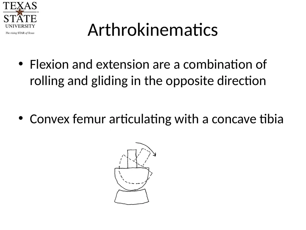
Arthrokinematics Flexion and extension are a combination of rolling and gliding in the opposite direction
Convex femur articulating with a concave tibia<br>
04
Flexion Arthrokinematics From full extension, the initial phase of flexion is pure rolling.
The degree of pure rolling varies per condyle
The medial condyle rolls for 10-15 degrees, the lateral condyle rolls for 20 degrees as it has a larger joint surface
The 15-20 degrees of rolling corresponds with the normal range of flexion/extension in walking.
The tibia glides posterior after the initial phase of flexion<br>
05
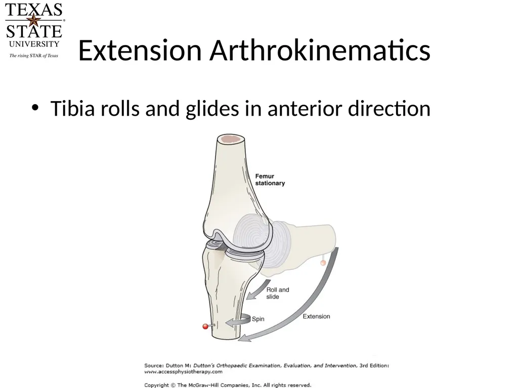
Extension Arthrokinematics Tibia rolls and glides in anterior direction<br>
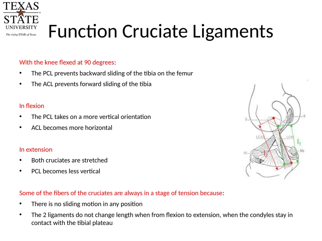
Function Cruciate Ligaments With the knee flexed at 90 degrees:
The PCL prevents backward sliding of the tibia on the femur
The ACL prevents forward sliding of the tibia
In flexion
The PCL takes on a more vertical orientation
ACL becomes more horizontal
In extension
Both cruciates are stretched
PCL becomes less vertical
Some of the fibers of the cruciates are always in a stage of tension because:
There is no sliding motion in any position
The 2 ligaments do not change length when from flexion to extension, when the condyles stay in contact with the tibial plateau<br>
08
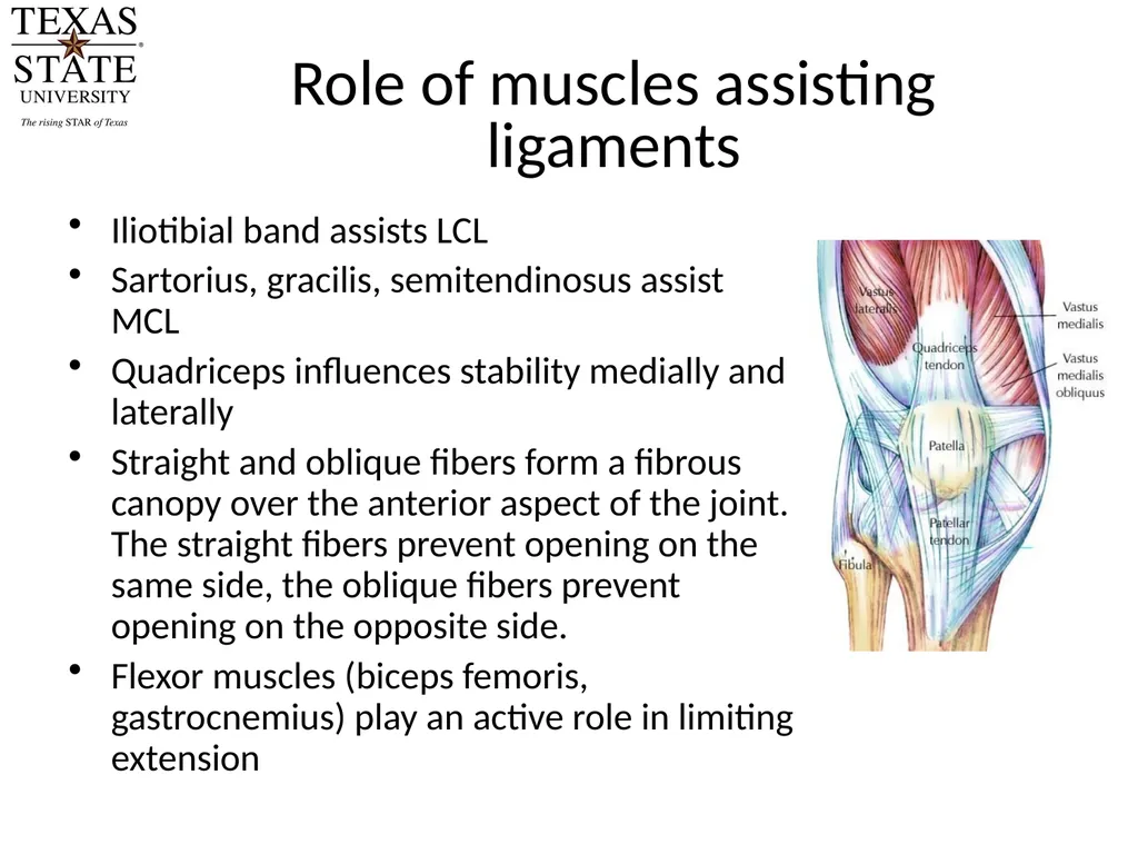
Role of muscles assisting ligaments Iliotibial band assists LCL
Sartorius, gracilis, semitendinosus assist MCL
Quadriceps influences stability medially and laterally
Straight and oblique fibers form a fibrous canopy over the anterior aspect of the joint. The straight fibers prevent opening on the same side, the oblique fibers prevent opening on the opposite side.
Flexor muscles (biceps femoris, gastrocnemius) play an active role in limiting extension<br>
09
Rotatory stability of the knee Axial rotation can only occur when the knee is flexed.
In full extension, rotation of the knee is prevented by tension in the collaterals and the cruciates
The cruciates are wound counter clockwise. Therefore if the tibia is rotated medially, they are wound up more, and will tighten. During lateral rotation of the tibia they unwind
The collaterals are wound clockwise. If the tibia is rotated laterally, the ligaments are wound up more and will resist further rotation
So, the collateral ligaments prevent lateral rotation, the cruciate ligaments prevent medial rotation.<br>
10
The “screw home” mechanism Terminal extension is associated with a small measure of external rotation.
Occurs automatically in the absence of any voluntary movement.
Flexion is associated with an automatic internal rotation of 20 degrees
This motion is conjunct, it cannot be performed independently<br>
11
The “screw home” mechanism Reasons
The lateral femur condyle is greater than the medial; therefore it rolls over a greater distance.
The lateral femur condyle glides more freely on the convex tibial surface.
During extension, the MCL is stretched more rapidly than the LCL, which allows the lateral femur condyle to recede farther.
Tension of the cruciates at end of extension produces external rotation
Slight lateral pull of quadriceps<br>
12
Patella Sesamoid bone in the quadriceps tendon
Function: increases efficiency of the quadriceps by shifting the line of its muscular pull anteriorly.
Extensor efficiency of the quadriceps is increased 1.5 times by the presence of the patella.<br>
13
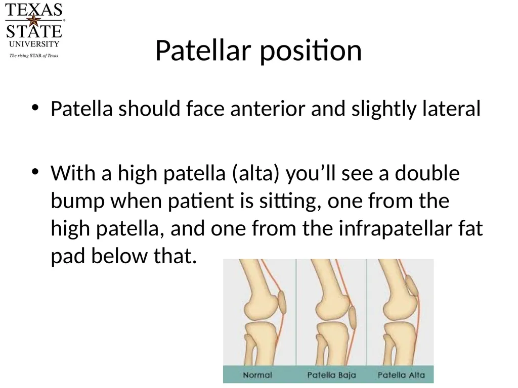
Patellar position Patella should face anterior and slightly lateral
With a high patella (alta) you’ll see a double bump when patient is sitting, one from the high patella, and one from the infrapatellar fat pad below that.<br>
14
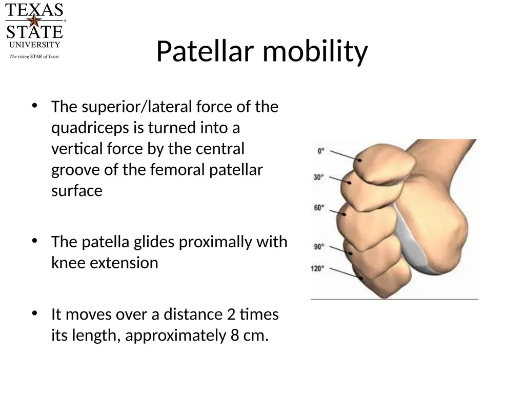
Patellar mobility The superior/lateral force of the quadriceps is turned into a vertical force by the central groove of the femoral patellar surface
The patella glides proximally with knee extension
It moves over a distance 2 times its length, approximately 8 cm.<br>
15
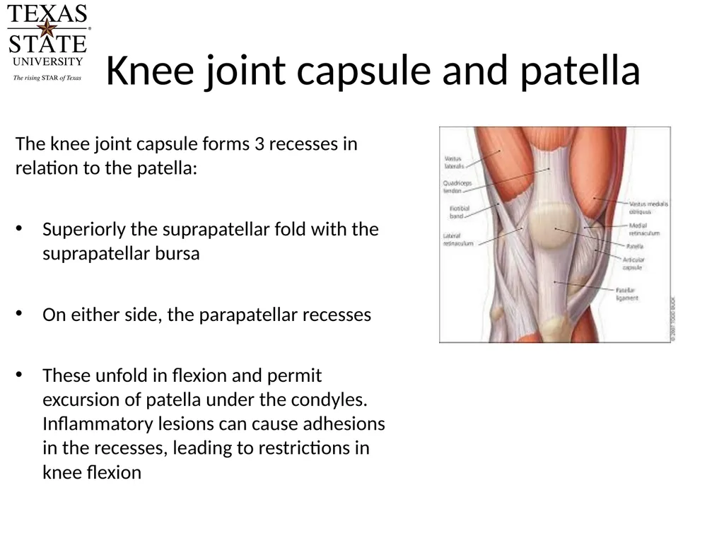
Knee joint capsule and patella The knee joint capsule forms 3 recesses in relation to the patella:
Superiorly the suprapatellar fold with the suprapatellar bursa
On either side, the parapatellar recesses
These unfold in flexion and permit excursion of patella under the condyles. Inflammatory lesions can cause adhesions in the recesses, leading to restrictions in knee flexion<br>
16
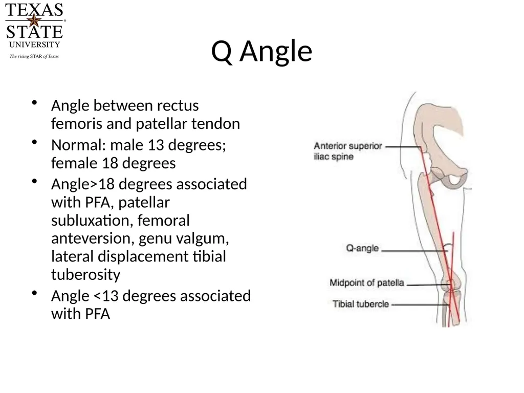
Q Angle Angle between rectus femoris and patellar tendon
Normal: male 13 degrees; female 18 degrees
Angle>18 degrees associated with PFA, patellar subluxation, femoral anteversion, genu valgum, lateral displacement tibial tuberosity
Angle <13 degrees associated with PFA<br>
17
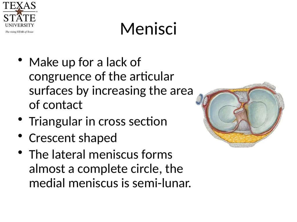
Menisci Make up for a lack of congruence of the articular surfaces by increasing the area of contact
Triangular in cross section
Crescent shaped
The lateral meniscus forms almost a complete circle, the medial meniscus is semi-lunar.<br>
18
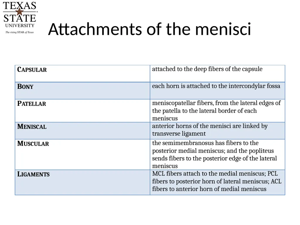
Attachments of the menisci<br>
19
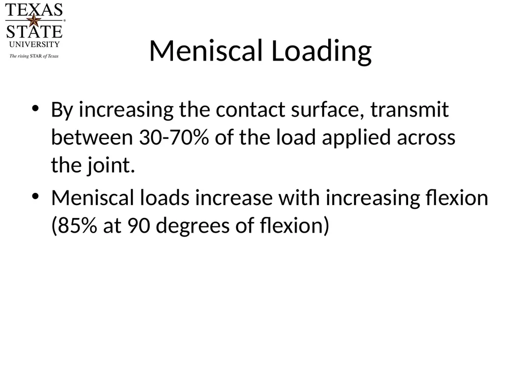
Meniscal Loading By increasing the contact surface, transmit between 30-70% of the load applied across the joint.
Meniscal loads increase with increasing flexion (85% at 90 degrees of flexion)<br>
20
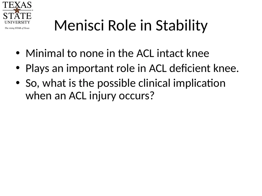
Menisci Role in Stability Minimal to none in the ACL intact knee
Plays an important role in ACL deficient knee.
So, what is the possible clinical implication when an ACL injury occurs?<br>