Methods In Medical Image Analysis Spring 2022 16-725 (CMU RI) : BioE 2630 (Pitt) Dr. John Galeotti What Are We Doing? Theoretical practical skills in medical image analysis Imaging modalities Segmentation Registration Image understanding
"Methods In Medical Image Analysis Spring 2022" is the property of its rightful owner. Permission is granted to
download and print the materials on this website for personal, non-commercial use only, and to display it
on your personal computer provided you do not modify the materials and that you retain all copyright
notices contained in the materials. By downloading content from our website, you accept the terms of this
agreement.
Presentation Transcript
01
Methods In Medical Image Analysis Spring 2022
16-725 (CMU RI) : BioE 2630 (Pitt)
Dr. John Galeotti<br>
02
What Are We Doing? Theoretical & practical skills in medical image analysis
Imaging modalities
Segmentation
Registration
Image understanding
Visualization
Established methods and current research
Focus on understanding & using algorithms 2<br>
03
Why Is Medical Image Analysis Special? Because of the patient
Computer Vision:
Good at detecting irregulars, e.g. on the factory floor
But no two patients are alike—everyone is “irregular”
Medicine is war
Radiology is primarily for reconnaissance
Surgeons are the marines
Life/death decisions made on insufficient information
Success measured by patient recovery
You’re not in “theory land” anymore 3<br>
04
What Do I Mean by Analysis? Different from “Image Processing”
Results in identification, measurement, &/or judgment
Produces numbers, words, & actions
Holy Grail: complete image understanding automated within a computer to perform diagnosis & control robotic intervention
State of the art: segmentation & registration 4<br>
05
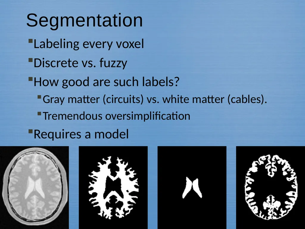
Segmentation Labeling every voxel
Discrete vs. fuzzy
How good are such labels?
Gray matter (circuits) vs. white matter (cables).
Tremendous oversimplification
Requires a model 5<br>
06
Registration Image to Image
same vs. different imaging modality
same vs. different patient
topological variation
Image to Model
deformable models
Model to Model
matching graphs 6<br>
07
Visualization Visualization used to mean to picture in the mind.
Retina is a 2D device
Analysis needed to visualize surfaces
Doctors prefer slices to renderings
Visualization is required to reach visual cortex
Computers have an advantage over humans in 3D 7<br>
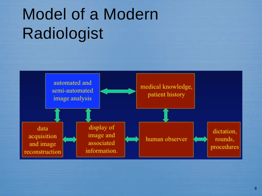
08
Model of a Modern Radiologist 8<br>
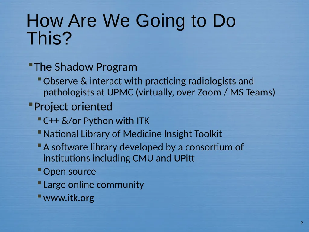
09
How Are We Going to Do This? The Shadow Program
Observe & interact with practicing radiologists and pathologists at UPMC (virtually, over Zoom / MS Teams)
Project oriented
C++ &/or Python with ITK
National Library of Medicine Insight Toolkit
A software library developed by a consortium of institutions including CMU and UPitt
Open source
Large online community
www.itk.org 9<br>
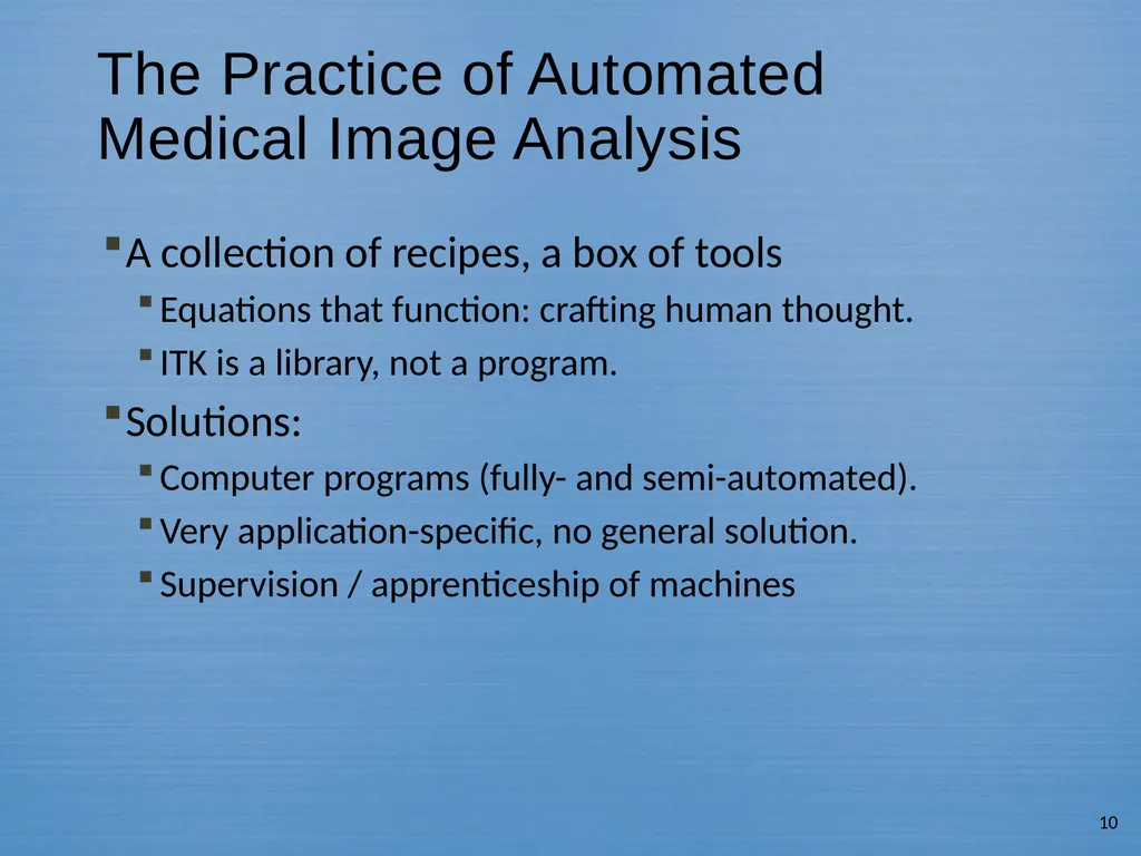
10
The Practice of Automated Medical Image Analysis A collection of recipes, a box of tools
Equations that function: crafting human thought.
ITK is a library, not a program.
Solutions:
Computer programs (fully- and semi-automated).
Very application-specific, no general solution.
Supervision / apprenticeship of machines 10<br>
11
Who Are We? Personal introductions
Name
Academic Background (ECE, Biology, etc.)
Research Interest
Why you’re here
Homework 1 (not yet assigned): submit the requested info about yourself, and a photo.
(photo is optional, but requested; please crop to your head and shoulders)
Details will be posted on the website 11<br>
12
Syllabus On the course website
http://www.cs.cmu.edu/~galeotti/methods_course/
Prerequisites
Vector calculus
Basic probability
Knowledge of C++ and/or Python
Including command-line usage and command-line argument passing to your code
Helpful but not required:
Knowledge of C++ templates & inheritance 12<br>
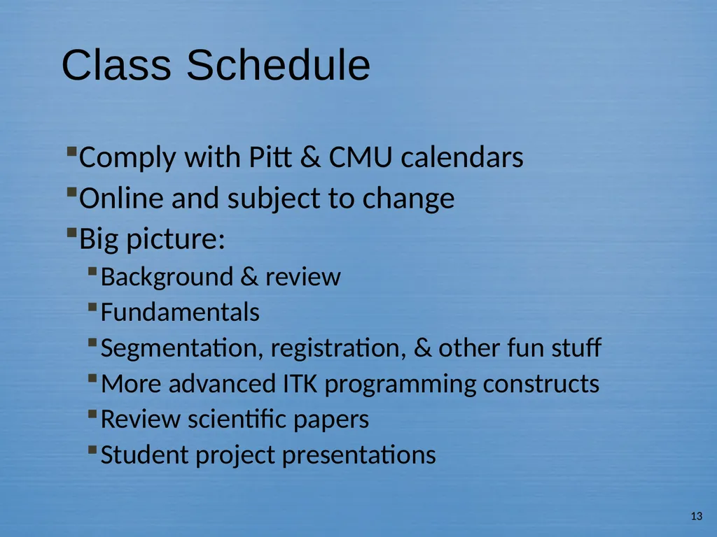
13
Class Schedule Comply with Pitt & CMU calendars
Online and subject to change
Big picture:
Background & review
Fundamentals
Segmentation, registration, & other fun stuff
More advanced ITK programming constructs
Review scientific papers
Student project presentations 13<br>
Textbooks Required: Machine Vision, Wesley E. Snyder & Hairong Qi
Recommended: Insight into Images: Principles and Practice for Segmentation, Registration and Image Analysis, Terry S. Yoo (Editor)
Others (build your bookshelf) 15<br>
16
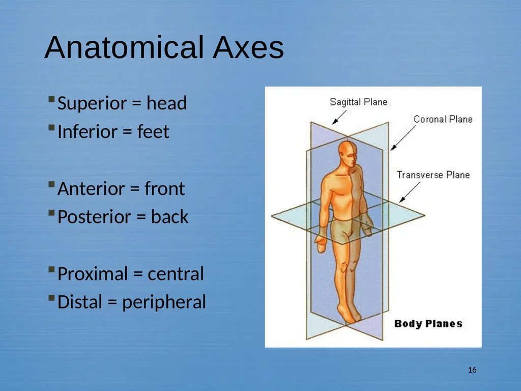
Superior = head
Inferior = feet
Anterior = front
Posterior = back
Proximal = central
Distal = peripheral 16 Anatomical Axes<br>