
NEPHROLITHIASIS AYURVEDIC AND MODERN CONCEPT BY DR
PC
Published · 29 slides · 0 views

1 / 1


Description
NEPHROLITHIASIS AYURVEDIC AND MODERN CONCEPT BY DR H S RAI ,BSc,B.A.M.M.S.,M.D.(Ay),kayachiktsa Rohilkhand Ayurvedic Medical College Bareilly DEFINITION Nephrolithiasis is a common disease specifically refers to calculi in the kidneys, but
Related Topics
Share
Embed code
Download this presentation From Below
"NEPHROLITHIASIS AYURVEDIC AND MODERN CONCEPT BY DR" is the property of its rightful owner. Permission is granted to download and print the materials on this website for personal, non-commercial use only, and to display it on your personal computer provided you do not modify the materials and that you retain all copyright notices contained in the materials. By downloading content from our website, you accept the terms of this agreement.
Presentation Transcript
01
NEPHROLITHIASIS AYURVEDIC AND MODERN CONCEPT
BY
DR H S RAI ,BSc,B.A.M.M.S.,M.D.(Ay),kayachiktsa Rohilkhand Ayurvedic Medical College Bareilly<br>
BY
DR H S RAI ,BSc,B.A.M.M.S.,M.D.(Ay),kayachiktsa Rohilkhand Ayurvedic Medical College Bareilly<br>
02
DEFINITION Nephrolithiasis is a common disease specifically refers to calculi in the kidneys, but this article discusses both renal calculi (Image 1) and ureteral calculi (ureterolithiasis). Ureteral calculi almost always originate in the kidneys, although they may continue to grow once they lodge in the ureter.<br>
03
AETIOLOGY DIETETIC
ALTERED URINARY SOLUTES AND COLLOIDS VITAMINE -A -desquamationof epithilium.form a nidus where stone are deposited
SUPERSATURATION OF URINARY SOLUTES<br>
ALTERED URINARY SOLUTES AND COLLOIDS VITAMINE -A -desquamationof epithilium.form a nidus where stone are deposited
SUPERSATURATION OF URINARY SOLUTES<br>
04
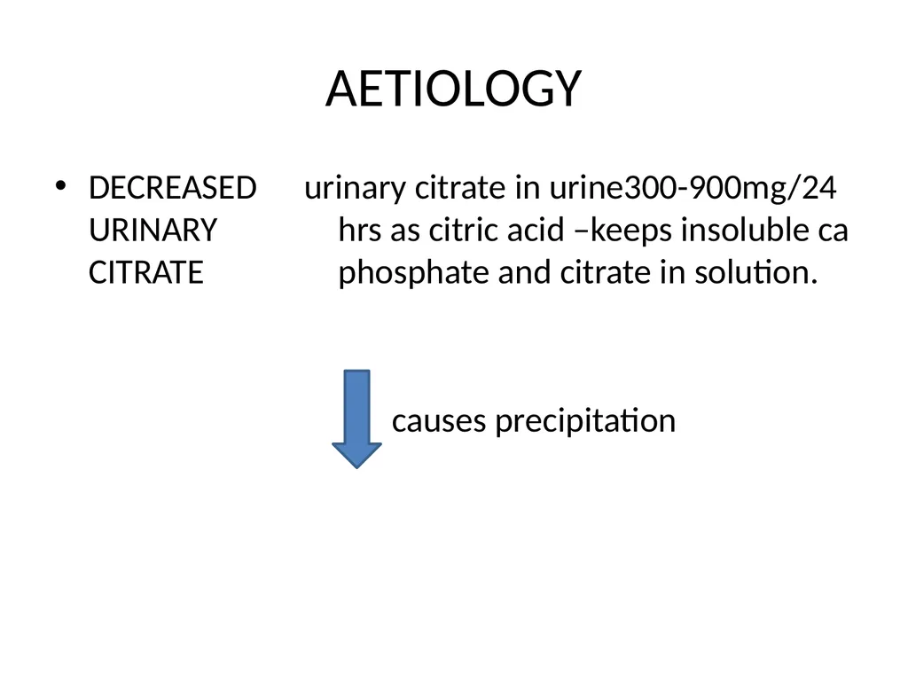
AETIOLOGY urinary citrate in urine300-900mg/24 hrs as citric acid –keeps insoluble ca phosphate and citrate in solution.
causes precipitation DECREASED URINARY CITRATE<br>
causes precipitation DECREASED URINARY CITRATE<br>
05
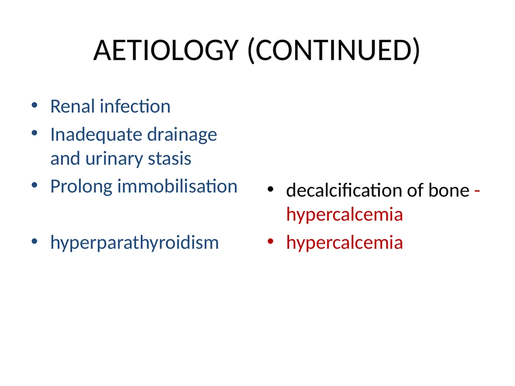
AETIOLOGY (CONTINUED) Renal infection
Inadequate drainage and urinary stasis
Prolong immobilisation
hyperparathyroidism decalcification of bone -hypercalcemia
hypercalcemia<br>
Inadequate drainage and urinary stasis
Prolong immobilisation
hyperparathyroidism decalcification of bone -hypercalcemia
hypercalcemia<br>
06
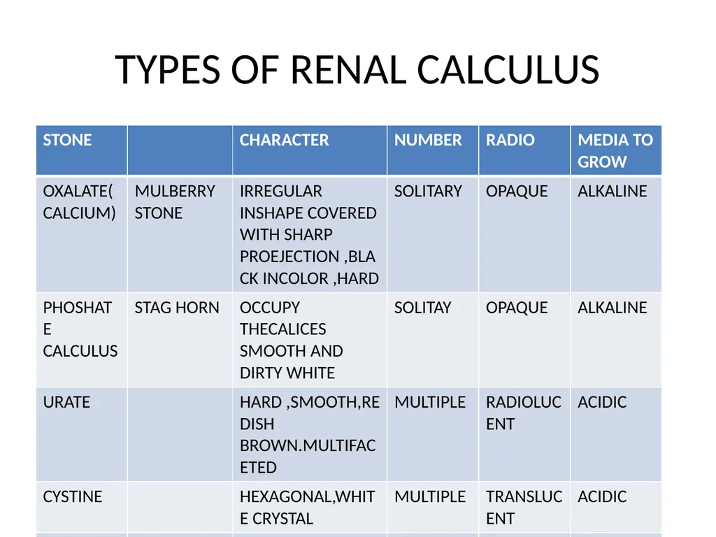
TYPES OF RENAL CALCULUS<br>
07
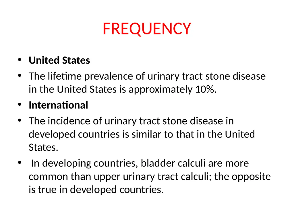
FREQUENCY United States
The lifetime prevalence of urinary tract stone disease in the United States is approximately 10%.
International
The incidence of urinary tract stone disease in developed countries is similar to that in the United States.
In developing countries, bladder calculi are more common than upper urinary tract calculi; the opposite is true in developed countries.<br>
The lifetime prevalence of urinary tract stone disease in the United States is approximately 10%.
International
The incidence of urinary tract stone disease in developed countries is similar to that in the United States.
In developing countries, bladder calculi are more common than upper urinary tract calculi; the opposite is true in developed countries.<br>
08
RACE MORE COMMON IN ASIANS
AREA-HOT AND DRY CLIMATES<br>
AREA-HOT AND DRY CLIMATES<br>
09
SEX MORE COMMON IN MALES THAN FEMALS(3:1)
Stones due to infection (struvite calculi) are more common in women than in men.<br>
Stones due to infection (struvite calculi) are more common in women than in men.<br>
10
AGE Most urinary calculi develop in persons aged 20-49 years.
An initial stone attack after age 50 years is relatively uncommon.<br>
An initial stone attack after age 50 years is relatively uncommon.<br>
11
CLINICAL
SYMPTOMS Renal colic is characterized by undulating cramps and severe pain and is often associated with nausea and vomiting.
As the stone travels through the ureter, the pain moves from the flank to the lower abdomen, down to the groin, and eventually to the scrotal or labial areas.
Associated irritative bladder symptoms are common when the stone is located in the distal or intramural ureter.
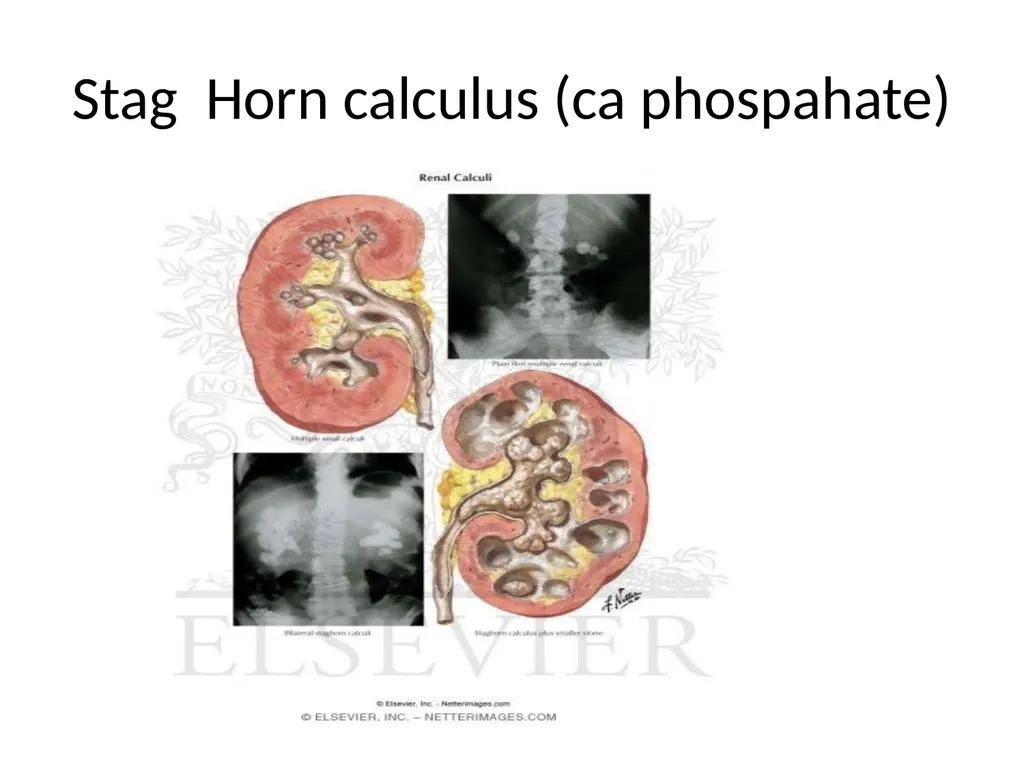
Patients with large renal stones known as staghorn calculi are often relatively asymptomatic.Staghorn refers to the presence of a branched kidney stone occupying the renal pelvis and at least one calyceal system. Such calculi usually manifest as infection and hematuria rather than as acute pain.
Hematurea-is an early feature in oxalate stone
Asymptomatic bilateral obstruction, which is uncommon, manifests as symptoms of renal failure.<br>
As the stone travels through the ureter, the pain moves from the flank to the lower abdomen, down to the groin, and eventually to the scrotal or labial areas.
Associated irritative bladder symptoms are common when the stone is located in the distal or intramural ureter.
Patients with large renal stones known as staghorn calculi are often relatively asymptomatic.Staghorn refers to the presence of a branched kidney stone occupying the renal pelvis and at least one calyceal system. Such calculi usually manifest as infection and hematuria rather than as acute pain.
Hematurea-is an early feature in oxalate stone
Asymptomatic bilateral obstruction, which is uncommon, manifests as symptoms of renal failure.<br>
12
Physical findings Dramatic costovertebral angle tenderness is common; this pain can move to the upper or lower abdominal quadrant as a ureteral stone migrates distally.
Hydronephrosis-apparent swelling on costovertibral angle
RIGIDITY of lateral abdominal muscles,,but not as rule rectus abdominis<br>
Hydronephrosis-apparent swelling on costovertibral angle
RIGIDITY of lateral abdominal muscles,,but not as rule rectus abdominis<br>
13
Lab Investigation Urinalysis-Approximately 85% of patients with urinary calculi exhibit gross or microscopic hematuria.
An absence of hematuria does not rule out urinary calculi; in fact, approximately 15% of patients with urinary stones do not exhibit hematuria.
Complete blood cell counts-leucocytosis-infection
Serum for uric acid,s.phosphate,serum calciumand oxalate.<br>
An absence of hematuria does not rule out urinary calculi; in fact, approximately 15% of patients with urinary stones do not exhibit hematuria.
Complete blood cell counts-leucocytosis-infection
Serum for uric acid,s.phosphate,serum calciumand oxalate.<br>
14
Imaging studies Plane x-ray
Ultrasound
Pylography
Helical CT Scanning
Tomography<br>
Ultrasound
Pylography
Helical CT Scanning
Tomography<br>
15
Medical care Three parts Emergency management
Medical therapy-short and long form
Prevention of stone formation<br>
Medical therapy-short and long form
Prevention of stone formation<br>
16
Emergency management General guidlines After diagnosing renal (ureteral) colic, determine the presence or absence of obstruction or infection.
Obstruction in the absence of infection can be initially managed with analgesics and with other medical measures to facilitate passage of the stone. Infection in the absence of obstruction can be initially managed with antimicrobial therapy. In either case, promptly refer the patient to a urologist.
If neither obstruction nor infection is present, analgesics and other medical measures to facilitate passage of the stone can be initiated with the expectation that the stone will likely pass from the upper urinary tract if its diameter is smaller than 5-6 mm (larger stones are more likely to require surgical measures).
If both obstruction and infection are present, emergent decompression of the upper urinary collecting system is required (see Surgical Care). Immediately consult with a urologist for patients whose pain fails to respond to ED management.<br>
Obstruction in the absence of infection can be initially managed with analgesics and with other medical measures to facilitate passage of the stone. Infection in the absence of obstruction can be initially managed with antimicrobial therapy. In either case, promptly refer the patient to a urologist.
If neither obstruction nor infection is present, analgesics and other medical measures to facilitate passage of the stone can be initiated with the expectation that the stone will likely pass from the upper urinary tract if its diameter is smaller than 5-6 mm (larger stones are more likely to require surgical measures).
If both obstruction and infection are present, emergent decompression of the upper urinary collecting system is required (see Surgical Care). Immediately consult with a urologist for patients whose pain fails to respond to ED management.<br>
17
Specific management Supranormal hydration
Analgesic
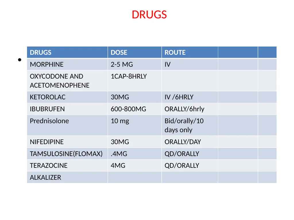
Morphine-drug of choice
NSAID-Ketorolac tromethamine -PARENTAL USE
ANTIEMETICS-METACHLORPROMDE AND PROCHLORPERAZINE
ORAL NORCOTICS IF TOLERATED-, codeine, oxycodone, hydrocodone, usually in a combination form with acetaminophen), NSAIDs, and antiemetics, as needed,<br>
Analgesic
Morphine-drug of choice
NSAID-Ketorolac tromethamine -PARENTAL USE
ANTIEMETICS-METACHLORPROMDE AND PROCHLORPERAZINE
ORAL NORCOTICS IF TOLERATED-, codeine, oxycodone, hydrocodone, usually in a combination form with acetaminophen), NSAIDs, and antiemetics, as needed,<br>
18
MEDICAL EXPULSIVE THERAPY (MET) URETERIC RELAXING DRUG Many randomized trials have confirmed the efficacy of MET in reducing the pain of stone passage, increasing the frequency of stone passage, and reducing the need for surgery. MET should be considered in any patient with a reasonable probability of stone passage. Stones smaller than 3 mm are already associated with an 85% chance of spontaneous passage, and, as such, MET is probably most useful for stones 3-10 mm in size. Overall, MET is associated with a 65% greater likelihood of stone passage.11<br>
19
MET AGENTS CORTICOSTEROIDS(PREDNISOLONE)
CALCUM CHANNEL BLOCKERS
ALPHA –BLOCKERS-TERAZOCINEAND TAMSULOSINE JUDICIOUS USE ,CARE ABOUT IT SIDE EFFECTS
The calcium channel blocker nifedipine relaxes ureteral smooth muscle and enhances stone passage.
The alpha-blockers, such as terazosin, and the alpha-1 selective blockers, such as tamsulosin, also relax musculature of the ureter and lower urinary tract, markedly facilitating passage of ureteral stones. Some literature suggests that the alpha-blockers are more effective in this setting than the calcium channel blockers.
MET with calcium channel blockers and alpha-blockers also appear to improve the results of extracorporeal shock-wave lithotripsy (ESWL; see Extracorporeal shockwave lithotripsy) inasmuch as the stone fragments resulting from treatment appear to clear the system more effectively.<br>
CALCUM CHANNEL BLOCKERS
ALPHA –BLOCKERS-TERAZOCINEAND TAMSULOSINE JUDICIOUS USE ,CARE ABOUT IT SIDE EFFECTS
The calcium channel blocker nifedipine relaxes ureteral smooth muscle and enhances stone passage.
The alpha-blockers, such as terazosin, and the alpha-1 selective blockers, such as tamsulosin, also relax musculature of the ureter and lower urinary tract, markedly facilitating passage of ureteral stones. Some literature suggests that the alpha-blockers are more effective in this setting than the calcium channel blockers.
MET with calcium channel blockers and alpha-blockers also appear to improve the results of extracorporeal shock-wave lithotripsy (ESWL; see Extracorporeal shockwave lithotripsy) inasmuch as the stone fragments resulting from treatment appear to clear the system more effectively.<br>
20
TYPICAL REGIME Analgesic therapy combined with MET dramatically improves the passage of stones, addresses pain, and reduces the need for surgical treatment. A typical regimen for this aggressive management is 1-2 oral narcotic/acetaminophen tablets every 4 hours as needed for pain, 600-800 mg ibuprofen every 8 hours, and MET with 30 mg nifedipine extended-release tablet once daily, 0.4 mg tamsulosin once daily, or 4 mg of terazosin once daily. Limit MET to a 10- to 14-day course, as most stones that pass during this regimen do so in that time frame. If outpatient treatment fails, promptly consult a urologist.<br>
21
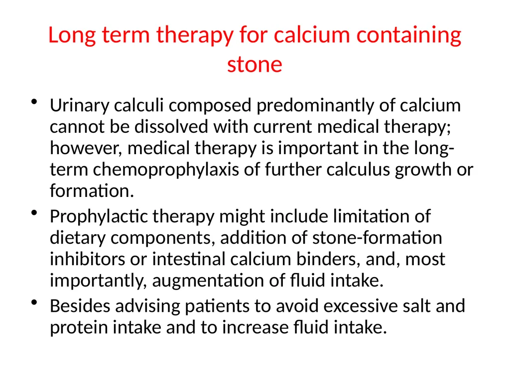
Long term therapy for calcium containing stone Urinary calculi composed predominantly of calcium cannot be dissolved with current medical therapy; however, medical therapy is important in the long-term chemoprophylaxis of further calculus growth or formation.
Prophylactic therapy might include limitation of dietary components, addition of stone-formation inhibitors or intestinal calcium binders, and, most importantly, augmentation of fluid intake.
Besides advising patients to avoid excessive salt and protein intake and to increase fluid intake.<br>
Prophylactic therapy might include limitation of dietary components, addition of stone-formation inhibitors or intestinal calcium binders, and, most importantly, augmentation of fluid intake.
Besides advising patients to avoid excessive salt and protein intake and to increase fluid intake.<br>
22
Uric acid acid and urate calculi Uric acid and cystine calculi can be dissolved with medical therapy. Patients with uric acid stones who do not require urgent surgical intervention for reasons of pain, obstruction, or infection can often have their stones dissolved with alkalization of the urine.
Sodium bicarbonate can be used as the alkalizing agent, but potassium citrate is usually preferred because of the availability of slow-release tablets and the avoidance of a high sodium load.
The dosage of the alkalizing agent should be adjusted to maintain the urinary pH between 6.5 and 7.0. Urinary pH of more than 7.5 should be avoided because of the potential deposition of calcium phosphate around the uric acid calculus, which would make it undissolvable.
Both uric acid and cystine calculi form in acidic environments.Even very large uric acid calculi can be dissolved in patients who comply with therapy. Roughly 1 cm per month dissolution can be achieved.<br>
Sodium bicarbonate can be used as the alkalizing agent, but potassium citrate is usually preferred because of the availability of slow-release tablets and the avoidance of a high sodium load.
The dosage of the alkalizing agent should be adjusted to maintain the urinary pH between 6.5 and 7.0. Urinary pH of more than 7.5 should be avoided because of the potential deposition of calcium phosphate around the uric acid calculus, which would make it undissolvable.
Both uric acid and cystine calculi form in acidic environments.Even very large uric acid calculi can be dissolved in patients who comply with therapy. Roughly 1 cm per month dissolution can be achieved.<br>
23
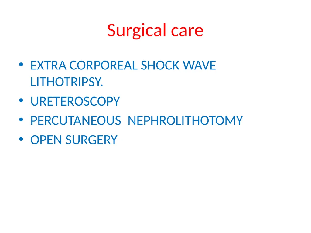
Surgical care EXTRA CORPOREAL SHOCK WAVE LITHOTRIPSY.
URETEROSCOPY
PERCUTANEOUS NEPHROLITHOTOMY
OPEN SURGERY<br>
URETEROSCOPY
PERCUTANEOUS NEPHROLITHOTOMY
OPEN SURGERY<br>
24
DRUGS MORPHINE<br>
25
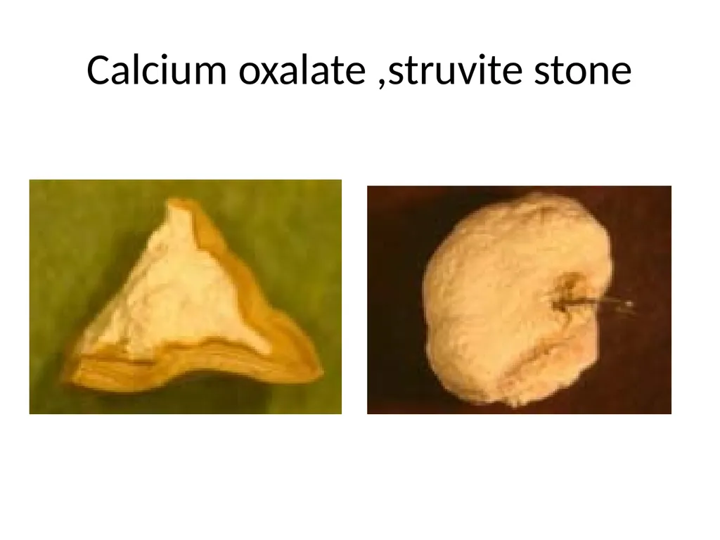
Calcium oxalate ,struvite stone<br>
26
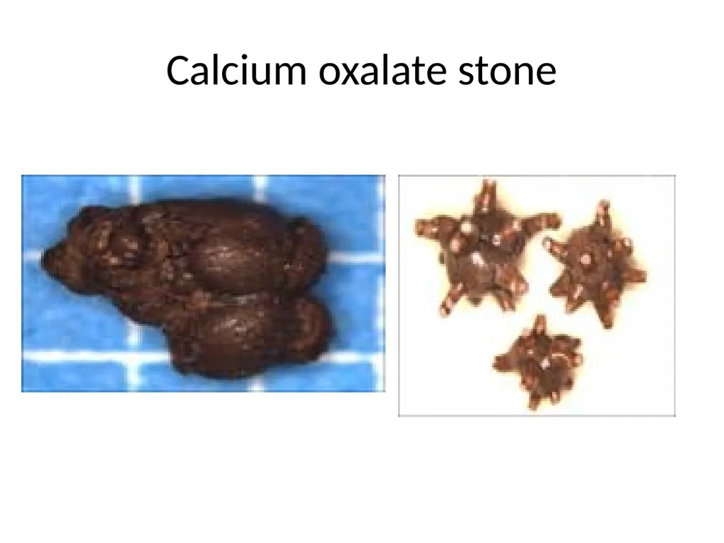
Calcium oxalate stone<br>
27
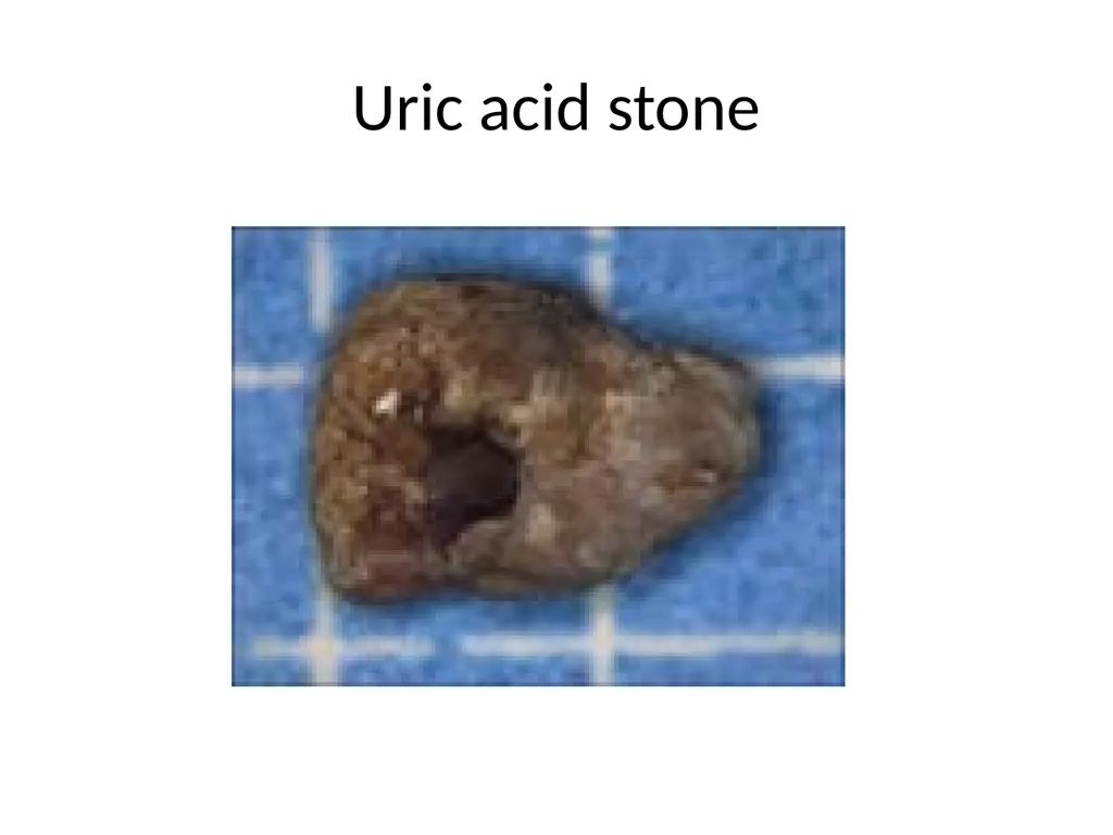
Uric acid stone<br>
28
Stag Horn calculus (ca phospahate)<br>
29
THANK YOU<br>