poultry diseases 2 fourth stage Dr.Harith Abdulla Department of Pathology and Poultry Disease College of veterinary medicine university of basrah chapter four Parasitic Diseases lecture 8 Avian Coccidiosis شعار الكلية Acute gut infection
"poultry diseases 2 fourth stage Dr.Harith Abdulla" is the property of its rightful owner. Permission is granted to
download and print the materials on this website for personal, non-commercial use only, and to display it
on your personal computer provided you do not modify the materials and that you retain all copyright
notices contained in the materials. By downloading content from our website, you accept the terms of this
agreement.
Presentation Transcript
01
poultry diseases 2
fourth stage Dr.Harith Abdulla
Department of Pathology and Poultry Disease
College of veterinary medicine university of basrah chapter four – Parasitic Diseases
lecture 8
Avian Coccidiosis شعار الكلية<br>
02
Acute gut infection in chickens and turkeys.
All avian species are affected.
Host specific.
Etiology : Protozoa.
Genus : Eimeria .
Poultry raised on the floor are highly susceptible
than those raised on wire floor.
Mature Oocyst is the infective stage of the parasite.
Oocysts are resistant to the environmental conditions. Avian Coccidiosis<br>
03
1-Mortality.
2-Retarded growth.
3-Cost of drug.
Conditions needed for maturation of oocyst.
1-Temperature ( 28 C° ) .
2-Moisture ( High humidity ).
3-Oxygen( O₂) . Economic importance:<br>
04
Species affecting chickens Post-mortem lesions
1-Eimeria necatrix a-Ballooning in mid gut.
b-White spots and petechial
hemorrhages (“salt and
and pepper” appearance
on serosal surface.
c-Gut filled with mucoid
bloody exudates.
d-Sausage-like intestine.
e- Severe necrosis. Species affecting chickens:<br>
05
2-Eimeria tenella a- Erosion of cecal wall
( Cecal Coccidiosis) b- Free blood and bloody cores in ceca
c- Caseous cores in old cases.
d- Petechiae.
e- Thickening and ecchymoses.
3- Eimeria brunetti a- Infection in lower small intestine
(Ileorectal coccidiosis) and rectum.
b-White area with thickening of lower small
intestine and rectal walls.
c-Coagulative necrosis and mucoid bloody
enteritis in the lower gut .
d-Petechiae.
e- Severe necrosis.<br>
06
4-Eimeria maxima a- Faint hemorrhage and
wall ballooning.
b-Blood –tinged mucous or
orange exudates.
c-Petechiae and thickening
of middle third of intestine.
d- Mild to severe enteritis.
5-Eimeria acervulina a-”Ladder like”-lesions
produced by white plaques
oocyst in the duodenum.
b- Thickening and petechiae.<br>
07
6-Eimeria mivati a- Occurs in epithelium of
the upper gut (duodenum).
b- Lesions similar to Eimeria
acervulina .
7-Eimeria mitis a- Occurs in upper third of
small intestine.
b- Difficult to see any lesion.
c- Slight mucoid appearance.
8-Eimeria praecox a- Causing diarrhea.
b-Excess liquid and mucus in
duodenal loop.
9-Eimeria hagani =<br>
08
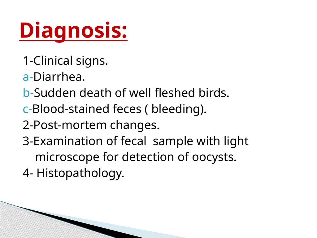
1-Clinical signs.
a-Diarrhea.
b-Sudden death of well fleshed birds.
c-Blood-stained feces ( bleeding).
2-Post-mortem changes.
3-Examination of fecal sample with light
microscope for detection of oocysts.
4- Histopathology. Diagnosis:<br>
09
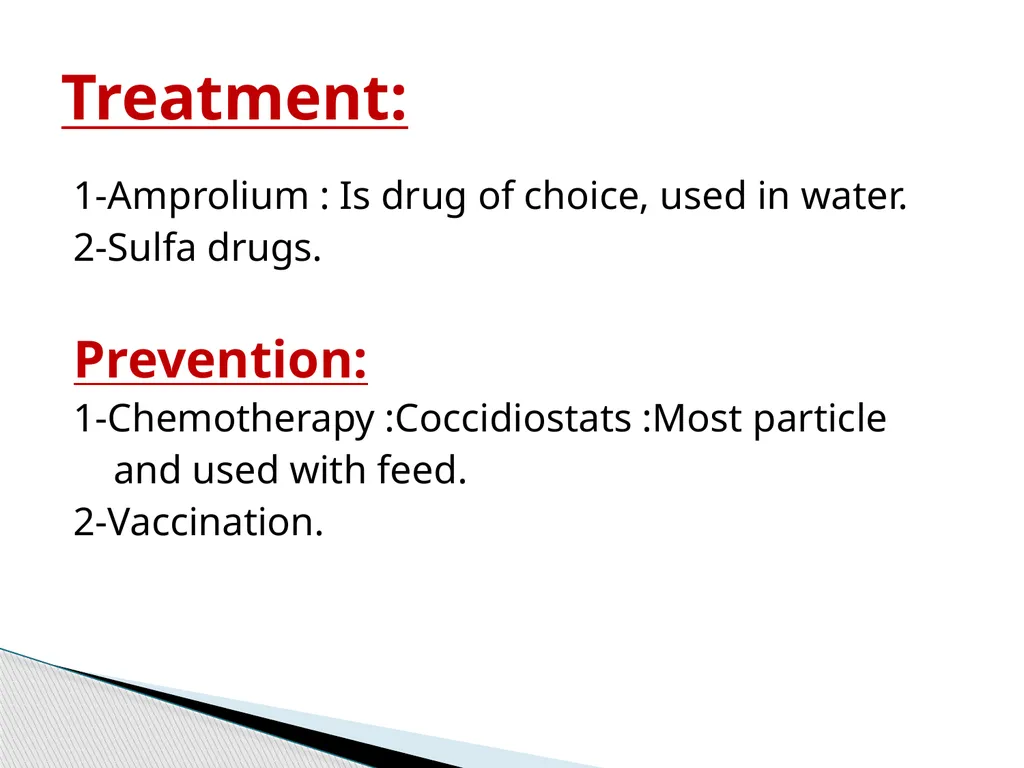
1-Amprolium : Is drug of choice, used in water.
2-Sulfa drugs.
Prevention:
1-Chemotherapy :Coccidiostats :Most particle
and used with feed.
2-Vaccination. Treatment:<br>