"Practical parasitology Platyhelminthes (flat" is the property of its rightful owner. Permission is granted to
download and print the materials on this website for personal, non-commercial use only, and to display it
on your personal computer provided you do not modify the materials and that you retain all copyright
notices contained in the materials. By downloading content from our website, you accept the terms of this
agreement.
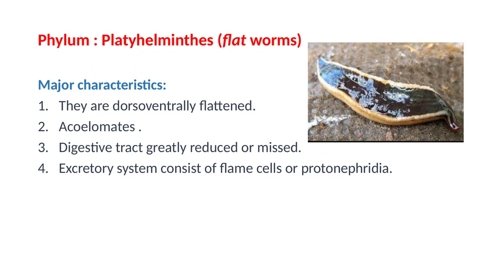
Phylum : Platyhelminthes (flat worms) Major characteristics:
They are dorsoventrally flattened.
Acoelomates .
Digestive tract greatly reduced or missed.
Excretory system consist of flame cells or protonephridia.<br>
03
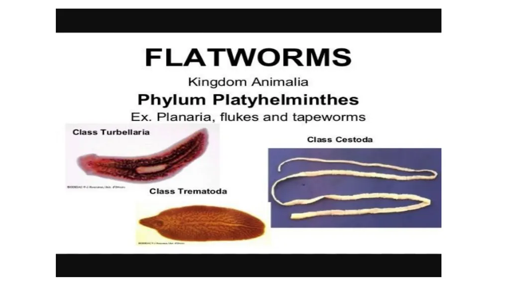
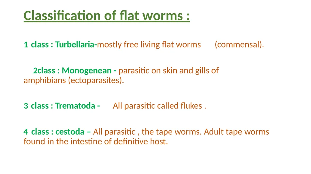
Classification of flat worms : class : Turbellaria-mostly free living flat worms (commensal).
class : Monogenean - parasitic on skin and gills of amphibians (ectoparasites).
class : Trematoda - All parasitic called flukes .
class : cestoda – All parasitic , the tape worms. Adult tape worms found in the intestine of definitive host.<br>
04
Class : Trematoda
The adult members of this class are called flukes and parasitic in vertebrate hosts. They have an intermediate host “usually snails”.
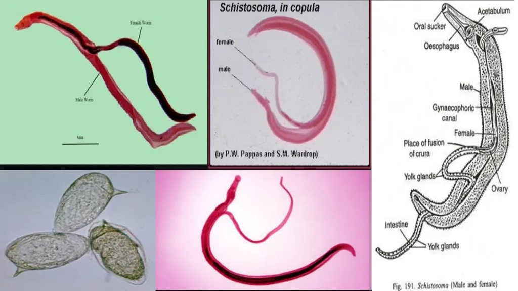
Schistosoma sp. ( blood fluke).
The adult dioecious worms live in the blood stream of humans.
Male fluke is shorter and thicker than female , and the sides of males body are curved under to form a canal along the ventral surface.
Female fluke is long and slender , and is carried in the canal of the male.
Cercaria is the infective stage that penetrate the skin of human.<br>
05
Schistosoma spp. Schistosoma mansoni
S. haematobium
S. japonicum<br>
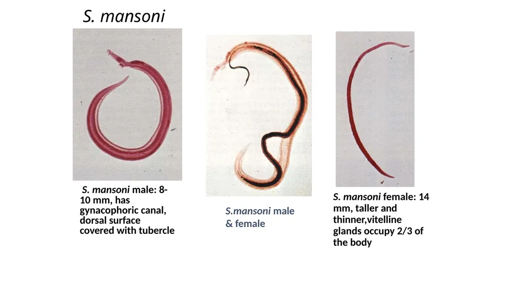
S. mansoni S. mansoni male: 8- 10 mm, has gynacophoric canal, dorsal surface covered with tubercle S. mansoni female: 14 mm, taller and thinner,vitelline glands occupy 2/3 of the body S.mansoni male
& female<br>
08
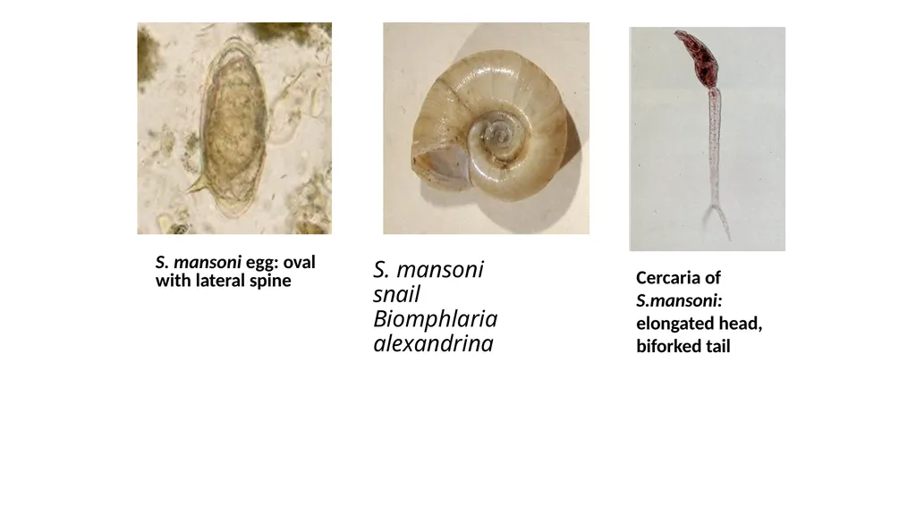
S. mansoni snail Biomphlaria alexandrina Cercaria of S.mansoni: elongated head, biforked tail S. mansoni egg: oval with lateral spine<br>
09
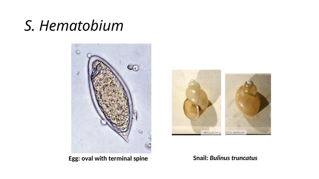
S. Hematobium Egg: oval with terminal spine Snail: Bulinus truncatus<br>
10
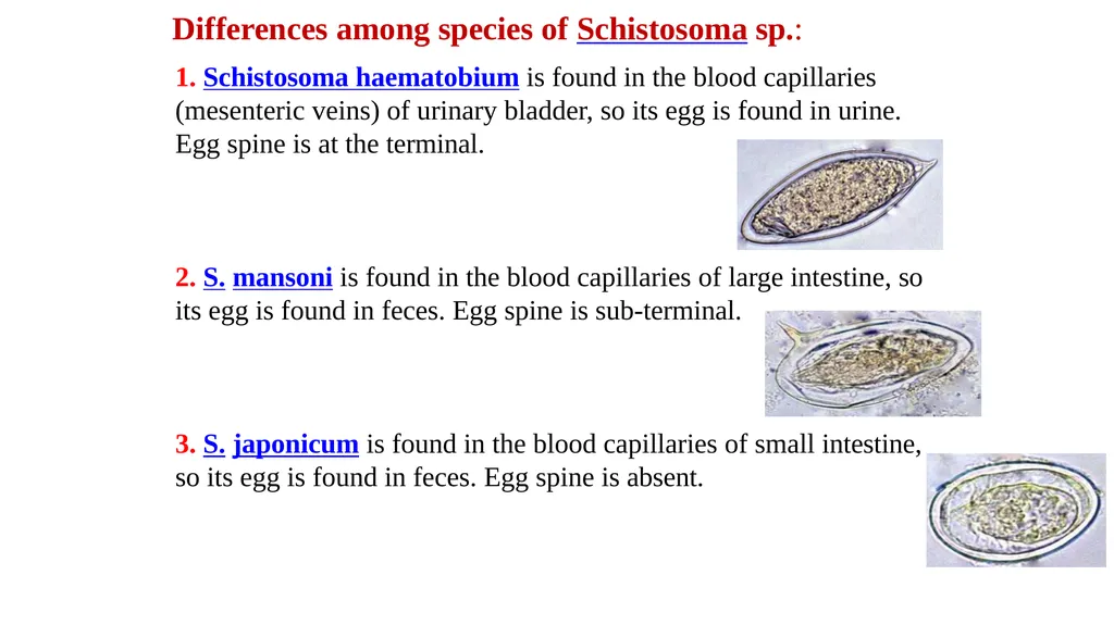
Differences among species of Schistosoma sp.: 1. Schistosoma haematobium is found in the blood capillaries (mesenteric veins) of urinary bladder, so its egg is found in urine. Egg spine is at the terminal. 2. S. mansoni is found in the blood capillaries of large intestine, so its egg is found in feces. Egg spine is sub-terminal. 3. S. japonicum is found in the blood capillaries of small intestine, so its egg is found in feces. Egg spine is absent.<br>
11
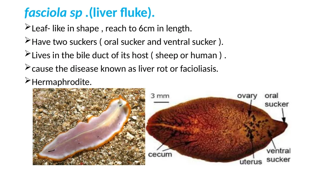
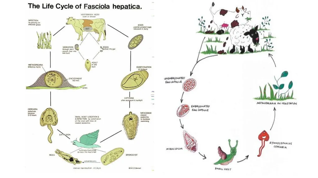
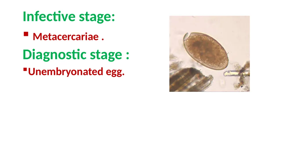
fasciola sp .(liver fluke). Leaf- like in shape , reach to 6cm in length.
Have two suckers ( oral sucker and ventral sucker ).
Lives in the bile duct of its host ( sheep or human ) .
cause the disease known as liver rot or facioliasis.
Hermaphrodite.<br>
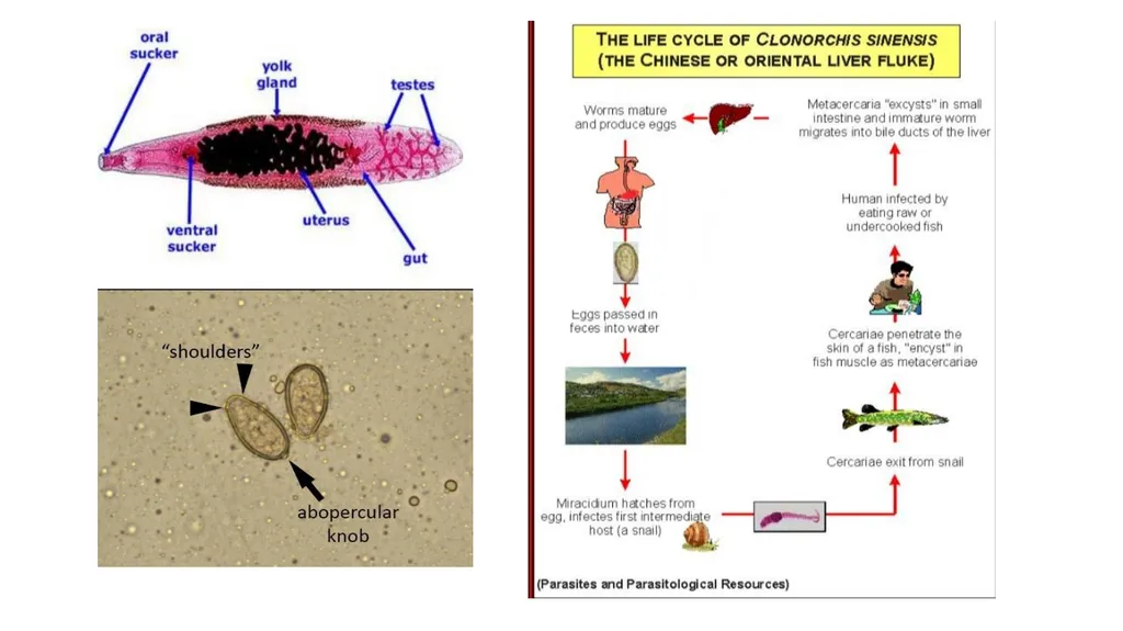
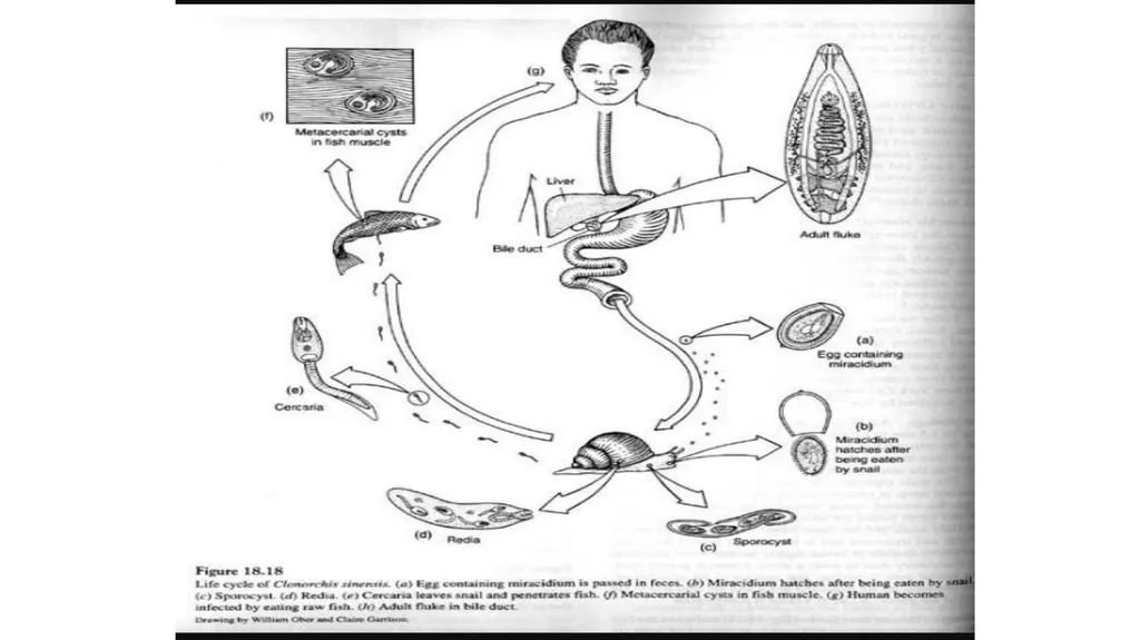
Clonorosis sinesis sp.(chines liver fluke) The chines or oriental liver fluke, the adult resides in the bile duct of human host inside the liver, varies in size 12 mm to 20 mm in length and 3 to 5 mm in width.
The body is tapered interiorly and blunt posteriorly.
When metacercaria reaches the duodenum develops to adult and migrates to the bile duct.
Eggs are passed in the host's feces and the first intermediate host is a snail. Cercariae emerge from the first intermediate host and infect the second intermediate host. The second intermediate host is a fish, The definitive host is infected when it eats raw or undercooked fish.
species of fish are susceptible to infection.<br>
14
Transmission : fecal-oral (ingestion of contaminated raw fish).
Infective stage : metacercaria . *Final host :carnivorous animals and humans.
*Intermediate host:
1-snail (miracidium , sporocyst ,rediae , cercariae). 2-fish.<br>
15
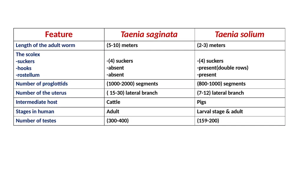
Class : cestoda consist of two species
Taenia saginata
Taenia solium<br>
16
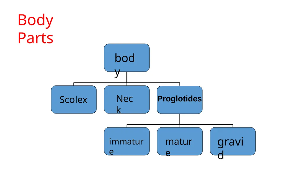
Body Parts body Scolex Neck Proglotides immature mature gravid<br>
17
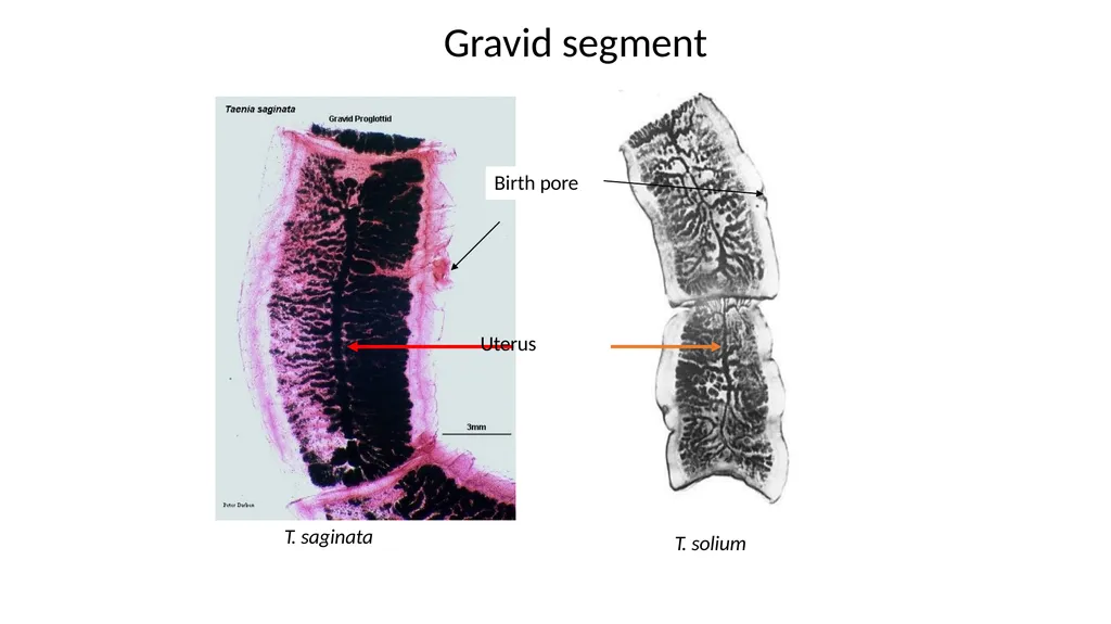
T. saginata T. solium Gravid segment Birth pore Uterus<br>
18
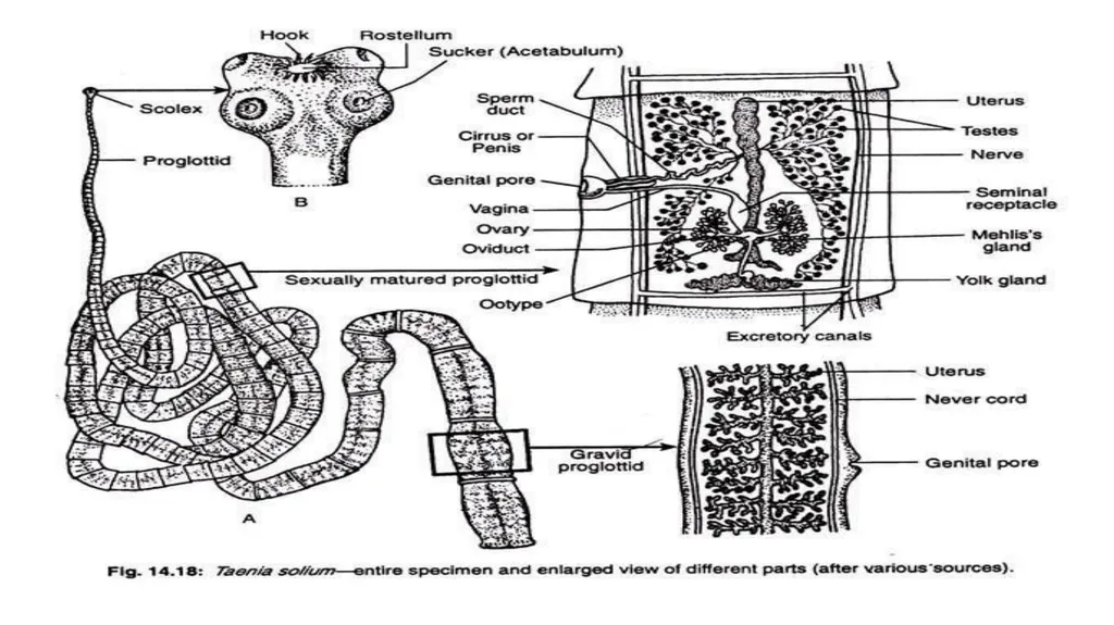
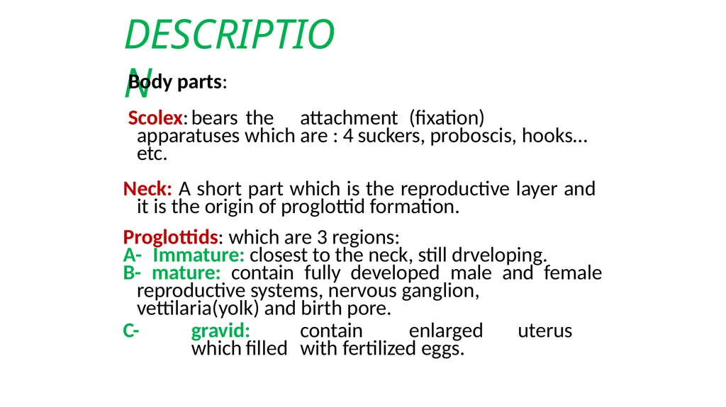
DESCRIPTION Body parts:
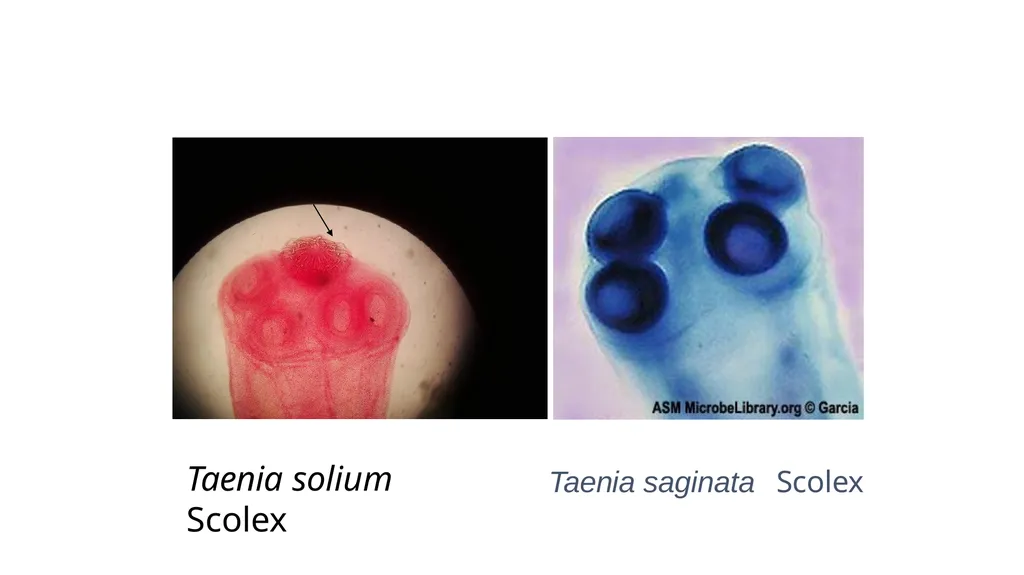
Scolex: bears the attachment (fixation) apparatuses which are : 4 suckers, proboscis, hooks… etc.
Neck: A short part which is the reproductive layer and it is the origin of proglottid formation.
Proglottids: which are 3 regions:
A- Immature: closest to the neck, still drveloping.
B- mature: contain fully developed male and female
reproductive systems, nervous ganglion, vettilaria(yolk) and birth pore.
C- gravid: contain enlarged uterus which filled with fertilized eggs.<br>
19
Taenia spp. Egg ( taenia saginata and taenia solium)<br>
20
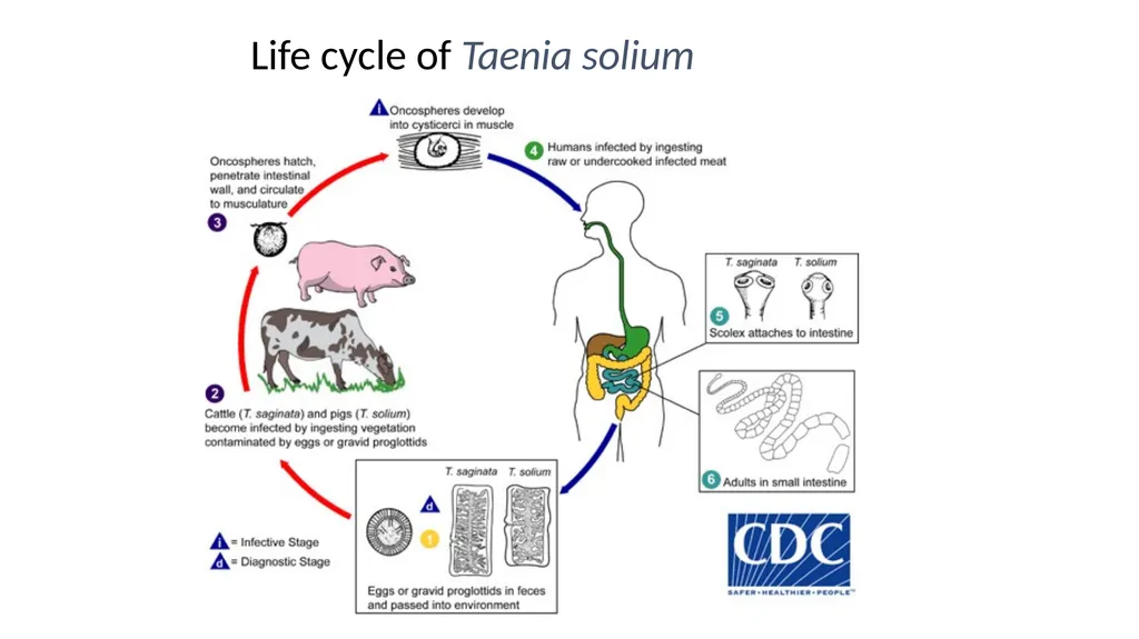
Life cycle of Taenia solium<br>
21
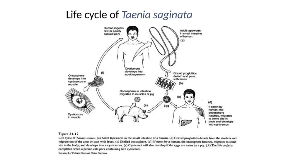
Life cycle of Taenia saginata<br>
22
Taenia solium Scolex Taenia saginata Scolex<br>
23
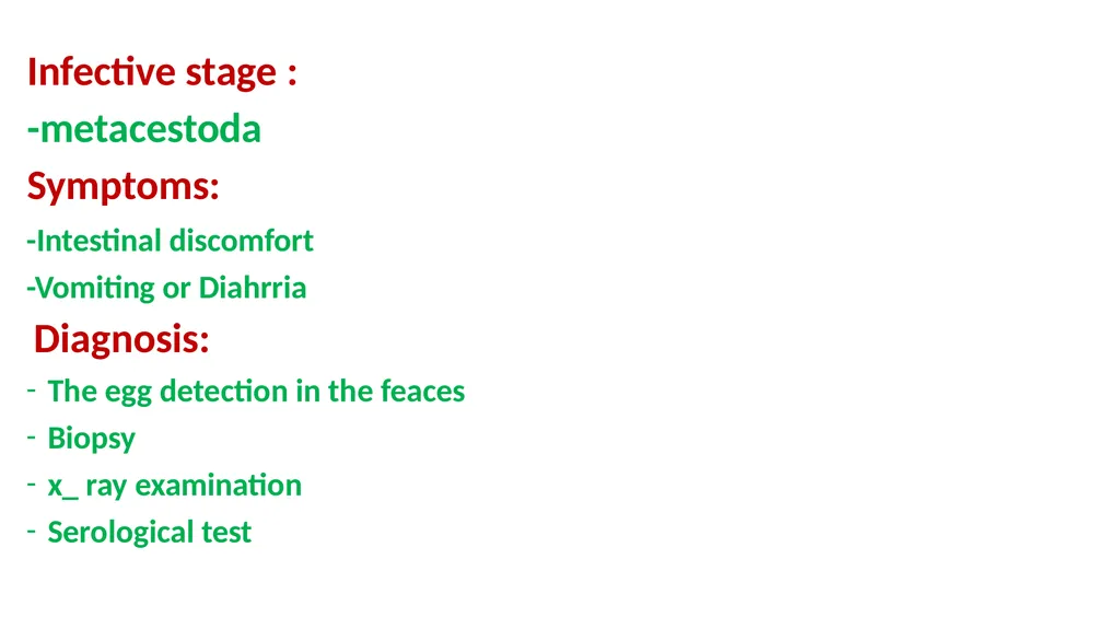
Infective stage :
-metacestoda Symptoms: -Intestinal discomfort
-Vomiting or Diahrria
Diagnosis:
The egg detection in the feaces
Biopsy
x_ ray examination
Serological test<br>