
Respiratory Emergencies CHAPTER 17 Introduction
GB
Published · 179 slides · 0 views

1 / 1




Description
Respiratory Emergencies CHAPTER 17 Introduction Respiratory disease Common pathologic condition Common EMS dispatch Epidemiology Asthma and COPD Among leading causes of death and disability 16 million American with COPD 25 million with
Related Topics
Share
Embed code
Download this presentation From Below
"Respiratory Emergencies CHAPTER 17 Introduction" is the property of its rightful owner. Permission is granted to download and print the materials on this website for personal, non-commercial use only, and to display it on your personal computer provided you do not modify the materials and that you retain all copyright notices contained in the materials. By downloading content from our website, you accept the terms of this agreement.
Presentation Transcript
01
Respiratory Emergencies CHAPTER 17<br>
02
Introduction Respiratory disease
Common pathologic condition
Common EMS dispatch<br>
Common pathologic condition
Common EMS dispatch<br>
03
Epidemiology Asthma and COPD
Among leading causes of death and disability
16 million American with COPD
25 million with asthma
Pneumonia
One of the most common fatal illnesses in developing countries
Genetic or caused by external factors<br>
Among leading causes of death and disability
16 million American with COPD
25 million with asthma
Pneumonia
One of the most common fatal illnesses in developing countries
Genetic or caused by external factors<br>
04
Anatomy and Physiology Review<br>
05
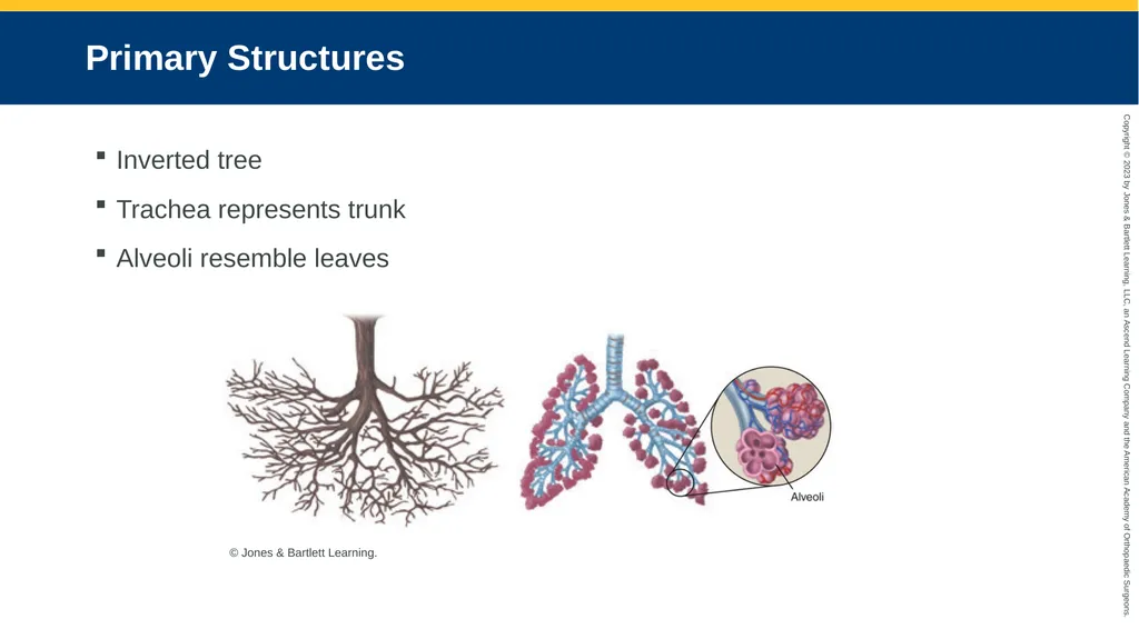
Primary Structures Inverted tree
Trachea represents trunk
Alveoli resemble leaves © Jones & Bartlett Learning.<br>
Trachea represents trunk
Alveoli resemble leaves © Jones & Bartlett Learning.<br>
06
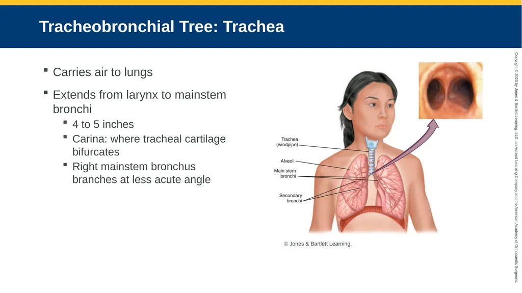
Tracheobronchial Tree: Trachea Carries air to lungs
Extends from larynx to mainstem bronchi
4 to 5 inches
Carina: where tracheal cartilage bifurcates
Right mainstem bronchus branches at less acute angle © Jones & Bartlett Learning.<br>
Extends from larynx to mainstem bronchi
4 to 5 inches
Carina: where tracheal cartilage bifurcates
Right mainstem bronchus branches at less acute angle © Jones & Bartlett Learning.<br>
07
Tracheobronchial Tree: Bronchi Mainstem bronchi branch into lobes of the lungs, then into:
Tertiary (segmental) bronchi
Subsegmental bronchi
Bronchioles
Dead space
Airways do not participate in gas exchange
Greater in patients with respiratory disease<br>
Tertiary (segmental) bronchi
Subsegmental bronchi
Bronchioles
Dead space
Airways do not participate in gas exchange
Greater in patients with respiratory disease<br>
08
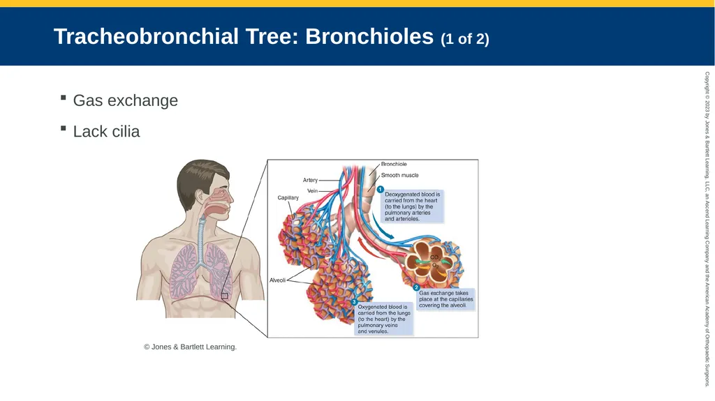
Tracheobronchial Tree: Bronchioles (1 of 2) Gas exchange
Lack cilia © Jones & Bartlett Learning.<br>
Lack cilia © Jones & Bartlett Learning.<br>
09
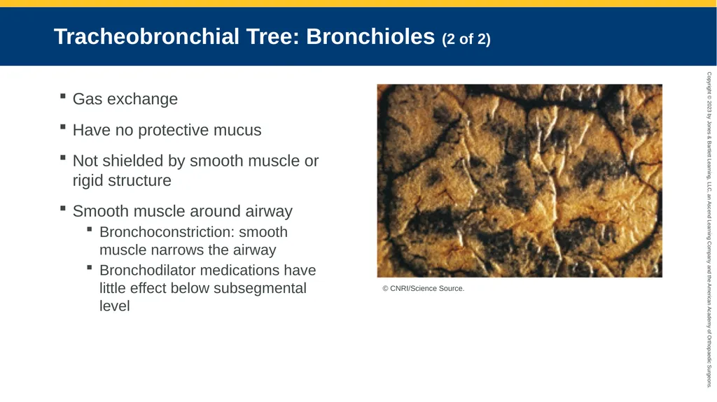
Tracheobronchial Tree: Bronchioles (2 of 2) Gas exchange
Have no protective mucus
Not shielded by smooth muscle or rigid structure
Smooth muscle around airway
Bronchoconstriction: smooth muscle narrows the airway
Bronchodilator medications have little effect below subsegmental level © CNRI/Science Source.<br>
Have no protective mucus
Not shielded by smooth muscle or rigid structure
Smooth muscle around airway
Bronchoconstriction: smooth muscle narrows the airway
Bronchodilator medications have little effect below subsegmental level © CNRI/Science Source.<br>
10
Tracheobronchial Tree: Alveoli Branches 16 to 24
Terminal airways
Alveoli
Entire surface covered in capillaries
Participate in gas exchange<br>
Terminal airways
Alveoli
Entire surface covered in capillaries
Participate in gas exchange<br>
11
Mediastinum Space in middle of chest
Consists of:
Heart
Large blood vessels
Large conducting airways
Other organs<br>
Consists of:
Heart
Large blood vessels
Large conducting airways
Other organs<br>
12
Pulmonary Blood Flow From heart to lungs via pulmonary artery
Artery branches into smaller vessels
More gas exchange between lung bases and circulatory system
Pulmonary capillaries are narrow
Polycythemia
Cor pulmonale<br>
Artery branches into smaller vessels
More gas exchange between lung bases and circulatory system
Pulmonary capillaries are narrow
Polycythemia
Cor pulmonale<br>
13
Perfusion Circulatory component of respiratory system
Blood must keep flowing through pulmonary vessels
Large embolus can block blood flow<br>
Blood must keep flowing through pulmonary vessels
Large embolus can block blood flow<br>
14
Mechanisms of Respiratory Control (1 of 6) Cardiovascular regulation
Lungs closely linked to cardiac function
Heart changes have pulmonary consequences
Left-sided heart failure progresses faster than right-sided heart failure
Mild hypoxia increases in heart rate
Severe hypoxia causes bradycardia
Uncorrected hypoxic insults may trigger lethal cardiac arrhythmia<br>
Lungs closely linked to cardiac function
Heart changes have pulmonary consequences
Left-sided heart failure progresses faster than right-sided heart failure
Mild hypoxia increases in heart rate
Severe hypoxia causes bradycardia
Uncorrected hypoxic insults may trigger lethal cardiac arrhythmia<br>
15
Mechanisms of Respiratory Control (2 of 6) Heath failure caused by changes in:
Fluid balance
Right-sided heart pumping pressure
Left-sided heart pumping pressure<br>
Fluid balance
Right-sided heart pumping pressure
Left-sided heart pumping pressure<br>
16
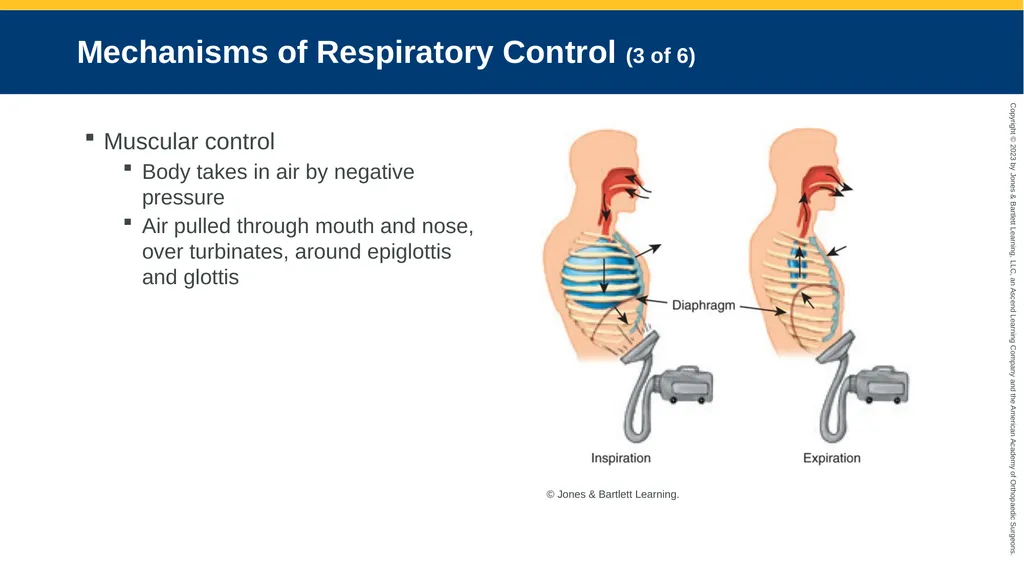
Mechanisms of Respiratory Control (3 of 6) Muscular control
Body takes in air by negative pressure
Air pulled through mouth and nose, over turbinates, around epiglottis and glottis © Jones & Bartlett Learning.<br>
Body takes in air by negative pressure
Air pulled through mouth and nose, over turbinates, around epiglottis and glottis © Jones & Bartlett Learning.<br>
17
Mechanisms of Respiratory Control (4 of 6) Muscular control
Thorax
Airtight box
Diaphragm at bottom
Trachea at top
Diaphragm flattens during quiet breathing<br>
Thorax
Airtight box
Diaphragm at bottom
Trachea at top
Diaphragm flattens during quiet breathing<br>
18
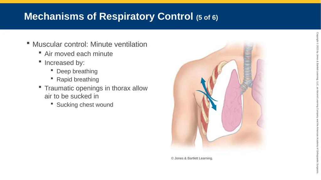
Mechanisms of Respiratory Control (5 of 6) Muscular control: Minute ventilation
Air moved each minute
Increased by:
Deep breathing
Rapid breathing
Traumatic openings in thorax allow air to be sucked in
Sucking chest wound © Jones & Bartlett Learning.<br>
Air moved each minute
Increased by:
Deep breathing
Rapid breathing
Traumatic openings in thorax allow air to be sucked in
Sucking chest wound © Jones & Bartlett Learning.<br>
19
Mechanisms of Respiratory Control (6 of 6) Renal Status
Kidneys help control
Fluid balance
Acid-base balance
Blood pressure
Factor into pulmonary mechanics and oxygen delivery to body tissues<br>
Kidneys help control
Fluid balance
Acid-base balance
Blood pressure
Factor into pulmonary mechanics and oxygen delivery to body tissues<br>
20
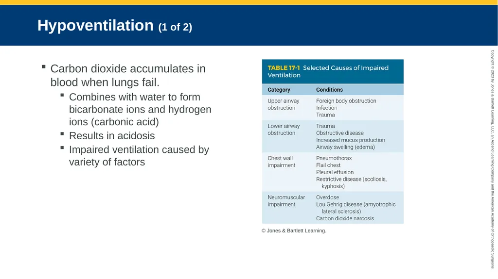
Hypoventilation (1 of 2) Carbon dioxide accumulates in blood when lungs fail.
Combines with water to form bicarbonate ions and hydrogen ions (carbonic acid)
Results in acidosis
Impaired ventilation caused by variety of factors © Jones & Bartlett Learning.<br>
Combines with water to form bicarbonate ions and hydrogen ions (carbonic acid)
Results in acidosis
Impaired ventilation caused by variety of factors © Jones & Bartlett Learning.<br>
21
Hypoventilation (2 of 2) Carbon dioxide level directly related to pH
As carbon dioxide rises, pH drops
Patients usually have respiratory acidosis
Ultimate manifestation
Respiratory arrest followed by cardiac arrest<br>
As carbon dioxide rises, pH drops
Patients usually have respiratory acidosis
Ultimate manifestation
Respiratory arrest followed by cardiac arrest<br>
22
Hypoventilation: Causes (1 of 4) Conditions that impair lung function
Atelectasis
Pneumonia
Pulmonary edema
Asthma
COPD<br>
Atelectasis
Pneumonia
Pulmonary edema
Asthma
COPD<br>
23
Hypoventilation: Causes (2 of 4) Conditions that impair mechanics of breathing
Flail chest
Diaphragmatic rupture
Severe retractions
Air- or blood-filled abdomen
Abdominal or chest binding
Obesity hypoventilation syndrome<br>
Flail chest
Diaphragmatic rupture
Severe retractions
Air- or blood-filled abdomen
Abdominal or chest binding
Obesity hypoventilation syndrome<br>
24
Hypoventilation: Causes (3 of 4) Conditions that impair neuromuscular apparatus
Head trauma, intracranial infections, or brain tumors
Serious spinal cord injury
Guillain-Barré syndrome
Amyotrophic lateral sclerosis (Lou Gehrig disease)
Botulism<br>
Head trauma, intracranial infections, or brain tumors
Serious spinal cord injury
Guillain-Barré syndrome
Amyotrophic lateral sclerosis (Lou Gehrig disease)
Botulism<br>
25
Hypoventilation: Causes (4 of 4) Conditions that reduce respiratory drive
Intoxication
Head injury
Hypoxic drive
Asphyxia<br>
Intoxication
Head injury
Hypoxic drive
Asphyxia<br>
26
Hyperventilation (1 of 2) When people breathe in excess of need
From increasing rate and/or depth of respiration
Releases more carbon dioxide than normal
Results in alkalosis
Triggered by emotional distress or panic
Hysterical hyperventilation
Hyperventilation syndrome<br>
From increasing rate and/or depth of respiration
Releases more carbon dioxide than normal
Results in alkalosis
Triggered by emotional distress or panic
Hysterical hyperventilation
Hyperventilation syndrome<br>
27
Hyperventilation (2 of 2) Respiratory alkalosis
Causes numbness in hands, feet, and mouth
Ultimately leads to carpopedal spasm
Having patients rebreathe carbon dioxide can be dangerous.
Patients quickly exhaust oxygen
Hyperventilation may be body’s attempt to raise pH levels
Kussmaul respirations
Sepsis
Shock<br>
Causes numbness in hands, feet, and mouth
Ultimately leads to carpopedal spasm
Having patients rebreathe carbon dioxide can be dangerous.
Patients quickly exhaust oxygen
Hyperventilation may be body’s attempt to raise pH levels
Kussmaul respirations
Sepsis
Shock<br>
28
Hyperventilation: Treatments Sedation (extreme measure)
Psychological support:
Breathing with patient
Having patient count to two between breaths
Distraction techniques
Having patient sing a song<br>
Psychological support:
Breathing with patient
Having patient count to two between breaths
Distraction techniques
Having patient sing a song<br>
29
Patient Assessment<br>
30
Respiratory Assessment Thorough assessment
Many respiratory ailments are life threatening
More than listening to patient’s lungs
Early in patient assessment<br>
Many respiratory ailments are life threatening
More than listening to patient’s lungs
Early in patient assessment<br>
31
Scene Size-Up (1 of 3) Take standard precautions
Use proper PPE
Evaluate scene safety for:
Diminished oxygen concentrations
Carbon monoxide
Irritant gases
Highly contagious respiratory illness<br>
Use proper PPE
Evaluate scene safety for:
Diminished oxygen concentrations
Carbon monoxide
Irritant gases
Highly contagious respiratory illness<br>
32
Scene Size-Up (2 of 3) Respiratory diseases can impair:
Ventilation
Diffusion
Perfusion
Combination of all three
Rapid-onset dyspnea may be caused by:
Acute bronchospasm
Anaphylaxis
Pulmonary embolism
Pneumothorax<br>
Ventilation
Diffusion
Perfusion
Combination of all three
Rapid-onset dyspnea may be caused by:
Acute bronchospasm
Anaphylaxis
Pulmonary embolism
Pneumothorax<br>
33
Scene Size-Up (3 of 3) Paroxysmal nocturnal dyspnea
Sudden
Middle of the night
May signal left-sided heart failure
Ability to move air may be hindered by factors that:
Limit diaphragm movement
Restrict chest wall movement
Disrupt the integrity of the thoracic cage<br>
Sudden
Middle of the night
May signal left-sided heart failure
Ability to move air may be hindered by factors that:
Limit diaphragm movement
Restrict chest wall movement
Disrupt the integrity of the thoracic cage<br>
34
Primary Survey (1 of 22) Establish and maintain open airway
Form general impression
Body type may be associated with condition
Emphysema
Chronic bronchitis<br>
Form general impression
Body type may be associated with condition
Emphysema
Chronic bronchitis<br>
35
Primary Survey (2 of 22) Assess oxygen demand and work of breathing.
Observe condition during typical exertion.
Tachycardia, diaphoresis, and pallor triggers:
Increased work of breathing
Anxiety
Hypoxia<br>
Observe condition during typical exertion.
Tachycardia, diaphoresis, and pallor triggers:
Increased work of breathing
Anxiety
Hypoxia<br>
36
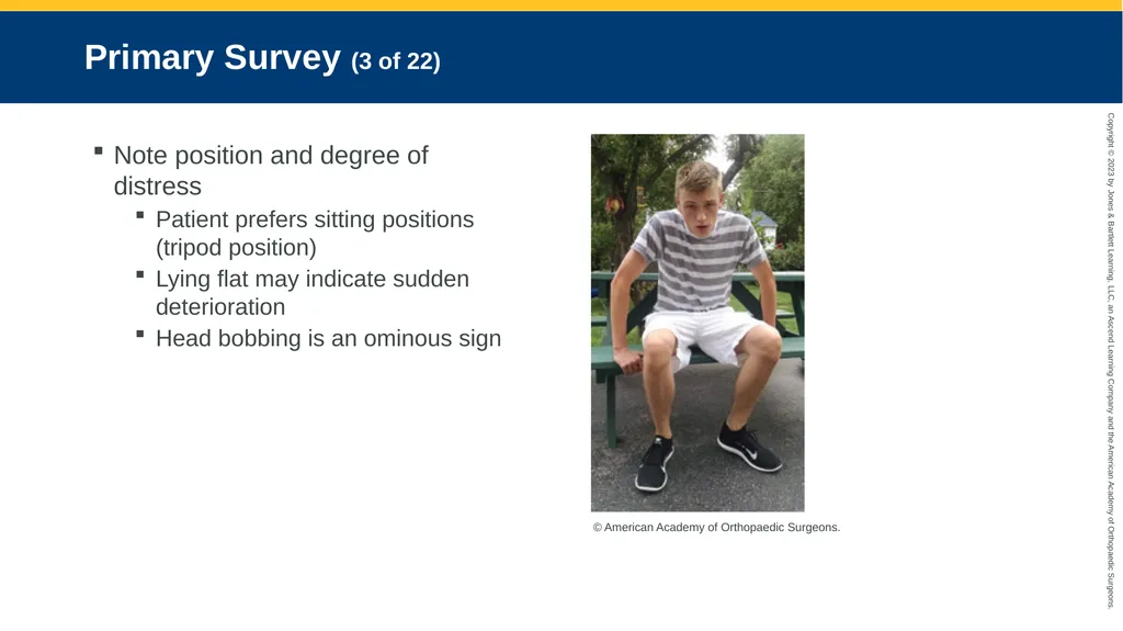
Primary Survey (3 of 22) Note position and degree of distress
Patient prefers sitting positions (tripod position)
Lying flat may indicate sudden deterioration
Head bobbing is an ominous sign © American Academy of Orthopaedic Surgeons.<br>
Patient prefers sitting positions (tripod position)
Lying flat may indicate sudden deterioration
Head bobbing is an ominous sign © American Academy of Orthopaedic Surgeons.<br>
37
Primary Survey (4 of 22) Breathing alterations can involve:
Conducting airways (trachea, bronchi and bronchioles)
Alveoli
Muscles and nerves involved in breathing
Rigid structure of thorax<br>
Conducting airways (trachea, bronchi and bronchioles)
Alveoli
Muscles and nerves involved in breathing
Rigid structure of thorax<br>
38
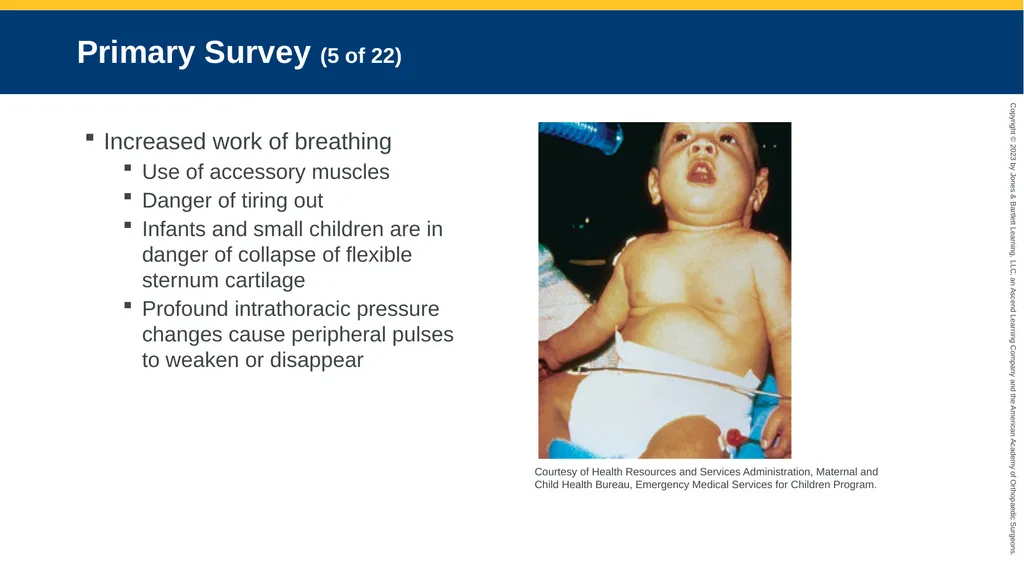
Primary Survey (5 of 22) Increased work of breathing
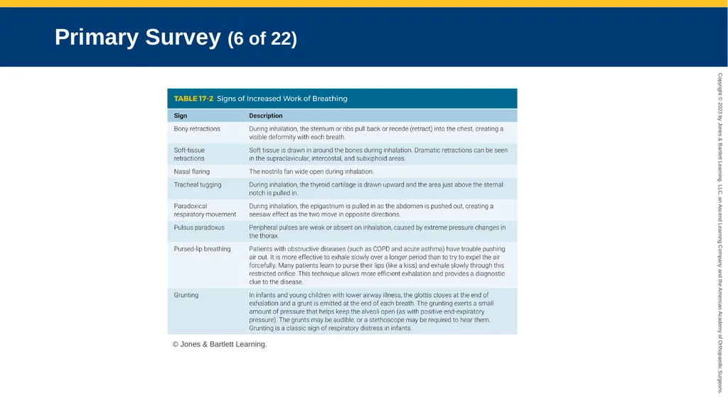
Use of accessory muscles
Danger of tiring out
Infants and small children are in danger of collapse of flexible sternum cartilage
Profound intrathoracic pressure changes cause peripheral pulses to weaken or disappear Courtesy of Health Resources and Services Administration, Maternal and Child Health Bureau, Emergency Medical Services for Children Program.<br>
Use of accessory muscles
Danger of tiring out
Infants and small children are in danger of collapse of flexible sternum cartilage
Profound intrathoracic pressure changes cause peripheral pulses to weaken or disappear Courtesy of Health Resources and Services Administration, Maternal and Child Health Bureau, Emergency Medical Services for Children Program.<br>
39
Primary Survey (6 of 22) © Jones & Bartlett Learning.<br>
40
Primary Survey (7 of 22) Altered rate and depth of respiration
Monitor without patient noticing
Patient with adequate rate but low volume will have inadequate minute volume
Monitor trends in respiratory rates
Note inspiratory-to-expiratory (I:E) ratio<br>
Monitor without patient noticing
Patient with adequate rate but low volume will have inadequate minute volume
Monitor trends in respiratory rates
Note inspiratory-to-expiratory (I:E) ratio<br>
41
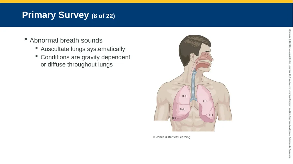
Primary Survey (8 of 22) Abnormal breath sounds
Auscultate lungs systematically
Conditions are gravity dependent or diffuse throughout lungs © Jones & Bartlett Learning.<br>
Auscultate lungs systematically
Conditions are gravity dependent or diffuse throughout lungs © Jones & Bartlett Learning.<br>
42
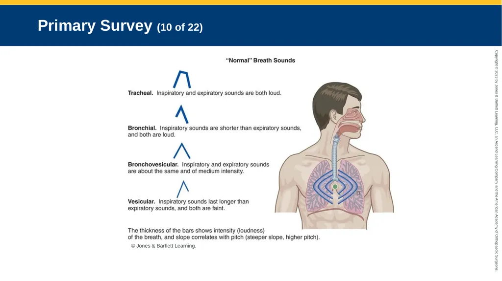
Primary Survey (9 of 22) Abnormal breath sounds (cont’d)
Breath sounds created by airflow in large airways
Tracheal
Bronchial
Bronchovesicular
Vesicular<br>
Breath sounds created by airflow in large airways
Tracheal
Bronchial
Bronchovesicular
Vesicular<br>
43
Primary Survey (10 of 22) © Jones & Bartlett Learning.<br>
44
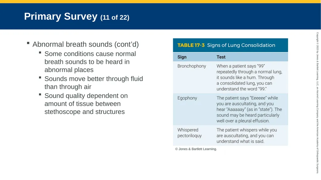
Primary Survey (11 of 22) Abnormal breath sounds (cont’d)
Some conditions cause normal breath sounds to be heard in abnormal places
Sounds move better through fluid than through air
Sound quality dependent on amount of tissue between stethoscope and structures © Jones & Bartlett Learning.<br>
Some conditions cause normal breath sounds to be heard in abnormal places
Sounds move better through fluid than through air
Sound quality dependent on amount of tissue between stethoscope and structures © Jones & Bartlett Learning.<br>
45
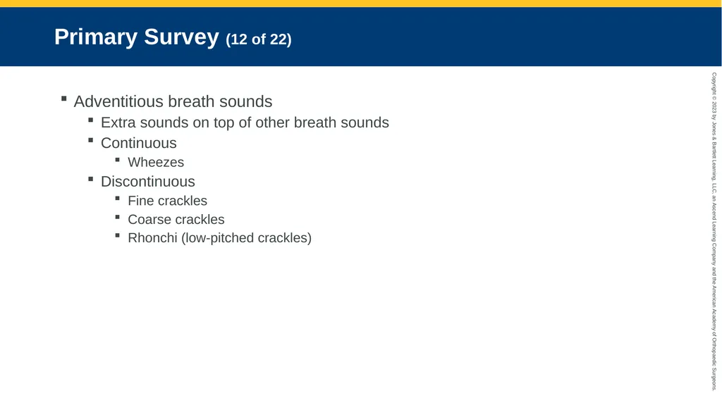
Primary Survey (12 of 22) Adventitious breath sounds
Extra sounds on top of other breath sounds
Continuous
Wheezes
Discontinuous
Fine crackles
Coarse crackles
Rhonchi (low-pitched crackles)<br>
Extra sounds on top of other breath sounds
Continuous
Wheezes
Discontinuous
Fine crackles
Coarse crackles
Rhonchi (low-pitched crackles)<br>
46
Primary Survey (13 of 22) Abnormal breath sounds (cont’d)
Audible sounds include:
Stridor
Grunting
Death rattle
Most ominous: no sounds<br>
Audible sounds include:
Stridor
Grunting
Death rattle
Most ominous: no sounds<br>
47
Primary Survey (14 of 22) Abnormal breath sounds (cont’d)
Noisy breathing
Snoring
Gurgling
Stridor
Quiet breathing
Hyperventilation
Shock
Acidosis<br>
Noisy breathing
Snoring
Gurgling
Stridor
Quiet breathing
Hyperventilation
Shock
Acidosis<br>
48
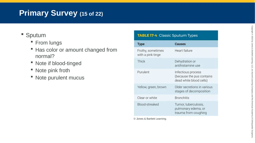
Primary Survey (15 of 22) Sputum
From lungs
Has color or amount changed from normal?
Note if blood-tinged
Note pink froth
Note purulent mucus © Jones & Bartlett Learning.<br>
From lungs
Has color or amount changed from normal?
Note if blood-tinged
Note pink froth
Note purulent mucus © Jones & Bartlett Learning.<br>
49
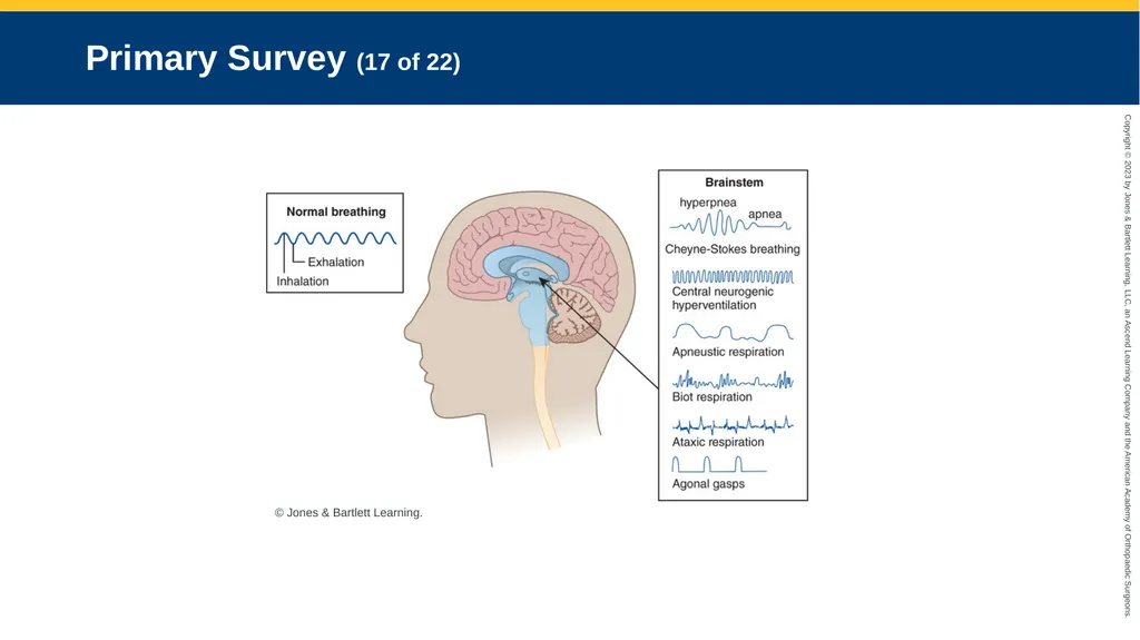
Primary Survey (16 of 22) Abnormal breathing patterns
May indicate neurologic insults
Brain trauma or any disturbance
Depress respiratory control centers in medulla
Damage or deprive blood flow
Most respiratory centers are in and around brainstem
Apneustic breathing
Biot respirations
Cheyne-Stoke respiration<br>
May indicate neurologic insults
Brain trauma or any disturbance
Depress respiratory control centers in medulla
Damage or deprive blood flow
Most respiratory centers are in and around brainstem
Apneustic breathing
Biot respirations
Cheyne-Stoke respiration<br>
50
Primary Survey (17 of 22) © Jones & Bartlett Learning.<br>
51
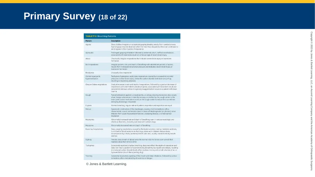
Primary Survey (18 of 22) [ © Jones & Bartlett Learning.<br>
52
Primary Survey (19 of 22) Injury to spinal cord and polio
May disable respiratory muscles from functioning normally
Tidal volume is shallow, and minute volume decreases
Patients often need assisted ventilation<br>
May disable respiratory muscles from functioning normally
Tidal volume is shallow, and minute volume decreases
Patients often need assisted ventilation<br>
53
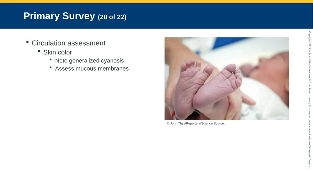
Primary Survey (20 of 22) Circulation assessment
Skin color
Note generalized cyanosis
Assess mucous membranes © John Thys/Reporters/Science Source.<br>
Skin color
Note generalized cyanosis
Assess mucous membranes © John Thys/Reporters/Science Source.<br>
54
Primary Survey (21 of 22) Circulation assessment (cont’d)
Cyanosis
Healthy hemoglobin levels are 12 to 14 g/dL.
Cyanosis begins at about 5 g/dL desaturation.
Dark brown skin
High levels of methemoglobin
More apparent in venous blood
Pallor
Caused by a blood flow reduction to small vessels
Possible sources: Shock, hypoxia, frostbite, lack of sun, anemia, catecholamine release<br>
Cyanosis
Healthy hemoglobin levels are 12 to 14 g/dL.
Cyanosis begins at about 5 g/dL desaturation.
Dark brown skin
High levels of methemoglobin
More apparent in venous blood
Pallor
Caused by a blood flow reduction to small vessels
Possible sources: Shock, hypoxia, frostbite, lack of sun, anemia, catecholamine release<br>
55
Primary Survey (22 of 22) Circulation assessment (cont’d)
Check for dehydration:
Dry, cracked lips
Dry, furrowed tongue
Dry, sunken eyes<br>
Check for dehydration:
Dry, cracked lips
Dry, furrowed tongue
Dry, sunken eyes<br>
56
Primary Survey: Transport Decisions Usually transported to closest hospital
Patient experiencing renal failure
Consider facility that can provide emergency dialysis
Consider taking patient to his or her preferred facility
If distance allows
Easy access to records
Access to previous physician<br>
Patient experiencing renal failure
Consider facility that can provide emergency dialysis
Consider taking patient to his or her preferred facility
If distance allows
Easy access to records
Access to previous physician<br>
57
History Taking<br>
58
Investigate Chief Complaint (1 of 2) Have patients explain what they are feeling in their own words.
Common complaints
Increased cough
Change in amount or color of sputum
Fever
Wheezing
Dyspnea
Chest pain<br>
Common complaints
Increased cough
Change in amount or color of sputum
Fever
Wheezing
Dyspnea
Chest pain<br>
59
Investigate Chief Complaint (2 of 2) Patient may know problem
Asthma with fever
Nondelivery of medication
Travel-related conditions
Dyspnea triggers
Seasonal conditions
Noncompliance with therapy<br>
Asthma with fever
Nondelivery of medication
Travel-related conditions
Dyspnea triggers
Seasonal conditions
Noncompliance with therapy<br>
60
SAMPLE History Signs and symptoms
Allergies
Medications
Pertinent past medical history
Last oral intake
Events preceding the onset of the complaint<br>
Allergies
Medications
Pertinent past medical history
Last oral intake
Events preceding the onset of the complaint<br>
61
Secondary Assessment<br>
62
Physical Examination (1 of 5) Neurologic assessment
Note level of consciousness
Lungs not functioning correctly
Oxygen may not be delivered
Carbon dioxide may not be removed<br>
Note level of consciousness
Lungs not functioning correctly
Oxygen may not be delivered
Carbon dioxide may not be removed<br>
63
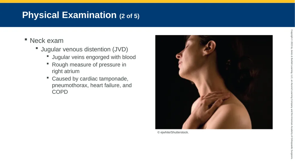
Physical Examination (2 of 5) Neck exam
Jugular venous distention (JVD)
Jugular veins engorged with blood
Rough measure of pressure in right atrium
Caused by cardiac tamponade, pneumothorax, heart failure, and COPD © ejwhite/Shutterstock.<br>
Jugular venous distention (JVD)
Jugular veins engorged with blood
Rough measure of pressure in right atrium
Caused by cardiac tamponade, pneumothorax, heart failure, and COPD © ejwhite/Shutterstock.<br>
64
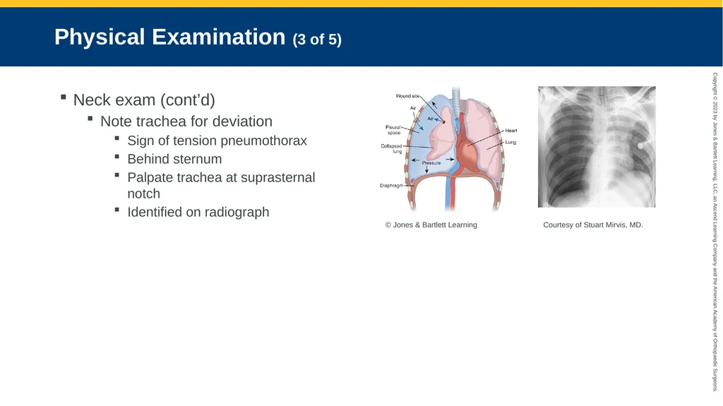
Physical Examination (3 of 5) Neck exam (cont’d)
Note trachea for deviation
Sign of tension pneumothorax
Behind sternum
Palpate trachea at suprasternal notch
Identified on radiograph © Jones & Bartlett Learning Courtesy of Stuart Mirvis, MD.<br>
Note trachea for deviation
Sign of tension pneumothorax
Behind sternum
Palpate trachea at suprasternal notch
Identified on radiograph © Jones & Bartlett Learning Courtesy of Stuart Mirvis, MD.<br>
65
Physical Examination (4 of 5) Chest and abdominal exam
Pressing on liver when patient is in respiratory distress and semi-Fowler’s position will cause jugular veins to bulge.
Feel for vibrations in the chest as patient breathes.
Chest or abdominal trauma can cause respiratory distress.<br>
Pressing on liver when patient is in respiratory distress and semi-Fowler’s position will cause jugular veins to bulge.
Feel for vibrations in the chest as patient breathes.
Chest or abdominal trauma can cause respiratory distress.<br>
66
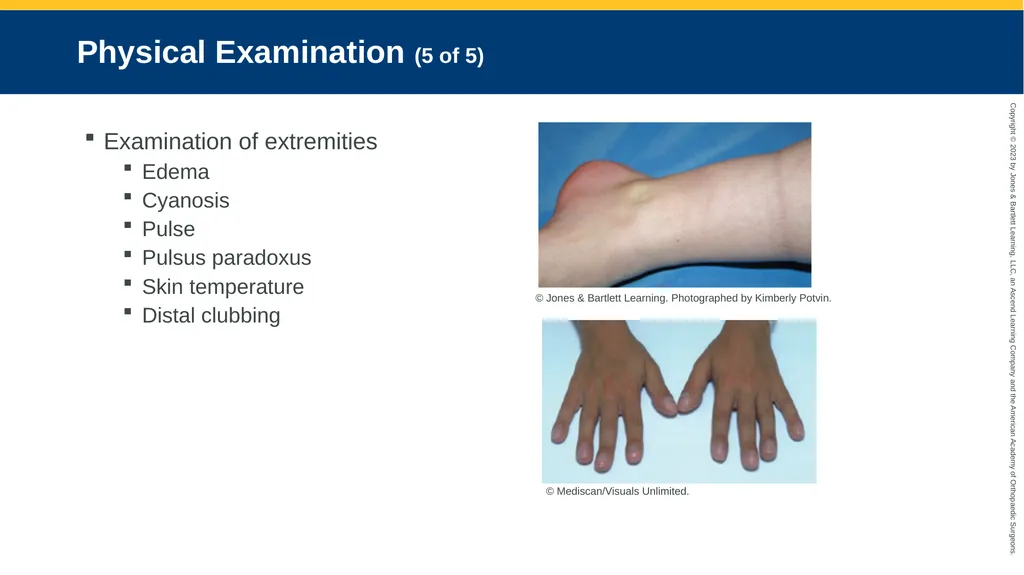
Physical Examination (5 of 5) Examination of extremities
Edema
Cyanosis
Pulse
Pulsus paradoxus
Skin temperature
Distal clubbing © Jones & Bartlett Learning. Photographed by Kimberly Potvin. © Mediscan/Visuals Unlimited.<br>
Edema
Cyanosis
Pulse
Pulsus paradoxus
Skin temperature
Distal clubbing © Jones & Bartlett Learning. Photographed by Kimberly Potvin. © Mediscan/Visuals Unlimited.<br>
67
Vital Signs and Monitoring Devices (1 of 5) Vital signs
Patients under stress
Tachycardia
Hypertension
Ominous signs:
Bradycardia
Hypotension
Falling respiratory rates<br>
Patients under stress
Tachycardia
Hypertension
Ominous signs:
Bradycardia
Hypotension
Falling respiratory rates<br>
68
Vital Signs and Monitoring Devices (2 of 5) Stethoscope
Frequent use
Requires regular cleaning and maintenance
Buy best within budget<br>
Frequent use
Requires regular cleaning and maintenance
Buy best within budget<br>
69
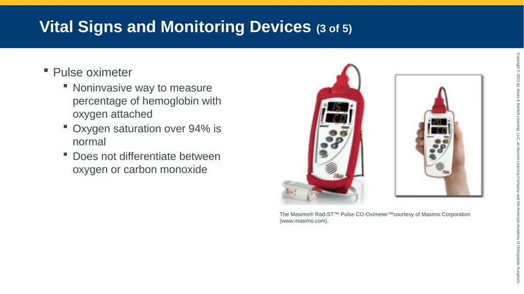
Vital Signs and Monitoring Devices (3 of 5) Pulse oximeter
Noninvasive way to measure percentage of hemoglobin with oxygen attached
Oxygen saturation over 94% is normal
Does not differentiate between oxygen or carbon monoxide The Masimo® Rad-ST™ Pulse CO-Oximeter™courtesy of Masimo Corporation (www.masimo.com).<br>
Noninvasive way to measure percentage of hemoglobin with oxygen attached
Oxygen saturation over 94% is normal
Does not differentiate between oxygen or carbon monoxide The Masimo® Rad-ST™ Pulse CO-Oximeter™courtesy of Masimo Corporation (www.masimo.com).<br>
70
Vital Signs and Monitoring Devices (4 of 5) Pulse oximeter (cont’d)
Oxygen saturation should match patient’s palpated heart rate.
If hemoglobin level is low, pulse oximetry result will be high.
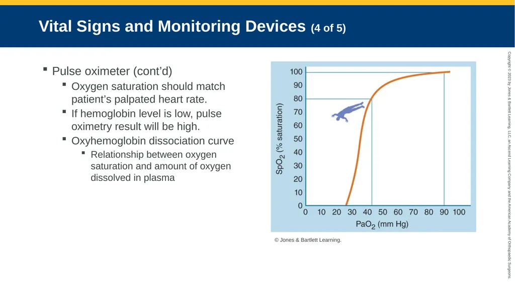
Oxyhemoglobin dissociation curve
Relationship between oxygen saturation and amount of oxygen dissolved in plasma © Jones & Bartlett Learning.<br>
Oxygen saturation should match patient’s palpated heart rate.
If hemoglobin level is low, pulse oximetry result will be high.
Oxyhemoglobin dissociation curve
Relationship between oxygen saturation and amount of oxygen dissolved in plasma © Jones & Bartlett Learning.<br>
71
Vital Signs and Monitoring Devices (5 of 5) End-tidal carbon dioxide monitor
Peak expiratory flow
Maximum rate at which patient can expel air
Normal values: 350 to 700 L/min
Varies by age, sex, and height
Inadequate level: 150 L/min<br>
Peak expiratory flow
Maximum rate at which patient can expel air
Normal values: 350 to 700 L/min
Varies by age, sex, and height
Inadequate level: 150 L/min<br>
72
Reassessment Report changes in patient’s LOC or increased difficulty breathing.
Contact medical control before assisting with prescribed medications.
Document changes and orders from medical control.<br>
Contact medical control before assisting with prescribed medications.
Document changes and orders from medical control.<br>
73
Emergency Medical Care<br>
74
Emergency Medical Care Provide supportive care.
Administer supplemental oxygen.
Provide monitoring and transport.<br>
Administer supplemental oxygen.
Provide monitoring and transport.<br>
75
Perform Standard Interventions Oxygen (keep saturations above 94%)
IV line
Psychological support
Position of comfort<br>
IV line
Psychological support
Position of comfort<br>
76
Decrease the Work of Breathing (1 of 2) Muscles work harder during respiratory distress.
Substantial energy required to compensate for respiratory distress
Require more oxygen and ventilation
Patient cannot eat or drink normally
May fatigue to point of decompensation<br>
Substantial energy required to compensate for respiratory distress
Require more oxygen and ventilation
Patient cannot eat or drink normally
May fatigue to point of decompensation<br>
77
Decrease the Work of Breathing (2 of 2) Help patient sit up.
Remove restrictive clothing.
Do not make patient walk.
Relieve gastric distention.
Do not bind chest or have patient lie on unaffected lung.<br>
Remove restrictive clothing.
Do not make patient walk.
Relieve gastric distention.
Do not bind chest or have patient lie on unaffected lung.<br>
78
Provide Supplemental Oxygen Administer in effective concentrations
If necessary
Reassess, then adjust as needed
Pulse oximetry is good guide to oxygenation
Concentrations higher than 50%
Use only with hypoxia that does not respond to lower concentrations.
Most patients with good oxygen saturation (at least 94%) do not benefit from supplemental oxygen.<br>
If necessary
Reassess, then adjust as needed
Pulse oximetry is good guide to oxygenation
Concentrations higher than 50%
Use only with hypoxia that does not respond to lower concentrations.
Most patients with good oxygen saturation (at least 94%) do not benefit from supplemental oxygen.<br>
79
Administer a Bronchodilator (1 of 11) Those without bronchospasms benefit only slightly
Bronchodilators ineffective in cases of:
Pneumonia
Pulmonary edema
Heart disease<br>
Bronchodilators ineffective in cases of:
Pneumonia
Pulmonary edema
Heart disease<br>
80
Administer a Bronchodilator (2 of 11) Fast-acting bronchodilators
Most stimulate beta-2 receptors in lung
Ipratropium now available as aerosol or inhaler
Sometimes prescribed with ipratropium in premixed “cocktail”
Improve quality of life<br>
Most stimulate beta-2 receptors in lung
Ipratropium now available as aerosol or inhaler
Sometimes prescribed with ipratropium in premixed “cocktail”
Improve quality of life<br>
81
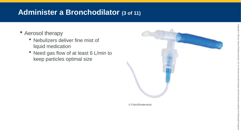
Administer a Bronchodilator (3 of 11) Aerosol therapy
Nebulizers deliver fine mist of liquid medication
Need gas flow of at least 6 L/min to keep particles optimal size © Chas/Shutterstock.<br>
Nebulizers deliver fine mist of liquid medication
Need gas flow of at least 6 L/min to keep particles optimal size © Chas/Shutterstock.<br>
82
Administer a Bronchodilator (4 of 11) Nebulizer can be attached to:
A mouthpiece
A face mask
A tracheostomy collar
Nebulizer can also be held in front of patient’s face (blow-by technique)<br>
A mouthpiece
A face mask
A tracheostomy collar
Nebulizer can also be held in front of patient’s face (blow-by technique)<br>
83
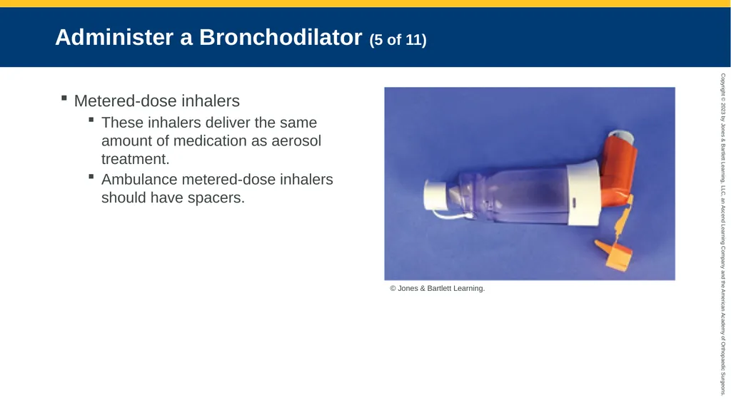
Administer a Bronchodilator (5 of 11) Metered-dose inhalers
These inhalers deliver the same amount of medication as aerosol treatment.
Ambulance metered-dose inhalers should have spacers. © Jones & Bartlett Learning.<br>
These inhalers deliver the same amount of medication as aerosol treatment.
Ambulance metered-dose inhalers should have spacers. © Jones & Bartlett Learning.<br>
84
Administer a Bronchodilator (6 of 11) To avoid common errors with metered-dose inhalers:
Inhale deeply at discharge
Suck medication out of the bottom
Flow should be smooth and low pressure
Inhale deeply; hold breath for a few seconds
Make sure inhaler contains medication
Keep spacer and canister holder clean
Rinse mouth after using a corticosteroid inhaler<br>
Inhale deeply at discharge
Suck medication out of the bottom
Flow should be smooth and low pressure
Inhale deeply; hold breath for a few seconds
Make sure inhaler contains medication
Keep spacer and canister holder clean
Rinse mouth after using a corticosteroid inhaler<br>
85
Administer a Bronchodilator (7 of 11) Failure of a metered-dose inhaler
Usually user error
Patient cannot move enough air into lungs
Patient may not realize inhaler is empty
Patient may inhale at wrong time
Limited by spacers<br>
Usually user error
Patient cannot move enough air into lungs
Patient may not realize inhaler is empty
Patient may inhale at wrong time
Limited by spacers<br>
86
Administer a Bronchodilator (8 of 11) Dry powder inhalers
Fine powder
Medication may be dispensed by means of plastic disk
Other devices require patient to insert a capsule of powdered medication
Rarely used during emergency care
Convenient and easy to use
Expensive<br>
Fine powder
Medication may be dispensed by means of plastic disk
Other devices require patient to insert a capsule of powdered medication
Rarely used during emergency care
Convenient and easy to use
Expensive<br>
87
Administer a Bronchodilator (9 of 11) Leukotriene modifiers
Block bronchoconstricting chemicals (leukotrienes)
Help with allergic reactions
Electrolytes
IV Magnesium for severe asthma attacks
Can cause hypotension<br>
Block bronchoconstricting chemicals (leukotrienes)
Help with allergic reactions
Electrolytes
IV Magnesium for severe asthma attacks
Can cause hypotension<br>
88
Administer a Bronchodilator (10 of 11) Corticosteroids
Reduce bronchial edema
Adverse effects
Cushing syndrome
Rapid change in blood glucose levels
Blunt the immune system
Must be discontinued slowly<br>
Reduce bronchial edema
Adverse effects
Cushing syndrome
Rapid change in blood glucose levels
Blunt the immune system
Must be discontinued slowly<br>
89
Administer a Bronchodilator (11 of 11) Inhaled corticosteroids
Not the same adverse effects as oral versions
IV corticosteroids
No apparent negative long-term effects with single bolus
Methylprednisolone and hydrocortisone IV boluses
Acute asthma attacks
Acute COPD exacerbations<br>
Not the same adverse effects as oral versions
IV corticosteroids
No apparent negative long-term effects with single bolus
Methylprednisolone and hydrocortisone IV boluses
Acute asthma attacks
Acute COPD exacerbations<br>
90
Administer a Vasodilator Sequester more fluid in venous circulation and decrease preload
Nitrates used if patient:
Has adequate blood pressure
Does not take a phosphodiesterase inhibitor
Morphine sulfate not likely to increase venous capacity<br>
Nitrates used if patient:
Has adequate blood pressure
Does not take a phosphodiesterase inhibitor
Morphine sulfate not likely to increase venous capacity<br>
91
Restore Fluid Balance Common to give fluid bolus to dehydrated, younger patients
Elderly patients or patients with cardiac dysfunction could develop pulmonary edema.
Assess breath sounds before and after.
Hydrating patients with pneumonia may cause pneumonia to spread.
Hydration only in medical facility<br>
Elderly patients or patients with cardiac dysfunction could develop pulmonary edema.
Assess breath sounds before and after.
Hydrating patients with pneumonia may cause pneumonia to spread.
Hydration only in medical facility<br>
92
Administer a Diuretic (1 of 2) Patients with heart failure
Helps reduce blood pressure
Maintains fluid balance
Helps remove excess fluid from circulation
Keeps it out of lungs
Patients with pulmonary edema<br>
Helps reduce blood pressure
Maintains fluid balance
Helps remove excess fluid from circulation
Keeps it out of lungs
Patients with pulmonary edema<br>
93
Administer a Diuretic (2 of 2) May cause potassium loss
Regular supplements required
May lead to cardiac dysrhythmias and chronic muscle cramping
Not for patients with pneumonia or dehydration
Higher doses required for renal failure
Patient may not respond
Ineffective<br>
Regular supplements required
May lead to cardiac dysrhythmias and chronic muscle cramping
Not for patients with pneumonia or dehydration
Higher doses required for renal failure
Patient may not respond
Ineffective<br>
94
Support or Assist Ventilation (1 of 7) If patient becomes fatigued:
Breathing may need more aggressive support
CPAP and BPAP may preclude intubation
Patients may require bag-mask ventilation<br>
Breathing may need more aggressive support
CPAP and BPAP may preclude intubation
Patients may require bag-mask ventilation<br>
95
Support or Assist Ventilation (2 of 7) Continuous positive airway pressure
Used to treat:
Obstructive sleep apnea
Respiratory failure
Patients with obstructive sleep apnea wear CPAP unit to maintain airway while they sleep
Nasal pillows
Nasal mask
Face mask like bag-mask ventilation unit
Mask covering entire face<br>
Used to treat:
Obstructive sleep apnea
Respiratory failure
Patients with obstructive sleep apnea wear CPAP unit to maintain airway while they sleep
Nasal pillows
Nasal mask
Face mask like bag-mask ventilation unit
Mask covering entire face<br>
96
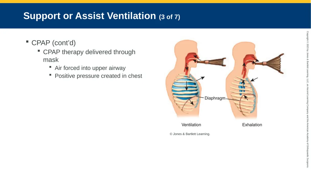
Support or Assist Ventilation (3 of 7) CPAP (cont’d)
CPAP therapy delivered through mask
Air forced into upper airway
Positive pressure created in chest © Jones & Bartlett Learning.<br>
CPAP therapy delivered through mask
Air forced into upper airway
Positive pressure created in chest © Jones & Bartlett Learning.<br>
97
Support or Assist Ventilation (4 of 7) Bag-mask ventilation:
Produces positive pressure in chest
Pressure that is too high may:
Cause tension pneumothorax
Cause subcutaneous air
Block venous returns
New guidelines emphasize:
Lower ventilation rates
Smaller volumes
Lower pressures<br>
Produces positive pressure in chest
Pressure that is too high may:
Cause tension pneumothorax
Cause subcutaneous air
Block venous returns
New guidelines emphasize:
Lower ventilation rates
Smaller volumes
Lower pressures<br>
98
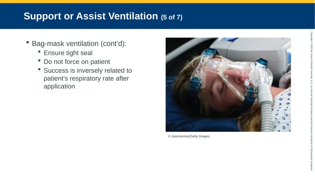
Support or Assist Ventilation (5 of 7) Bag-mask ventilation (cont’d):
Ensure tight seal
Do not force on patient
Success is inversely related to patient’s respiratory rate after application © Juanmonino/Getty Images.<br>
Ensure tight seal
Do not force on patient
Success is inversely related to patient’s respiratory rate after application © Juanmonino/Getty Images.<br>
99
Support or Assist Ventilation (6 of 7) Bilevel positive airway pressure (BPAP)
One pressure on inspiration and different pressure during exhalation
More like normal breathing
More complex and expensive<br>
One pressure on inspiration and different pressure during exhalation
More like normal breathing
More complex and expensive<br>
100
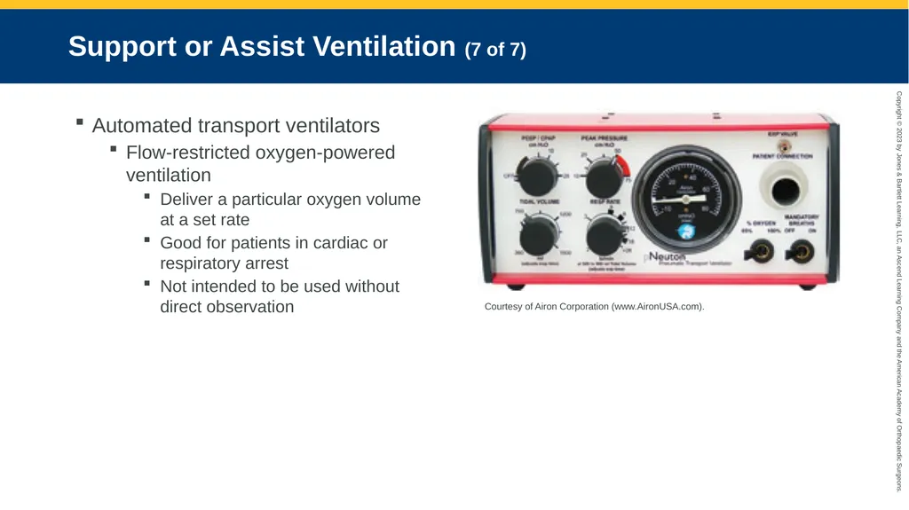
Support or Assist Ventilation (7 of 7) Automated transport ventilators
Flow-restricted oxygen-powered ventilation
Deliver a particular oxygen volume at a set rate
Good for patients in cardiac or respiratory arrest
Not intended to be used without direct observation Courtesy of Airon Corporation (www.AironUSA.com).<br>
Flow-restricted oxygen-powered ventilation
Deliver a particular oxygen volume at a set rate
Good for patients in cardiac or respiratory arrest
Not intended to be used without direct observation Courtesy of Airon Corporation (www.AironUSA.com).<br>
101
Intubating the Adult Patient (1 of 2) Intubation is last option for patients with severe asthma.
Ventilate patients before cardiac arrest.
Patients who are severely intoxicated or have had a stroke may have no gag reflex.<br>
Ventilate patients before cardiac arrest.
Patients who are severely intoxicated or have had a stroke may have no gag reflex.<br>
102
Intubating the Adult Patient (2 of 2) With diabetes or overdose:
An ampule of 50% dextrose or naloxone may change need for intubation
Bag-mask ventilation for a few minutes to monitor effects
Slow administration<br>
An ampule of 50% dextrose or naloxone may change need for intubation
Bag-mask ventilation for a few minutes to monitor effects
Slow administration<br>
103
Inject a Beta-Adrenergic Receptor Agonist Subcutaneously Use if inhalation techniques are ineffective
May cause more tachycardia and hypertension
Be careful using in elderly patients<br>
May cause more tachycardia and hypertension
Be careful using in elderly patients<br>
104
Pathophysiology, Assessment, and Management of Obstructive Upper Airway Diseases<br>
105
Anatomic Obstruction: Pathophysiology Tongue is most common cause of airway obstruction if patient is unresponsive
Tongue obstructions lead to deaths
Trauma patients
Insulin shock
Seizure
Intoxicated<br>
Tongue obstructions lead to deaths
Trauma patients
Insulin shock
Seizure
Intoxicated<br>
106
Anatomic Obstruction: Assessment Risks include:
Decreased level of consciousness
Audible signs include:
Sonorous respirations
Gurgling
Squeaking and bubbling
Stridor<br>
Decreased level of consciousness
Audible signs include:
Sonorous respirations
Gurgling
Squeaking and bubbling
Stridor<br>
107
Anatomic Obstruction: Management Laying patients head on pillow worsens problem
Obstructive sleep apnea
May be caused by excess soft tissue in airway
Can be manually displaced
Recovery position reduces risk<br>
Obstructive sleep apnea
May be caused by excess soft tissue in airway
Can be manually displaced
Recovery position reduces risk<br>
108
Inflammation Caused by Infection: Pathophysiology (1 of 2) Infections can cause upper airway swelling.
Can lead to laryngotracheobronchitis
Common cause of croup
Stridor
Hoarseness
Barking cough<br>
Can lead to laryngotracheobronchitis
Common cause of croup
Stridor
Hoarseness
Barking cough<br>
109
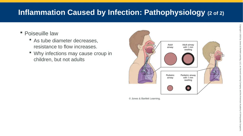
Inflammation Caused by Infection: Pathophysiology (2 of 2) Poiseuille law
As tube diameter decreases, resistance to flow increases.
Why infections may cause croup in children, but not adults © Jones & Bartlett Learning.<br>
As tube diameter decreases, resistance to flow increases.
Why infections may cause croup in children, but not adults © Jones & Bartlett Learning.<br>
110
Inflammation Caused by Infection: Assessment (1 of 2) Croup and tonsillitis
Common
Other conditions are rare thanks to immunizations
Critical emergencies when they occur
Avoid manipulating airways<br>
Common
Other conditions are rare thanks to immunizations
Critical emergencies when they occur
Avoid manipulating airways<br>
111
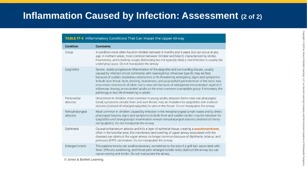
Inflammation Caused by Infection: Assessment (2 of 2) © Jones & Bartlett Learning.<br>
112
Inflammation Caused by Infection: Management Airway may be entirely obscured.
Laryngoscopy may worsen swelling
Have partner press on chest while you check for bubble stream.
ET tube at least two sizes smaller than typical
Cricothyrotomy may be necessary<br>
Laryngoscopy may worsen swelling
Have partner press on chest while you check for bubble stream.
ET tube at least two sizes smaller than typical
Cricothyrotomy may be necessary<br>
113
Aspiration Inhalation of anything other than breathable gases
Water
Blood
Vomitus
Food
Foreign bodies
Stomach contents (pneumonitis)<br>
Water
Blood
Vomitus
Food
Foreign bodies
Stomach contents (pneumonitis)<br>
114
Aspiration: Pathophysiology Patients at risk:
Tube-fed patients placed supine after large meal
Geriatric patients with impaired swallowing
Unresponsive patients
High mortality<br>
Tube-fed patients placed supine after large meal
Geriatric patients with impaired swallowing
Unresponsive patients
High mortality<br>
115
Aspiration: Assessment Determine scenario of sudden onset dyspnea
Immediately after eating?
Gastric feeding tube?
Particulate matter?<br>
Immediately after eating?
Gastric feeding tube?
Particulate matter?<br>
116
Aspiration: Management Avoid gastric distention when ventilating.
Monitor patient’s ability to protect airway.
Treat with suction and airway control.<br>
Monitor patient’s ability to protect airway.
Treat with suction and airway control.<br>
117
Pathophysiology, Assessment, and Management of Obstructive Lower Airway Diseases<br>
118
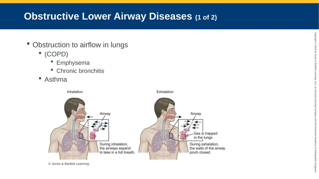
Obstructive Lower Airway Diseases (1 of 2) Obstruction to airflow in lungs
(COPD)
Emphysema
Chronic bronchitis
Asthma © Jones & Bartlett Learning.<br>
(COPD)
Emphysema
Chronic bronchitis
Asthma © Jones & Bartlett Learning.<br>
119
Obstructive Lower Airway Diseases (2 of 2) Physical findings:
Pursed lip breathing
Increased I:E ratio
Abdominal muscle use
Jugular venous distention (JVD)<br>
Pursed lip breathing
Increased I:E ratio
Abdominal muscle use
Jugular venous distention (JVD)<br>
120
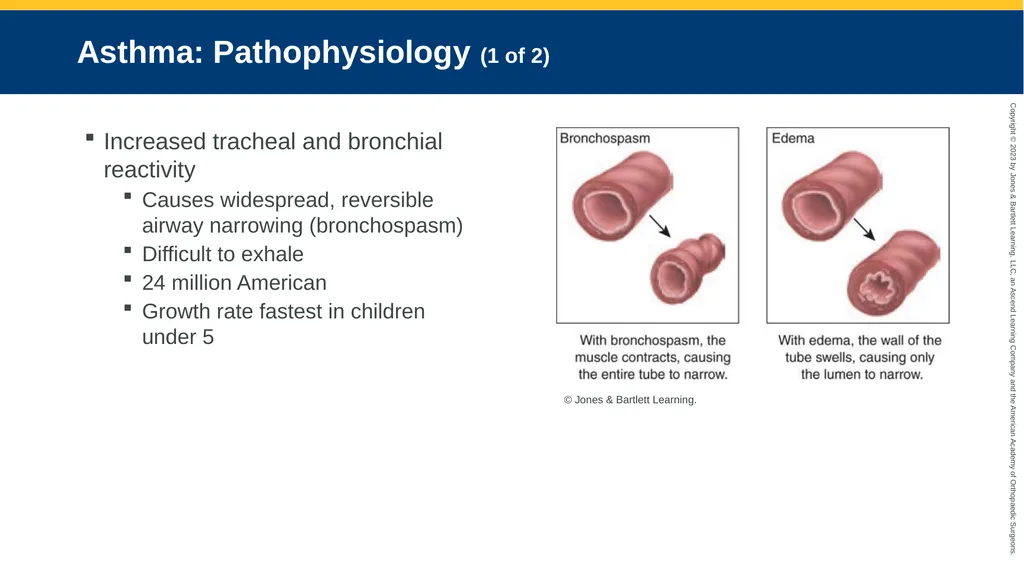
Asthma: Pathophysiology (1 of 2) Increased tracheal and bronchial reactivity
Causes widespread, reversible airway narrowing (bronchospasm)
Difficult to exhale
24 million American
Growth rate fastest in children under 5 © Jones & Bartlett Learning.<br>
Causes widespread, reversible airway narrowing (bronchospasm)
Difficult to exhale
24 million American
Growth rate fastest in children under 5 © Jones & Bartlett Learning.<br>
121
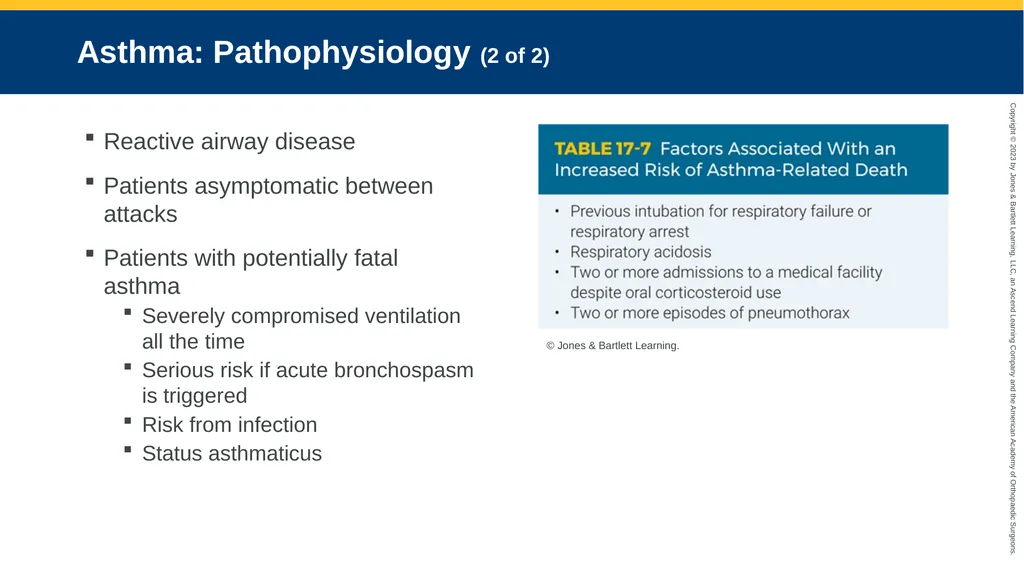
Asthma: Pathophysiology (2 of 2) Reactive airway disease
Patients asymptomatic between attacks
Patients with potentially fatal asthma
Severely compromised ventilation all the time
Serious risk if acute bronchospasm is triggered
Risk from infection
Status asthmaticus © Jones & Bartlett Learning.<br>
Patients asymptomatic between attacks
Patients with potentially fatal asthma
Severely compromised ventilation all the time
Serious risk if acute bronchospasm is triggered
Risk from infection
Status asthmaticus © Jones & Bartlett Learning.<br>
122
Asthma: Assessment (1 of 3) Symptoms return after inhaler use
Possible infection
Must remove/mitigate trigger
Patient in status asthmaticus
Struggling to move air through obstructed airways
Prominent use of accessory muscles
Hyperinflated chest
Inaudible breath sounds
Exhausted, severely acidotic, and dehydrated<br>
Possible infection
Must remove/mitigate trigger
Patient in status asthmaticus
Struggling to move air through obstructed airways
Prominent use of accessory muscles
Hyperinflated chest
Inaudible breath sounds
Exhausted, severely acidotic, and dehydrated<br>
123
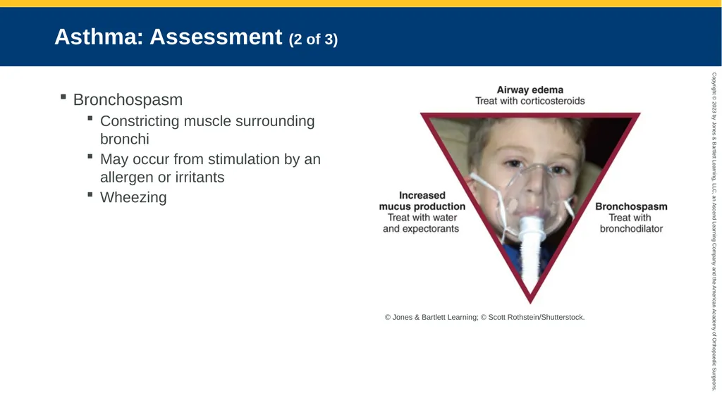
Asthma: Assessment (2 of 3) Bronchospasm
Constricting muscle surrounding bronchi
May occur from stimulation by an allergen or irritants
Wheezing © Jones & Bartlett Learning; © Scott Rothstein/Shutterstock.<br>
Constricting muscle surrounding bronchi
May occur from stimulation by an allergen or irritants
Wheezing © Jones & Bartlett Learning; © Scott Rothstein/Shutterstock.<br>
124
Asthma: Assessment (3 of 3) Bronchial edema
Swelling of bronchi and bronchioles
Turbulent airflow, wheezing, and air trapping
Bronchodilator medications do not work
Increased mucus production
Thick secretions contribute to air trapping
Dehydration makes secretions thicker<br>
Swelling of bronchi and bronchioles
Turbulent airflow, wheezing, and air trapping
Bronchodilator medications do not work
Increased mucus production
Thick secretions contribute to air trapping
Dehydration makes secretions thicker<br>
125
Asthma: Management (1 of 2) Bronchospasm
Nebulized bronchodilator medication
Magnesium sulfate
Epinephrine
Bronchial edema
Corticosteroids
Several hours to take effect
Excessive mucus secretion
Improve hydration
Mucolytic agents<br>
Nebulized bronchodilator medication
Magnesium sulfate
Epinephrine
Bronchial edema
Corticosteroids
Several hours to take effect
Excessive mucus secretion
Improve hydration
Mucolytic agents<br>
126
Asthma: Management (2 of 2) Transport considerations
Infection or continuous exposure to a trigger
No improvement in peak flow: consider corticosteroids
Undernourished or dehydrated: consider IV fluids
Advanced life support more than a few minutes away: consider transport to nearest ED<br>
Infection or continuous exposure to a trigger
No improvement in peak flow: consider corticosteroids
Undernourished or dehydrated: consider IV fluids
Advanced life support more than a few minutes away: consider transport to nearest ED<br>
127
COPD: Emphysema and Chronic Bronchitis Pathophysiology (1 of 2) Emphysema damages or destroys terminal bronchiole structures.
Restrictive lung diseases
Caused by trauma and diseases of bones/muscles
Impair ability to move air
Alveoli merge into large blebs (bullae)<br>
Restrictive lung diseases
Caused by trauma and diseases of bones/muscles
Impair ability to move air
Alveoli merge into large blebs (bullae)<br>
128
COPD: Emphysema and Chronic Bronchitis Pathophysiology (2 of 2) Chronic bronchitis
Sputum production most days of the month for 3 or more months of the year for more than 2 years
Excessive mucus production in bronchial tree
Accompanied by chronic or recurrent productive cough
Abnormal blood gas levels
Heavy smokers or overweight
Bluish complexion<br>
Sputum production most days of the month for 3 or more months of the year for more than 2 years
Excessive mucus production in bronchial tree
Accompanied by chronic or recurrent productive cough
Abnormal blood gas levels
Heavy smokers or overweight
Bluish complexion<br>
129
COPD: Emphysema and Chronic Bronchitis Assessment (1 of 8) Emphysema
Barrel chest from chronic lung hyperinflation
Tachypnea
Use muscle mass for energy to breathe<br>
Barrel chest from chronic lung hyperinflation
Tachypnea
Use muscle mass for energy to breathe<br>
130
COPD: Emphysema and Chronic Bronchitis Assessment (2 of 8) Causes of diffuse wheezing:
Left-sided heart failure (cardiac asthma)
Smoke inhalation
Chronic bronchitis
Acute pulmonary embolism
Cause of localized wheezing:
Obstruction from foreign body or tumor<br>
Left-sided heart failure (cardiac asthma)
Smoke inhalation
Chronic bronchitis
Acute pulmonary embolism
Cause of localized wheezing:
Obstruction from foreign body or tumor<br>
131
COPD: Emphysema and Chronic Bronchitis Assessment (3 of 8) COPD with pneumonia
Lung infection
Check for:
Fever
Change in sputum
Other infection signs
Breath sounds consistent with pneumonia<br>
Lung infection
Check for:
Fever
Change in sputum
Other infection signs
Breath sounds consistent with pneumonia<br>
132
COPD: Emphysema and Chronic Bronchitis Assessment (4 of 8) COPD with right-sided heart failure
Look for:
Peripheral edema
Jugular venous distention with hepatojugular reflux
End inspiratory crackles
Progressive increase in dyspnea
Greater-than-usual fluid intake
Improper use of diuretics<br>
Look for:
Peripheral edema
Jugular venous distention with hepatojugular reflux
End inspiratory crackles
Progressive increase in dyspnea
Greater-than-usual fluid intake
Improper use of diuretics<br>
133
COPD: Emphysema and Chronic Bronchitis Assessment (5 of 8) COPD with left-sided heart failure
Caused by any abrupt left ventricular dysfunction
Initial impression of COPD should not preclude swift identification of acute myocardial infarction.<br>
Caused by any abrupt left ventricular dysfunction
Initial impression of COPD should not preclude swift identification of acute myocardial infarction.<br>
134
COPD: Emphysema and Chronic Bronchitis Assessment (6 of 8) Acute exacerbation of COPD
Sudden decompensation with no co-pathologic conditions
Often from environmental change or inhalation of trigger substances<br>
Sudden decompensation with no co-pathologic conditions
Often from environmental change or inhalation of trigger substances<br>
135
COPD: Emphysema and Chronic Bronchitis Assessment (7 of 8) End-stage chronic COPD
Lungs no longer support oxygenation and ventilation
It is difficult to tell whether situation can be resolved
Secure documentation of patient’s wishes
Follow local protocol or contact medical control<br>
Lungs no longer support oxygenation and ventilation
It is difficult to tell whether situation can be resolved
Secure documentation of patient’s wishes
Follow local protocol or contact medical control<br>
136
COPD: Emphysema and Chronic Bronchitis Assessment (8 of 8) COPD and trauma
COPD lessens patient’s ability to tolerate trauma
Monitor closely
“Normal” oxygen saturation might be less than 90%
Saturation of 98% is unrealistic<br>
COPD lessens patient’s ability to tolerate trauma
Monitor closely
“Normal” oxygen saturation might be less than 90%
Saturation of 98% is unrealistic<br>
137
COPD: Emphysema and Chronic Bronchitis Management (1 of 3) Immediate help can help improve immediate distress.
Determine what caused situation to worsen.
Hypoxic drive
When breathing stimulus comes from decrease in PaO2 rather than increase in PaO2
Affects small percentage during end stage of disease process
Must decide whether to administer oxygen<br>
Determine what caused situation to worsen.
Hypoxic drive
When breathing stimulus comes from decrease in PaO2 rather than increase in PaO2
Affects small percentage during end stage of disease process
Must decide whether to administer oxygen<br>
138
COPD: Emphysema and Chronic Bronchitis Management (2 of 3) Hypoxic drive (cont’d)
Impossible to tell which patients breathe because of hypoxic drive
Verbal and physical stimulation to encourage breathing
Skin appearance may remain perfused if patient becomes apneic.
Provide artificial ventilation and consider intubation if patient becomes apneic.
Oxygen saturation values are less useful in patients with COPD.<br>
Impossible to tell which patients breathe because of hypoxic drive
Verbal and physical stimulation to encourage breathing
Skin appearance may remain perfused if patient becomes apneic.
Provide artificial ventilation and consider intubation if patient becomes apneic.
Oxygen saturation values are less useful in patients with COPD.<br>
139
COPD: Emphysema and Chronic Bronchitis Management (3 of 3) Auto-PEEP
Allow complete exhalation before next breath during ventilation.
Otherwise, pressure in thorax continues to rise (auto-PEEP)
Necessary restraint
Patients should be ventilated at four to six breaths/min.<br>
Allow complete exhalation before next breath during ventilation.
Otherwise, pressure in thorax continues to rise (auto-PEEP)
Necessary restraint
Patients should be ventilated at four to six breaths/min.<br>
140
Pathophysiology, Assessment, and Management of Common Respiratory Conditions<br>
141
Pulmonary Infections: Pathophysiology (1 of 3) Infections caused by:
Bacteria
Viruses
Fungi
Protozoa
Other organisms<br>
Bacteria
Viruses
Fungi
Protozoa
Other organisms<br>
142
Pulmonary Infections: Pathophysiology (2 of 3) Infectious diseases cause:
Swelling of the respiratory tissues
Increase in mucus production
Production of pus
Resistance to airflow increases as airway diameter is narrowed (Poiseuille law).
Alveoli can become nonfunctional if filled with pus.<br>
Swelling of the respiratory tissues
Increase in mucus production
Production of pus
Resistance to airflow increases as airway diameter is narrowed (Poiseuille law).
Alveoli can become nonfunctional if filled with pus.<br>
143
Pulmonary Infections: Pathophysiology (3 of 3) At greater risk of pneumonia:
Older people
People with chronic illnesses
Smokers
Anyone not ventilating efficiently
Those with excessive secretions
Immunocompromised people<br>
Older people
People with chronic illnesses
Smokers
Anyone not ventilating efficiently
Those with excessive secretions
Immunocompromised people<br>
144
Pulmonary Infections: Assessment (1 of 3) Patients report:
Several hours to days of weakness
Productive cough
Fever
Chest pains worsened by cough<br>
Several hours to days of weakness
Productive cough
Fever
Chest pains worsened by cough<br>
145
Pulmonary Infections: Assessment (2 of 3) May start abruptly or gradually
During physical examination, patient may:
Look grievously ill
May not be coughing
Present with crackles
Have increased tactile fremitus and sputum production
Have bronchial or bronchovesicular breath sounds over areas of consolidation<br>
During physical examination, patient may:
Look grievously ill
May not be coughing
Present with crackles
Have increased tactile fremitus and sputum production
Have bronchial or bronchovesicular breath sounds over areas of consolidation<br>
146
Pulmonary Infections: Assessment (3 of 3) Pneumonia occurs in lung bases
Patients often dehydrated
Supportive care
Oxygenation
Secretion management (suctioning)
Transport to the closest facility<br>
Patients often dehydrated
Supportive care
Oxygenation
Secretion management (suctioning)
Transport to the closest facility<br>
147
Pulmonary Infections: Management Upper airway infections
Require aggressive airway management
Lower airway infections
Supportive care
Transport to facility<br>
Require aggressive airway management
Lower airway infections
Supportive care
Transport to facility<br>
148
Atelectasis: Pathophysiology (1 of 2) Disorders of alveoli
Collapse from proximal airway obstruction or external pressure
Fill with pus, blood, or fluid
Smoke or toxin damage<br>
Collapse from proximal airway obstruction or external pressure
Fill with pus, blood, or fluid
Smoke or toxin damage<br>
149
Atelectasis: Pathophysiology (2 of 2) Common for some alveoli to collapse
Helps open closed alveoli
Sighing
Coughing
Sneezing
Changing positions
When alveoli do not reopen:
Entire lung segments eventually collapse
Atelectasis increases chance of pneumonia<br>
Helps open closed alveoli
Sighing
Coughing
Sneezing
Changing positions
When alveoli do not reopen:
Entire lung segments eventually collapse
Atelectasis increases chance of pneumonia<br>
150
Atelectasis: Assessment Affected area can harbor pathogens.
Result in pneumonia
Check if patient with fever has had recent chest or abdominal surgery.
Check for abnormal sputum.<br>
Result in pneumonia
Check if patient with fever has had recent chest or abdominal surgery.
Check for abnormal sputum.<br>
151
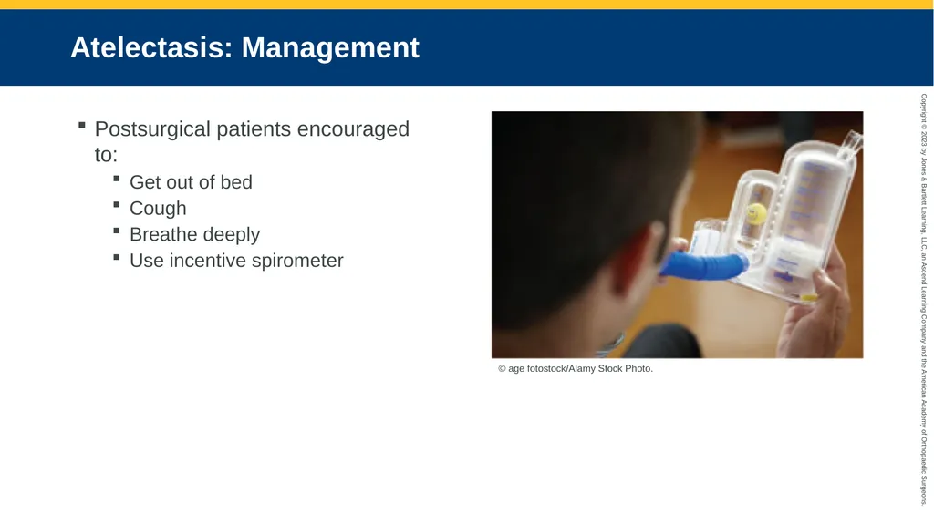
Atelectasis: Management Postsurgical patients encouraged to:
Get out of bed
Cough
Breathe deeply
Use incentive spirometer © age fotostock/Alamy Stock Photo.<br>
Get out of bed
Cough
Breathe deeply
Use incentive spirometer © age fotostock/Alamy Stock Photo.<br>
152
Cancer: Pathophysiology Lung cancer
One of most common forms of cancer
Cigarette smoking
Exposure to occupational lung hazards, secondhand smoke<br>
One of most common forms of cancer
Cigarette smoking
Exposure to occupational lung hazards, secondhand smoke<br>
153
Cancer: Assessment (1 of 2) Hemoptysis often first sign
Frequently accompanied by:
COPD
Impaired lung function
Often metastasizes in lung from other body sites<br>
Frequently accompanied by:
COPD
Impaired lung function
Often metastasizes in lung from other body sites<br>
154
Cancer: Assessment (2 of 2) Other cancers may invade lymph nodes in neck.
Radiation and chemotherapy might cause pulmonary complications.
Tumors or treatments may cause pleural effusion.<br>
Radiation and chemotherapy might cause pulmonary complications.
Tumors or treatments may cause pleural effusion.<br>
155
Cancer: Management Little prehospital treatment for pleural effusions or hemoptysis
Oxygenation and ventilation
Pain management
Sometimes called for end-of-life issues<br>
Oxygenation and ventilation
Pain management
Sometimes called for end-of-life issues<br>
156
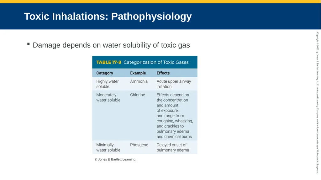
Toxic Inhalations: Pathophysiology Damage depends on water solubility of toxic gas © Jones & Bartlett Learning.<br>
157
Toxic Inhalations: Assessment (1 of 2) Highly water-soluble gases react with moist mucous membranes.
Causes upper airway swelling and irritation
Less water-soluble gases
Get deep in lower airway
More damage over time
Phosgene and nitrogen dioxide<br>
Causes upper airway swelling and irritation
Less water-soluble gases
Get deep in lower airway
More damage over time
Phosgene and nitrogen dioxide<br>
158
Toxic Inhalations: Assessment (2 of 2) Moderately water-soluble gases
Signs and symptoms between irritation and pulmonary edema
Mixing drain cleaner and chlorine bleach may produce an irritant chlorine gas.
Irritant gas-forming chemicals in industrial settings
Higher quantities
Greater concentrations<br>
Signs and symptoms between irritation and pulmonary edema
Mixing drain cleaner and chlorine bleach may produce an irritant chlorine gas.
Irritant gas-forming chemicals in industrial settings
Higher quantities
Greater concentrations<br>
159
Toxic Inhalations: Management Immediate removal from contact with gas
100% oxygen or assisted ventilation
May require aggressive airway management
Exposure to slightly water-soluble gases
Initially feel fine
Transfer to ED for observation
Patients may have acute dyspnea hours later<br>
100% oxygen or assisted ventilation
May require aggressive airway management
Exposure to slightly water-soluble gases
Initially feel fine
Transfer to ED for observation
Patients may have acute dyspnea hours later<br>
160
Pulmonary Edema: Pathophysiology Fluid buildup in lungs
Blood plasma fluid enters lung parenchyma
Compromises gas exchange
Before overt signs present
Classifications
High pressure (cardiogenic)
High permeability (noncardiogenic)<br>
Blood plasma fluid enters lung parenchyma
Compromises gas exchange
Before overt signs present
Classifications
High pressure (cardiogenic)
High permeability (noncardiogenic)<br>
161
Pulmonary Edema: Assessment (1 of 2) When crackles can be heard, fluid has:
Leaked out of capillaries
Increased diffusion space between capillaries and alveoli
Swollen alveolar walls
Begun to seep into alveoli<br>
Leaked out of capillaries
Increased diffusion space between capillaries and alveoli
Swollen alveolar walls
Begun to seep into alveoli<br>
162
Pulmonary Edema: Assessment (2 of 2) Listen to lower lobes through the back
Crackles heard higher in lungs as condition worsens
Severe cases
Watery sputum coughed up
Pink-tinged sputum from red blood cells<br>
Crackles heard higher in lungs as condition worsens
Severe cases
Watery sputum coughed up
Pink-tinged sputum from red blood cells<br>
163
Pulmonary Edema: Management Identify and treat underlying cause.
Maintain recommended oxygen saturation levels.
Noninvasive positive-pressure ventilation
Stents airway
Allows alveoli to inflate
Relieves preload and afterload
Nitrates
Lower afterload
Increase stroke volume
Improve cardiac output<br>
Maintain recommended oxygen saturation levels.
Noninvasive positive-pressure ventilation
Stents airway
Allows alveoli to inflate
Relieves preload and afterload
Nitrates
Lower afterload
Increase stroke volume
Improve cardiac output<br>
164
Acute Respiratory Distress Syndrome: Pathophysiology (1 of 2) ARDS, shock lung, or Da Nang lung
Seldom seen in field
Caused by diffuse damage to alveoli from:
Shock
Aspiration of gastric contents
Pulmonary edema
Barotrauma
Hypoxic event<br>
Seldom seen in field
Caused by diffuse damage to alveoli from:
Shock
Aspiration of gastric contents
Pulmonary edema
Barotrauma
Hypoxic event<br>
165
Acute Respiratory Distress Syndrome: Pathophysiology (2 of 2) Worse with direct damage to lungs
Alveoli become stiff and difficult to ventilate<br>
Alveoli become stiff and difficult to ventilate<br>
166
Acute Respiratory Distress Syndrome: Assessment Document
Oxygen saturation
Breath sounds
Sudden changes
Monitor ventilation pressures
Lung-protective strategies
Low tidal volume
Inverse I:E ratio
Permissive hypercapnia<br>
Oxygen saturation
Breath sounds
Sudden changes
Monitor ventilation pressures
Lung-protective strategies
Low tidal volume
Inverse I:E ratio
Permissive hypercapnia<br>
167
Pathophysiology, Assessment, and Management of Conditions Outside the Lung Parenchyma<br>
168
Pneumothorax: Pathophysiology Air collects between visceral and parietal pleura.
Weak spots (blebs)
Can rupture under stress
Predispose a person
Susceptible people
Asthma
Tall and thin
Smokers<br>
Weak spots (blebs)
Can rupture under stress
Predispose a person
Susceptible people
Asthma
Tall and thin
Smokers<br>
169
Pneumothorax: Assessment Patients may have:
Sharp pain after coughing
Increasing dyspnea in subsequent minutes or hours<br>
Sharp pain after coughing
Increasing dyspnea in subsequent minutes or hours<br>
170
Pneumothorax: Management Most will not require acute intervention
Oxygen and close monitoring of respiratory status
Tension pneumothorax
Immediate intervention with needle decompression
Relieve intrathoracic pressure<br>
Oxygen and close monitoring of respiratory status
Tension pneumothorax
Immediate intervention with needle decompression
Relieve intrathoracic pressure<br>
171
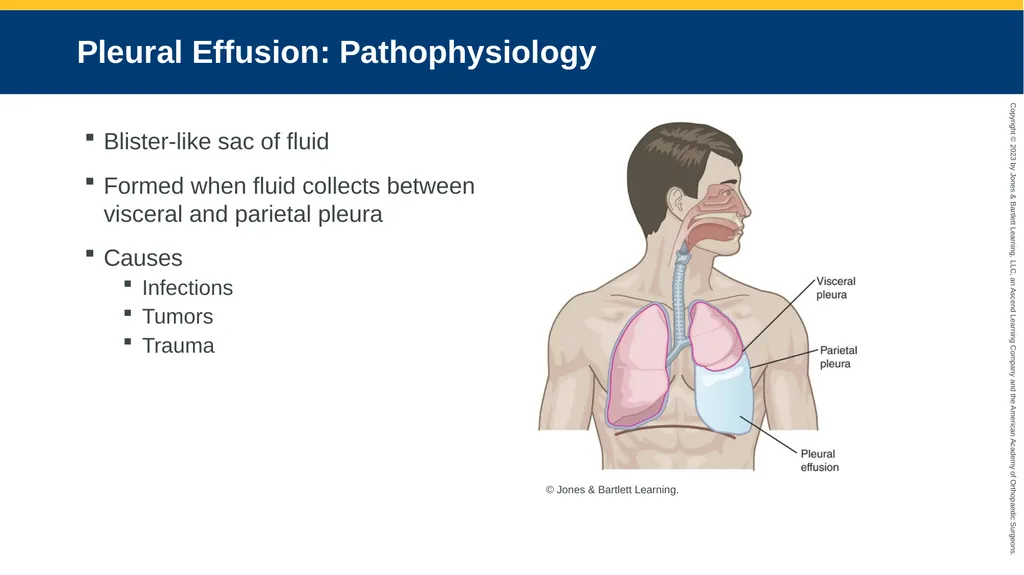
Pleural Effusion: Pathophysiology Blister-like sac of fluid
Formed when fluid collects between visceral and parietal pleura
Causes
Infections
Tumors
Trauma © Jones & Bartlett Learning.<br>
Formed when fluid collects between visceral and parietal pleura
Causes
Infections
Tumors
Trauma © Jones & Bartlett Learning.<br>
172
Pleural Effusion: Assessment Hard to hear breath sounds
Patient’s position affects ability to breathe<br>
Patient’s position affects ability to breathe<br>
173
Pleural Effusion: Management Fowler’s position likely most comfortable
Supportive care during transport to hospital
Thoracentesis at hospital<br>
Supportive care during transport to hospital
Thoracentesis at hospital<br>
174
Pulmonary Embolism: Pathophysiology Pulmonary circulation compromised by:
Blood clot
Fat embolism from broken bone
Amniotic fluid embolism during pregnancy
Air embolism from neck laceration or faulty IV
Large embolism
Lodges in major branch of pulmonary artery
Venous blood cannot reach alveoli<br>
Blood clot
Fat embolism from broken bone
Amniotic fluid embolism during pregnancy
Air embolism from neck laceration or faulty IV
Large embolism
Lodges in major branch of pulmonary artery
Venous blood cannot reach alveoli<br>
175
Pulmonary Embolism: Assessment (1 of 2) Early presentation
Normal breath sounds
Good peripheral aeration
Classic presentation
Sudden dyspnea and cyanosis
Sharp pain in chest
Cyanosis does not end with oxygen therapy<br>
Normal breath sounds
Good peripheral aeration
Classic presentation
Sudden dyspnea and cyanosis
Sharp pain in chest
Cyanosis does not end with oxygen therapy<br>
176
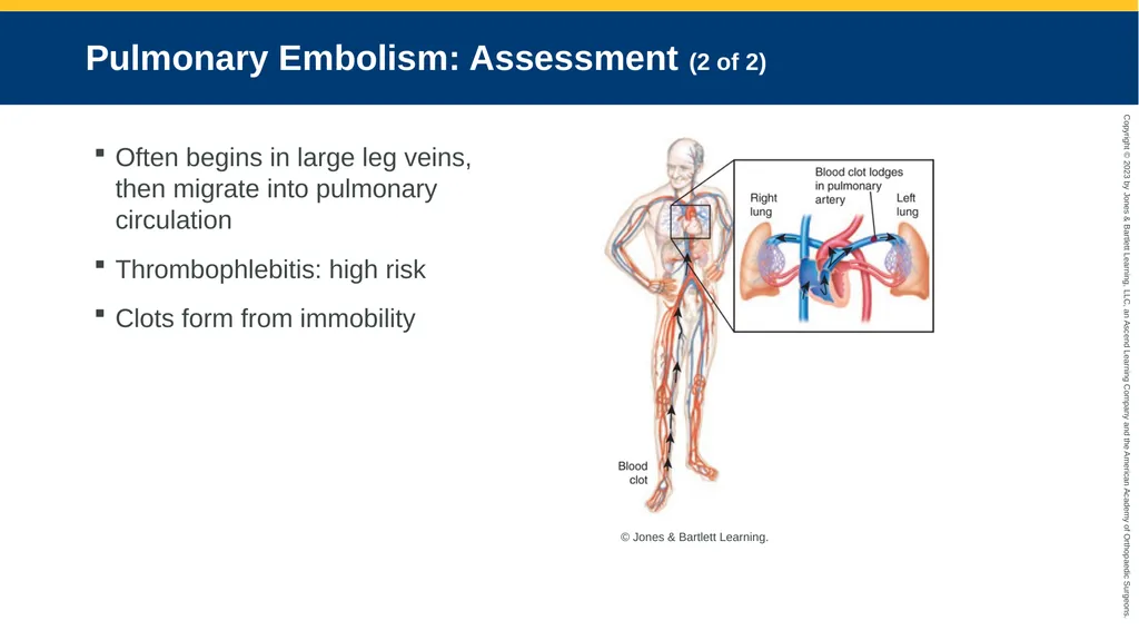
Pulmonary Embolism: Assessment (2 of 2) Often begins in large leg veins, then migrate into pulmonary circulation
Thrombophlebitis: high risk
Clots form from immobility © Jones & Bartlett Learning.<br>
Thrombophlebitis: high risk
Clots form from immobility © Jones & Bartlett Learning.<br>
177
Pulmonary Embolism: Management (1 of 3) Bedridden patients are often given:
Anticoagulants
Special stockings
Devices to reduce blood clot formation<br>
Anticoagulants
Special stockings
Devices to reduce blood clot formation<br>
178
Pulmonary Embolism: Management (2 of 3) Inferior vena cava filter
Patients with deep venous thrombosis
Opens like mesh umbrella
Catch clots traveling from legs in main vein
Recent shift to retrievable filters<br>
Patients with deep venous thrombosis
Opens like mesh umbrella
Catch clots traveling from legs in main vein
Recent shift to retrievable filters<br>
179
Pulmonary Embolism: Management (3 of 3) Saddle embolus
Exceptionally large embolus
Lodged at left/right pulmonary artery bifurcation
May be immediately fatal
Cape cyanosis despite CPR and ventilation<br>
Exceptionally large embolus
Lodged at left/right pulmonary artery bifurcation
May be immediately fatal
Cape cyanosis despite CPR and ventilation<br>