Rickets- radiology Dr jp,asst prof,ich,mch,kottayam The manifestations of rickets are most pronounced with rapid bone growth particularly the distal radius and ulna, distal femur, proximal tibia, proximal humerus, and anterior rib ends The
"Rickets- radiology Dr jp,asst" is the property of its rightful owner. Permission is granted to
download and print the materials on this website for personal, non-commercial use only, and to display it
on your personal computer provided you do not modify the materials and that you retain all copyright
notices contained in the materials. By downloading content from our website, you accept the terms of this
agreement.
Presentation Transcript
01
Rickets- radiology Dr jp,asst prof,ich,mch,kottayam<br>
02
The manifestations of rickets are most pronounced with rapid bone growth particularly the distal radius and ulna, distal femur, proximal tibia, proximal humerus, and anterior rib ends<br>
03
The initial radiographic finding in rickets is loss of mineralization of the zone of provisional calcification
loss of definition of the Laval-Jeantet collar, a short cylindricalsegment of the metaphysis adjacent to the growth plate that is an indicator of the most recently formed bone in young infants<br>
04
Deficient chondrocyte terminal differentiation and apoptosis causes accumulation of disorganized cartilage in the metaphysis in addition to nonmineralized osteoid, leading towidening of the distance between the epiphysis and metaphysis,metaphyseal fraying, and metaphyseal concavity (cupping).<br>
05
Metaphyseal concavity varies by site, being most pronounced in the distal forearm bones<br>
06
Distal ulnar metaphyseal concavity with no other abnormality should be recognized as a normal finding.
Metaphyseal findingsthat may be recognized on chest radiographs include involvement of the proximal humeral metaphyses and rib ends, producing the rachitic rosary<br>
07
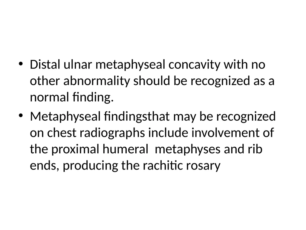
Vitamin D deficiency rickets.
The initial image (left) shows loss of definition of the zones of provisional calcification for the distal radial and ulnar metaphyses along with metaphyseal fraying and concavity (“cupping”) and physeal widening with an increased distance between the epiphysis and visualized portion of the metaphysis.Periosteal new bone also is present that is seen best along the metacarpals but also is present along the distal radius.
With healing (right), the zone of provisional calcification is well mineralized and the other findings have resolved<br>
08
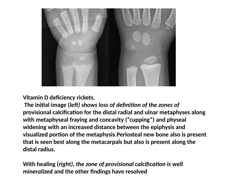
Anteroposterior (A) and lateral (B) chest radiograph views in a 5-month-old child with rickets from biliary atresia. Note rickets in the proximal humeral metaphyses and anterior rib ends. The rib findings are best seen on the lateral view<br>
09
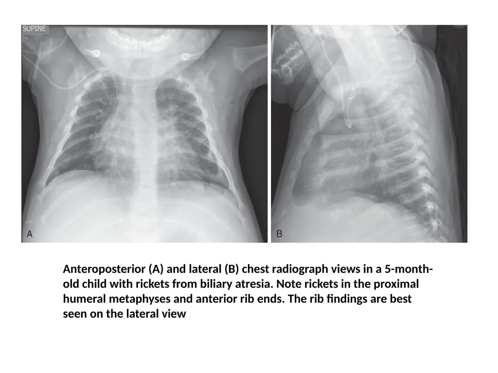
less severe case of Vitamin D deficiency rickets. At 9½ years of age (top), distal femoral physeal
widening and metaphyseal fraying is noted that resolved several months
later after treatment (bottom).<br>
10
Diaphyseal findings in a patient with severe vitamin D deficiency rickets. During the active phase (A), coarse demineralization and subperiosteal bone resorption are present, which are indicative of hyperparathyroidism as a result of rickets. Also note the severe rachitic findings in the metaphysis and poor mineralization of the distal radial epiphysis with loss of the zones of provisional calcification (arrow). With healing 3 months later (B), extensive periosteal new bone is seen (white
arrows) with calcification of previously nonmineralized osteoid (black arrows) produced by periosteal osteoblasts<br>
11
Key points The initial radiographic finding in rickets is loss of mineralization of the zone of provisional calcification
Isolated distal ulnar metaphyseal cupping is a normal variant in an infant and should not be confused with rickets
Ref: caffeys pediatric diagnostic radiology 2013.<br>