
Seizures & Epilepsy Dr Maria George Introduction
JO
Published · 69 slides · 0 views

1 / 1








Description
Seizures Epilepsy Dr Maria George Introduction Definition : SEIZURE : transient occurrence of signs or symptoms due to abnormal excessive hypersynchronous firing from an aggregate of neurons EPILEPSY: disorder of the brain characterized
Related Topics
Share
Embed code
Download this presentation From Below
"Seizures & Epilepsy Dr Maria George Introduction" is the property of its rightful owner. Permission is granted to download and print the materials on this website for personal, non-commercial use only, and to display it on your personal computer provided you do not modify the materials and that you retain all copyright notices contained in the materials. By downloading content from our website, you accept the terms of this agreement.
Presentation Transcript
01
Seizures & Epilepsy Dr Maria George<br>
02
Introduction Definition :
SEIZURE : transient occurrence of signs or symptoms due to abnormal excessive hypersynchronous firing from an aggregate of neurons
EPILEPSY: disorder of the brain characterized by an enduring predisposition to epileptic seizures.<br>
SEIZURE : transient occurrence of signs or symptoms due to abnormal excessive hypersynchronous firing from an aggregate of neurons
EPILEPSY: disorder of the brain characterized by an enduring predisposition to epileptic seizures.<br>
03
Epilepsy is said to occur when either of the following is present :
2 unprovoked seizures occurring more than 24 hours apart
1 unprovoked seizure and a probability of further seizures similar to general recurrence risk after 2 unprovoked seizures ( eg ≥ 60% ) occurring over next 10 years
Diagnosis of an epilepsy syndrome
Incidence of epilepsy : 0.3 to 0.5%<br>
2 unprovoked seizures occurring more than 24 hours apart
1 unprovoked seizure and a probability of further seizures similar to general recurrence risk after 2 unprovoked seizures ( eg ≥ 60% ) occurring over next 10 years
Diagnosis of an epilepsy syndrome
Incidence of epilepsy : 0.3 to 0.5%<br>
04
Classification Based on onset
Focal onset
Generalized onset
Unknown onset<br>
Focal onset
Generalized onset
Unknown onset<br>
05
Focal onset seizures
Originates within networks limited to one brain region - 2 types Intact awareness
Impaired awareness<br>
Impaired awareness<br>
06
Focal seizures with intact awareness Motor
Non motor
Somatic sensation – paraesthesia
Visual – flashing light, hallucination
equilibrium – sensation of falling/vertigo
Autonomic function – flushing, sweating, piloerection<br>
Non motor
Somatic sensation – paraesthesia
Visual – flashing light, hallucination
equilibrium – sensation of falling/vertigo
Autonomic function – flushing, sweating, piloerection<br>
07
3 features
Jacksonian march - the abnormal motor movements may begin in a very restricted region such as the fingers and gradually progress to include a larger portion of the extremity.
Todds paresis – localized paresis for minutes to many hours in involved region following seizure
Epilepsy partialis continua – seizures continuing for hours or days<br>
Jacksonian march - the abnormal motor movements may begin in a very restricted region such as the fingers and gradually progress to include a larger portion of the extremity.
Todds paresis – localized paresis for minutes to many hours in involved region following seizure
Epilepsy partialis continua – seizures continuing for hours or days<br>
08
Focal seizures with impaired awareness Aura
Arrest – characterised by motionless stare
Automatism - involuntary, automatic behaviours like lip smacking chewing
Amnesia – due to post ictal inhibition of cortical region most involved in seizure
Focal seizures can progress to generalised seizures<br>
Arrest – characterised by motionless stare
Automatism - involuntary, automatic behaviours like lip smacking chewing
Amnesia – due to post ictal inhibition of cortical region most involved in seizure
Focal seizures can progress to generalised seizures<br>
09
Generalised seizures Arise within and rapidly engage networks distributed across both cerebral hemispheres
2 types
Motor
Non motor<br>
2 types
Motor
Non motor<br>
10
Motor 1.Generalized tonic clonic seizures
Most common
Approximately 10% of all patients with epilepsy
1. Premonitary symptoms
2. Phase of tonic contraction : lasting 10 to 20s
EEG : progressive increase in generalized low voltage activity followed by generalized high amplitude polyspike discharge<br>
Most common
Approximately 10% of all patients with epilepsy
1. Premonitary symptoms
2. Phase of tonic contraction : lasting 10 to 20s
EEG : progressive increase in generalized low voltage activity followed by generalized high amplitude polyspike discharge<br>
11
3. Clonic phase : 1 min – due to superimposition of periods of relaxation onto the tonic muscle contraction
EEG : high amplitude activity is interrupted by slow waves – spike and slow wave pattern
4. Post ictal : muscle flaccidity, unresponsiveness, bowel and bladder incontinence
Prolonged in patients with underlying cns disease<br>
EEG : high amplitude activity is interrupted by slow waves – spike and slow wave pattern
4. Post ictal : muscle flaccidity, unresponsiveness, bowel and bladder incontinence
Prolonged in patients with underlying cns disease<br>
12
2. Clonic seizures
3. Tonic seizures
4. Atonic seizures :
characterized by sudden loss of postural muscle tone lasting 1–2s.
Consciousness is briefly impaired, but there is usually no postictal confusion
EEG: shows brief, generalized spike-and-wave discharges followed immediately by diffuse slow waves that correlate with the loss of muscle tone.
usually seen in association with known epilepsy syndromes.<br>
3. Tonic seizures
4. Atonic seizures :
characterized by sudden loss of postural muscle tone lasting 1–2s.
Consciousness is briefly impaired, but there is usually no postictal confusion
EEG: shows brief, generalized spike-and-wave discharges followed immediately by diffuse slow waves that correlate with the loss of muscle tone.
usually seen in association with known epilepsy syndromes.<br>
13
5. Myoclonic seizures
Sudden and brief muscle contraction that may involve one part of the body or the entire body
EEG: shows bilaterally synchronous spike-and-slow-wave discharges immediately prior to the movement and muscle artifact associated with the myoclonus.
Predominant feature of juvenile myoclonic epilepsy (JME).<br>
Sudden and brief muscle contraction that may involve one part of the body or the entire body
EEG: shows bilaterally synchronous spike-and-slow-wave discharges immediately prior to the movement and muscle artifact associated with the myoclonus.
Predominant feature of juvenile myoclonic epilepsy (JME).<br>
14
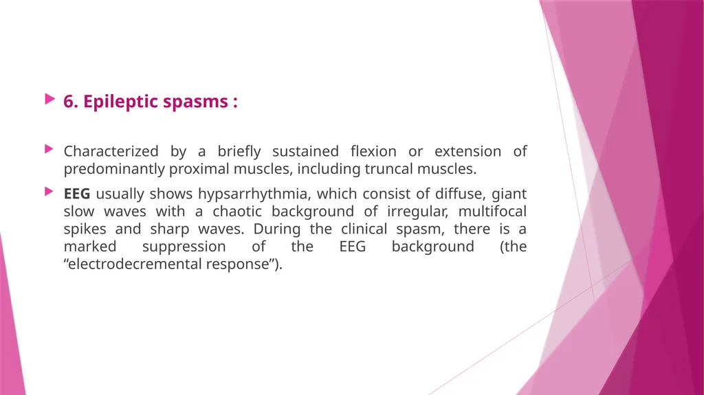
6. Epileptic spasms :
Characterized by a briefly sustained flexion or extension of predominantly proximal muscles, including truncal muscles.
EEG usually shows hypsarrhythmia, which consist of diffuse, giant slow waves with a chaotic background of irregular, multifocal spikes and sharp waves. During the clinical spasm, there is a marked suppression of the EEG background (the “electrodecremental response”).<br>
Characterized by a briefly sustained flexion or extension of predominantly proximal muscles, including truncal muscles.
EEG usually shows hypsarrhythmia, which consist of diffuse, giant slow waves with a chaotic background of irregular, multifocal spikes and sharp waves. During the clinical spasm, there is a marked suppression of the EEG background (the “electrodecremental response”).<br>
15
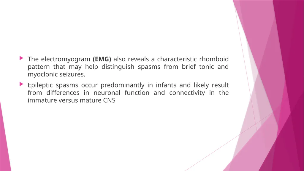
The electromyogram (EMG) also reveals a characteristic rhomboid pattern that may help distinguish spasms from brief tonic and myoclonic seizures.
Epileptic spasms occur predominantly in infants and likely result from differences in neuronal function and connectivity in the immature versus mature CNS<br>
Epileptic spasms occur predominantly in infants and likely result from differences in neuronal function and connectivity in the immature versus mature CNS<br>
16
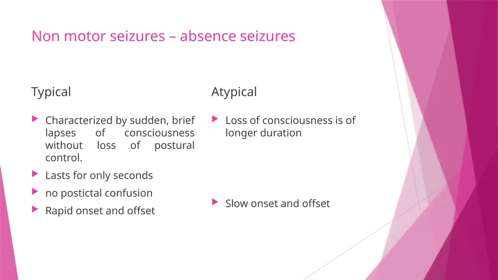
Non motor seizures – absence seizures Typical Characterized by sudden, brief lapses of consciousness without loss of postural control.
Lasts for only seconds
no postictal confusion
Rapid onset and offset Atypical Loss of consciousness is of longer duration
Slow onset and offset<br>
Lasts for only seconds
no postictal confusion
Rapid onset and offset Atypical Loss of consciousness is of longer duration
Slow onset and offset<br>
17
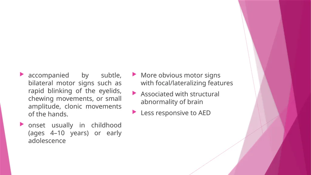
accompanied by subtle, bilateral motor signs such as rapid blinking of the eyelids, chewing movements, or small amplitude, clonic movements of the hands.
onset usually in childhood (ages 4–10 years) or early adolescence More obvious motor signs with focal/lateralizing features
Associated with structural abnormality of brain
Less responsive to AED<br>
onset usually in childhood (ages 4–10 years) or early adolescence More obvious motor signs with focal/lateralizing features
Associated with structural abnormality of brain
Less responsive to AED<br>
18
main seizure type in 15–20% of children with epilepsy.
occurs hundreds of times per day
EEG : generalized, symmetric, 3-Hz spike-and-slow-wave discharges that begins and ends suddenly, superimposed on a normal EEG background. EEG : generalized slow spike and slow wave pattern with a frequency less than 2.5Hz<br>
occurs hundreds of times per day
EEG : generalized, symmetric, 3-Hz spike-and-slow-wave discharges that begins and ends suddenly, superimposed on a normal EEG background. EEG : generalized slow spike and slow wave pattern with a frequency less than 2.5Hz<br>
19
Epilepsy syndromes Epilepsy syndromes are disorders in which epilepsy is a predominant feature, and there is sufficient evidence (e.g., through clinical, EEG, radiologic, or genetic observations) to suggest a common underlying mechanism<br>
20
Juvenile myoclonic epilepsy generalized seizure disorder
early adolescence
characterized by bilateral myoclonic jerks that may be single or repetitive, more frequent in the morning after awakening and provoked by sleep deprivation. Consciousness is preserved.
Can also present as gtcs or absence seizures
Associated with family history epilepsy, polygenic<br>
early adolescence
characterized by bilateral myoclonic jerks that may be single or repetitive, more frequent in the morning after awakening and provoked by sleep deprivation. Consciousness is preserved.
Can also present as gtcs or absence seizures
Associated with family history epilepsy, polygenic<br>
21
Lennox-Gastaut Syndrome multiple seizure types (usually including generalized tonic-clonic, atonic, and atypical absence seizures)
EEG showing slow (<3 Hz) spike-and-wave discharges and a variety of other abnormalities
impaired cognitive function in most cases
Associated with CNS disease or dysfunction<br>
EEG showing slow (<3 Hz) spike-and-wave discharges and a variety of other abnormalities
impaired cognitive function in most cases
Associated with CNS disease or dysfunction<br>
22
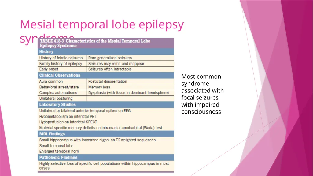
Mesial temporal lobe epilepsy syndrome Most common syndrome associated with focal seizures with impaired consciousness<br>
23
Causes of seizure and epilepsy Seizures are a result of a shift in the normal balance of excitation and inhibition within the CNS.
1. The normal brain is capable of having a seizure under the appropriate circumstances, and there are differences between individuals in the susceptibility or threshold for seizures.
2. There are a variety of conditions that have an extremely high likelihood of developing into a seizure : example : penetrating head trauma.
Process of transformation of a normal neuronal network into one that is hyper excitable – epileptogenesis
3. Seizures are episodic<br>
1. The normal brain is capable of having a seizure under the appropriate circumstances, and there are differences between individuals in the susceptibility or threshold for seizures.
2. There are a variety of conditions that have an extremely high likelihood of developing into a seizure : example : penetrating head trauma.
Process of transformation of a normal neuronal network into one that is hyper excitable – epileptogenesis
3. Seizures are episodic<br>
24
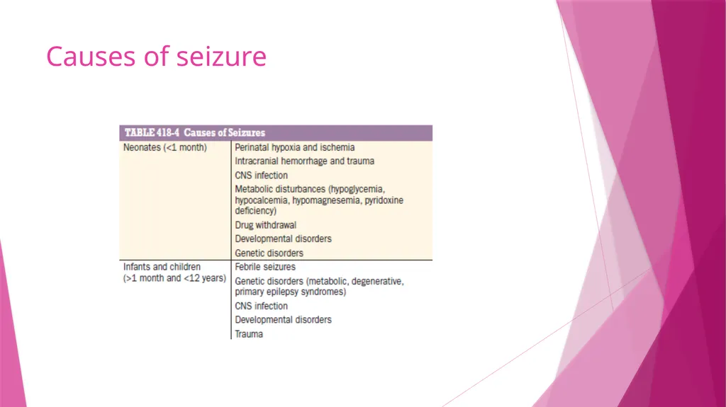
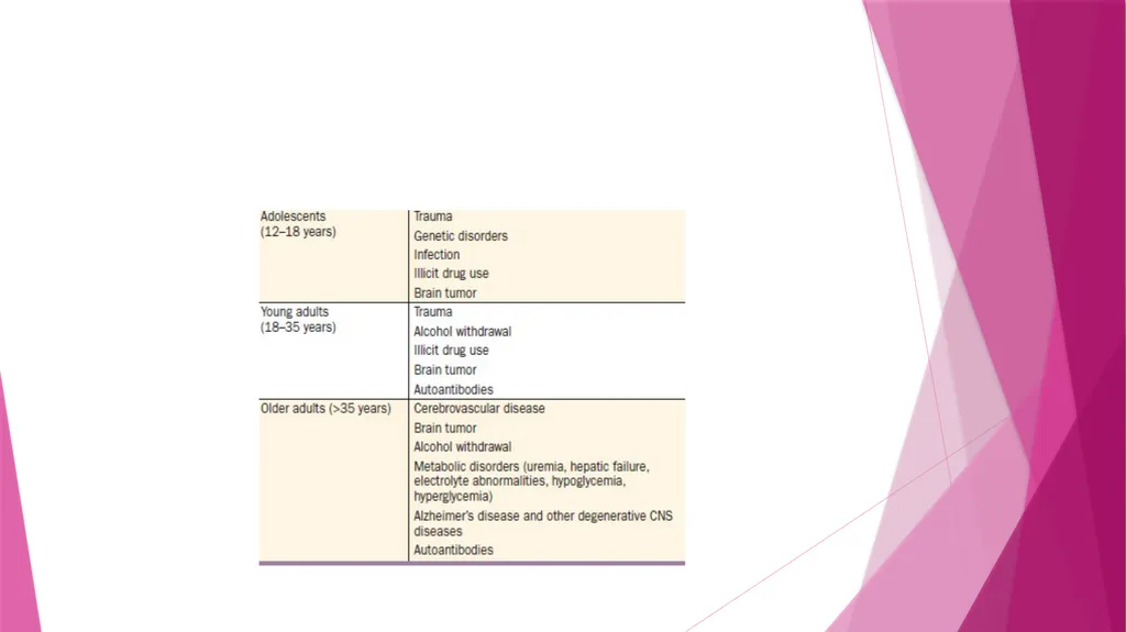
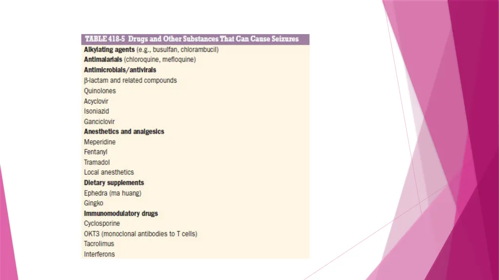
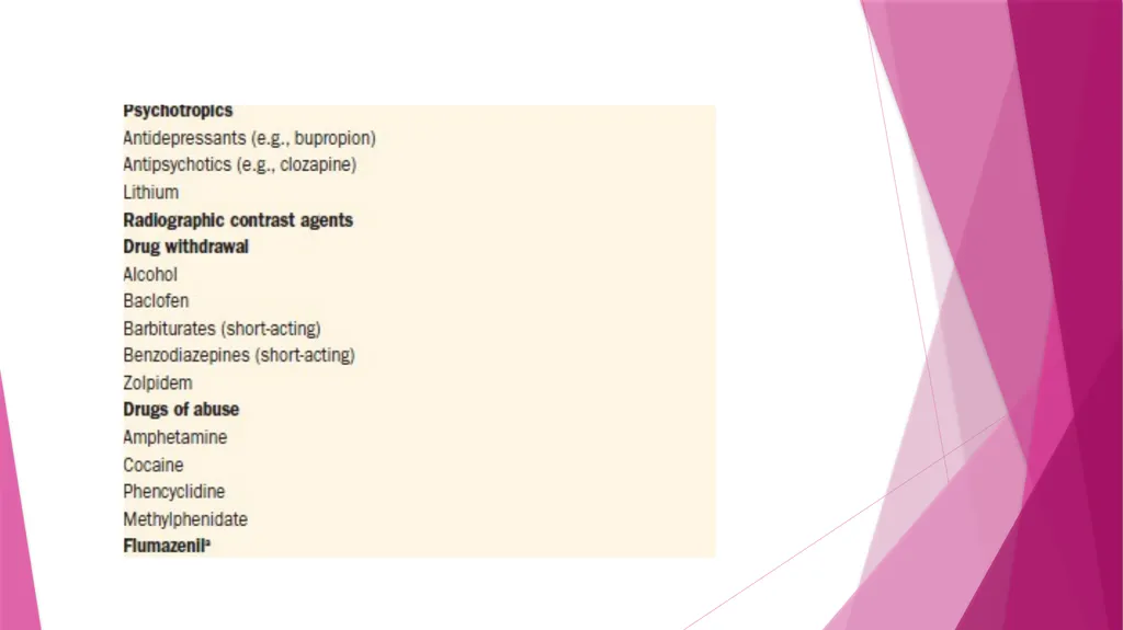
Causes of seizure<br>
25
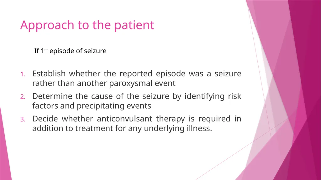
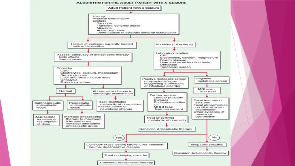
Approach to the patient Establish whether the reported episode was a seizure rather than another paroxysmal event
Determine the cause of the seizure by identifying risk factors and precipitating events
Decide whether anticonvulsant therapy is required in addition to treatment for any underlying illness. If 1st episode of seizure<br>
Determine the cause of the seizure by identifying risk factors and precipitating events
Decide whether anticonvulsant therapy is required in addition to treatment for any underlying illness. If 1st episode of seizure<br>
26
If patient has prior history of seizures:
Identification of the underlying cause and precipitating factors
Determination of the adequacy of the patient’s current therapy.<br>
Identification of the underlying cause and precipitating factors
Determination of the adequacy of the patient’s current therapy.<br>
27
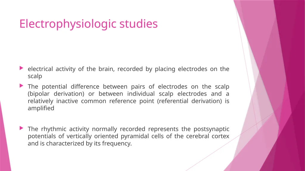
Electrophysiologic studies electrical activity of the brain, recorded by placing electrodes on the scalp
The potential difference between pairs of electrodes on the scalp (bipolar derivation) or between individual scalp electrodes and a relatively inactive common reference point (referential derivation) is amplified
The rhythmic activity normally recorded represents the postsynaptic potentials of vertically oriented pyramidal cells of the cerebral cortex and is characterized by its frequency.<br>
The potential difference between pairs of electrodes on the scalp (bipolar derivation) or between individual scalp electrodes and a relatively inactive common reference point (referential derivation) is amplified
The rhythmic activity normally recorded represents the postsynaptic potentials of vertically oriented pyramidal cells of the cerebral cortex and is characterized by its frequency.<br>
28
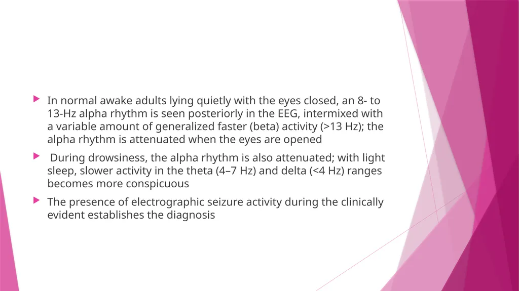
In normal awake adults lying quietly with the eyes closed, an 8- to 13-Hz alpha rhythm is seen posteriorly in the EEG, intermixed with a variable amount of generalized faster (beta) activity (>13 Hz); the alpha rhythm is attenuated when the eyes are opened
During drowsiness, the alpha rhythm is also attenuated; with light sleep, slower activity in the theta (4–7 Hz) and delta (<4 Hz) ranges becomes more conspicuous
The presence of electrographic seizure activity during the clinically evident establishes the diagnosis<br>
During drowsiness, the alpha rhythm is also attenuated; with light sleep, slower activity in the theta (4–7 Hz) and delta (<4 Hz) ranges becomes more conspicuous
The presence of electrographic seizure activity during the clinically evident establishes the diagnosis<br>
29
Epileptiform activity consists of bursts of abnormal discharges containing spikes or sharp waves
EEG is always abnormal during an episode of GTCS, maybe normal during a focal seizure
Normal EEG does not exclude a seizure disorder
Even in an individual who is known to have epilepsy, initial routine interictal EEG maybe normal in 60% cases.
Useful in assessing prognosis : normal EEG –better prognosis<br>
EEG is always abnormal during an episode of GTCS, maybe normal during a focal seizure
Normal EEG does not exclude a seizure disorder
Even in an individual who is known to have epilepsy, initial routine interictal EEG maybe normal in 60% cases.
Useful in assessing prognosis : normal EEG –better prognosis<br>
30
Magnetoencephalography : measures small magnetic fields generated by brain activity
Useful to localize potential seizure foci<br>
Useful to localize potential seizure foci<br>
31
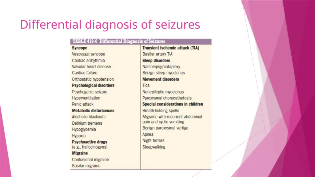
Differential diagnosis of seizures<br>
32
Psychogenic seizures Non epileptiform behaviors which resemble seizure
Part of conversion reaction
side-to-side turning of the head, asymmetric and large-amplitude shaking movements of the limbs, twitching of all four extremities without loss of consciousness, and pelvic thrusting are more commonly associated with psychogenic rather than epileptic seizures.
lasts longer than epileptic seizures and may wax and wane over minutes to hours
Video EEG and serum prolactin levels can be used to distinguish between the two<br>
Part of conversion reaction
side-to-side turning of the head, asymmetric and large-amplitude shaking movements of the limbs, twitching of all four extremities without loss of consciousness, and pelvic thrusting are more commonly associated with psychogenic rather than epileptic seizures.
lasts longer than epileptic seizures and may wax and wane over minutes to hours
Video EEG and serum prolactin levels can be used to distinguish between the two<br>
33
Treatment Treatment of underlying conditions which cause or contribute to seizures
Avoidance of precipitating factors
Suppression of recurrent seizures by prophylactic therapy with AED or surgery
Addressing the psycho – social issues associated with epilepsy<br>
Avoidance of precipitating factors
Suppression of recurrent seizures by prophylactic therapy with AED or surgery
Addressing the psycho – social issues associated with epilepsy<br>
34
1. Treatment of underlying condition Seizure due to metabolic derangement – correct metabolic parameter, AED is usually unnecessary
Seizure due to drug – avoid drug
Seizure due to brain tumor/vascular malformation/brain abscess – treatment of underlying condition, continue AED for at least 1 year then slowly withdrawn if patient is seizure free<br>
Seizure due to drug – avoid drug
Seizure due to brain tumor/vascular malformation/brain abscess – treatment of underlying condition, continue AED for at least 1 year then slowly withdrawn if patient is seizure free<br>
35
2. Avoidance of precipitating factors Sleep deprivation
Alcohol
Video game monitors, music
Stress<br>
Alcohol
Video game monitors, music
Stress<br>
36
3. Antiepileptic drug therapy GOAL:
To completely prevent seizures without causing any untoward side effects, preferably with a single medication and a dosing schedule that is easy to follow<br>
To completely prevent seizures without causing any untoward side effects, preferably with a single medication and a dosing schedule that is easy to follow<br>
37
When to initiate antiepileptic drug? Antiepileptic drug therapy should be started in any patient with recurrent seizures of unknown etiology or a known cause that cannot be reversed.
Patients with a single seizure due to an identified lesion such as a CNS tumor, infection, or trauma, in which there is strong evidence that the lesion is epileptogenic, should be treated.
The risk of seizure recurrence in a patient with an apparently unprovoked or idiopathic seizure is uncertain, and ranges from from 31 to 71% in the first 12 months after the initial seizure.<br>
Patients with a single seizure due to an identified lesion such as a CNS tumor, infection, or trauma, in which there is strong evidence that the lesion is epileptogenic, should be treated.
The risk of seizure recurrence in a patient with an apparently unprovoked or idiopathic seizure is uncertain, and ranges from from 31 to 71% in the first 12 months after the initial seizure.<br>
38
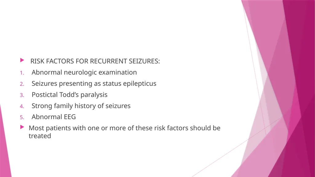
RISK FACTORS FOR RECURRENT SEIZURES:
Abnormal neurologic examination
Seizures presenting as status epilepticus
Postictal Todd’s paralysis
Strong family history of seizures
Abnormal EEG
Most patients with one or more of these risk factors should be treated<br>
Abnormal neurologic examination
Seizures presenting as status epilepticus
Postictal Todd’s paralysis
Strong family history of seizures
Abnormal EEG
Most patients with one or more of these risk factors should be treated<br>
39
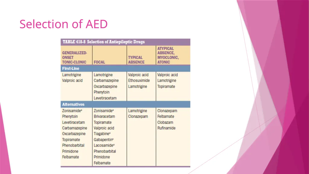
Selection of AED<br>
40
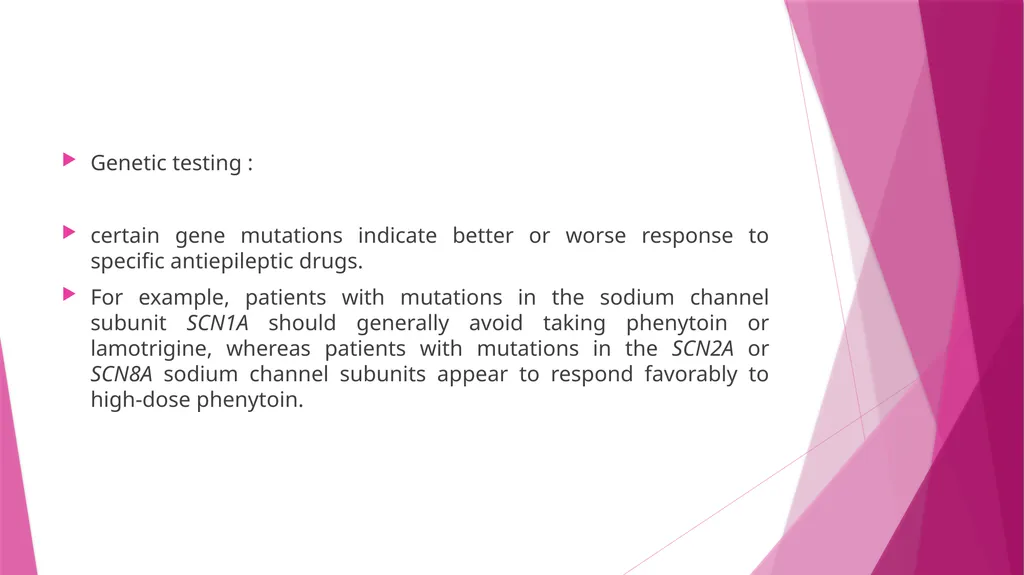
Genetic testing :
certain gene mutations indicate better or worse response to specific antiepileptic drugs.
For example, patients with mutations in the sodium channel subunit SCN1A should generally avoid taking phenytoin or lamotrigine, whereas patients with mutations in the SCN2A or SCN8A sodium channel subunits appear to respond favorably to high-dose phenytoin.<br>
certain gene mutations indicate better or worse response to specific antiepileptic drugs.
For example, patients with mutations in the sodium channel subunit SCN1A should generally avoid taking phenytoin or lamotrigine, whereas patients with mutations in the SCN2A or SCN8A sodium channel subunits appear to respond favorably to high-dose phenytoin.<br>
41
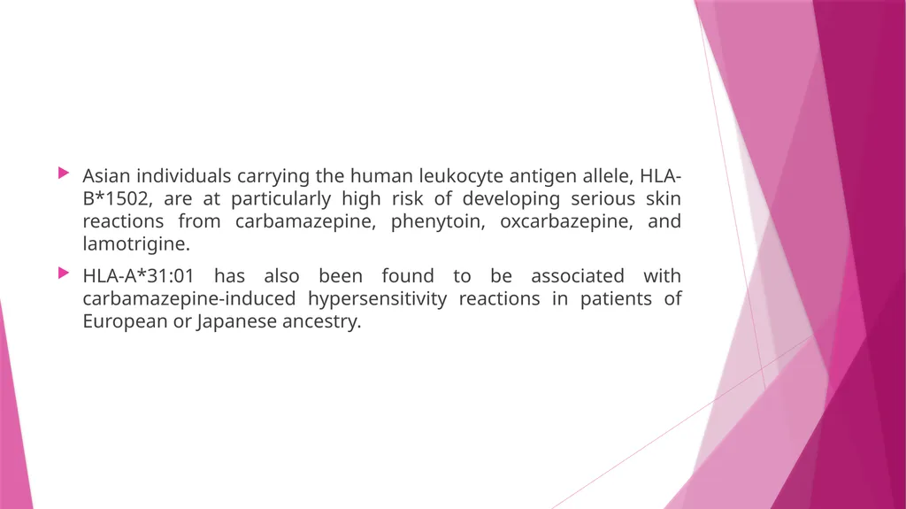
Asian individuals carrying the human leukocyte antigen allele, HLA-B*1502, are at particularly high risk of developing serious skin reactions from carbamazepine, phenytoin, oxcarbazepine, and lamotrigine.
HLA-A*31:01 has also been found to be associated with carbamazepine-induced hypersensitivity reactions in patients of European or Japanese ancestry.<br>
HLA-A*31:01 has also been found to be associated with carbamazepine-induced hypersensitivity reactions in patients of European or Japanese ancestry.<br>
42
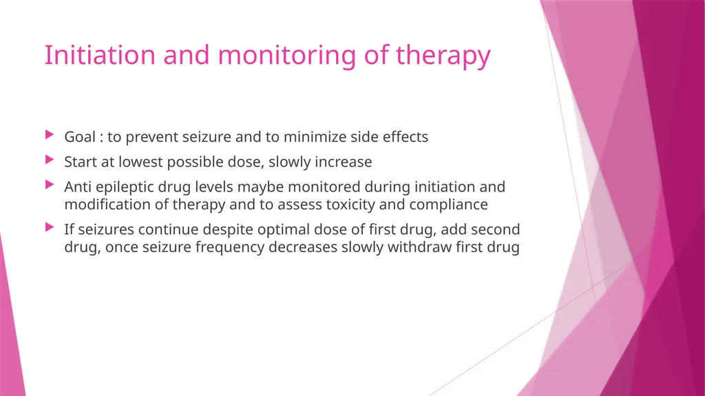
Initiation and monitoring of therapy Goal : to prevent seizure and to minimize side effects
Start at lowest possible dose, slowly increase
Anti epileptic drug levels maybe monitored during initiation and modification of therapy and to assess toxicity and compliance
If seizures continue despite optimal dose of first drug, add second drug, once seizure frequency decreases slowly withdraw first drug<br>
Start at lowest possible dose, slowly increase
Anti epileptic drug levels maybe monitored during initiation and modification of therapy and to assess toxicity and compliance
If seizures continue despite optimal dose of first drug, add second drug, once seizure frequency decreases slowly withdraw first drug<br>
43
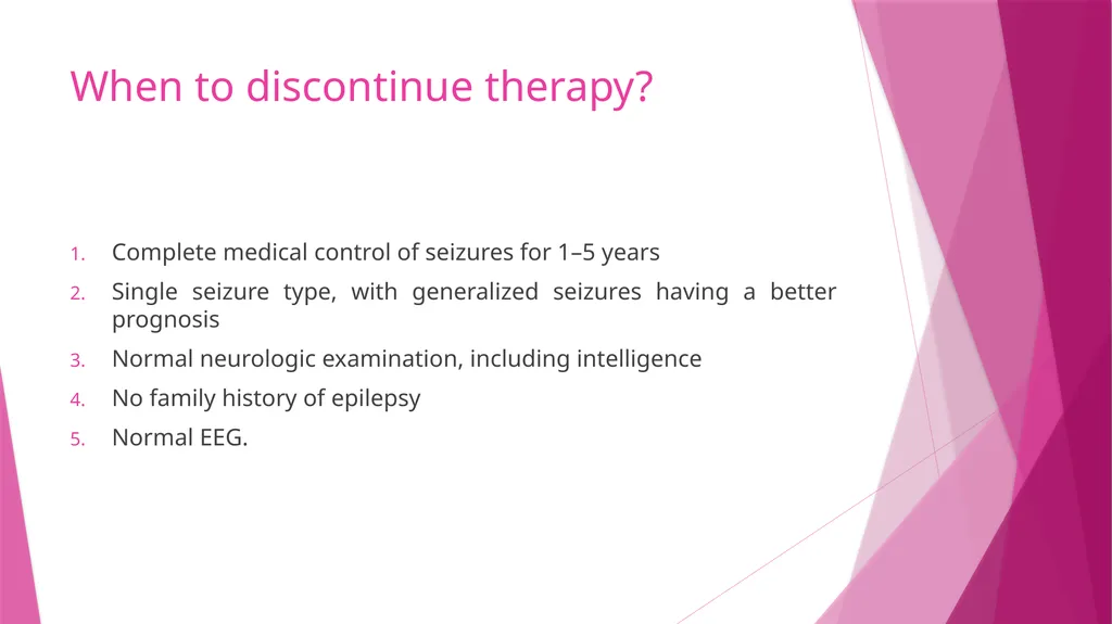
When to discontinue therapy? Complete medical control of seizures for 1–5 years
Single seizure type, with generalized seizures having a better prognosis
Normal neurologic examination, including intelligence
No family history of epilepsy
Normal EEG.<br>
Single seizure type, with generalized seizures having a better prognosis
Normal neurologic examination, including intelligence
No family history of epilepsy
Normal EEG.<br>
44
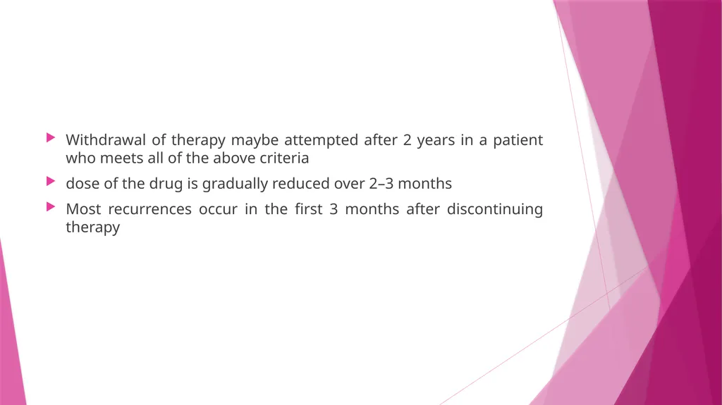
Withdrawal of therapy maybe attempted after 2 years in a patient who meets all of the above criteria
dose of the drug is gradually reduced over 2–3 months
Most recurrences occur in the first 3 months after discontinuing therapy<br>
dose of the drug is gradually reduced over 2–3 months
Most recurrences occur in the first 3 months after discontinuing therapy<br>
45
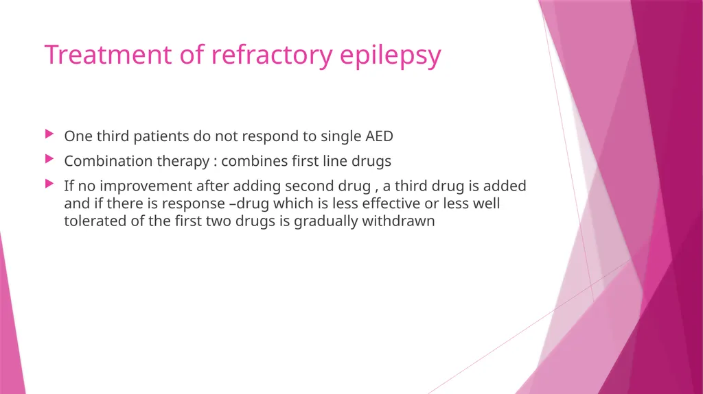
Treatment of refractory epilepsy One third patients do not respond to single AED
Combination therapy : combines first line drugs
If no improvement after adding second drug , a third drug is added and if there is response –drug which is less effective or less well tolerated of the first two drugs is gradually withdrawn<br>
Combination therapy : combines first line drugs
If no improvement after adding second drug , a third drug is added and if there is response –drug which is less effective or less well tolerated of the first two drugs is gradually withdrawn<br>
46
Surgical Treatment of Refractory Epilepsy 20 to 30% of patients with epilepsy continua continue to have seizures despite combination therapy with AED
The most common surgical procedure for patients with temporal lobe epilepsy involves resection of the anteromedial temporal lobe (temporal lobectomy) or a more limited removal of the underlying hippocampus and amygdala (amygdalohippocampectomy).
Focal seizures arising from extratemporal regions may be abolished by a focal neocortical resection with precise removal of an identified lesion (lesionectomy).<br>
The most common surgical procedure for patients with temporal lobe epilepsy involves resection of the anteromedial temporal lobe (temporal lobectomy) or a more limited removal of the underlying hippocampus and amygdala (amygdalohippocampectomy).
Focal seizures arising from extratemporal regions may be abolished by a focal neocortical resection with precise removal of an identified lesion (lesionectomy).<br>
47
When the cortical region cannot be removed, multiple subpial transection, which disrupts intracortical connections, is sometimes used to prevent seizure spread.
Hemispherectomy or multilobar resection is useful for some patients with severe seizures due to hemispheric abnormalities such as hemimegalencephaly or other dysplastic abnormalities
Corpus callosotomy has been shown to be effective for disabling tonic or atonic seizures, usually when they are part of a mixed-seizure syndrome (e.g., Lennox-Gastaut syndrome<br>
Hemispherectomy or multilobar resection is useful for some patients with severe seizures due to hemispheric abnormalities such as hemimegalencephaly or other dysplastic abnormalities
Corpus callosotomy has been shown to be effective for disabling tonic or atonic seizures, usually when they are part of a mixed-seizure syndrome (e.g., Lennox-Gastaut syndrome<br>
48
Vagus nerve stimulation has been used in patients in patients in whom seizure arises from more than one location
Responsive Neuro-Stimulation : it is an implantable device which detects the onset of seizure and delivers an electrical stimulation to abort the seizure<br>
Responsive Neuro-Stimulation : it is an implantable device which detects the onset of seizure and delivers an electrical stimulation to abort the seizure<br>
49
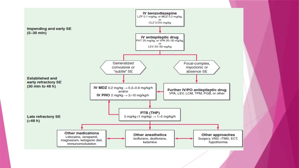
Status Epilepticus Continuous seizure or repetitive discrete seizures with impaired consciousness in the inter ictal period
Maybe convulsive or non convulsive satus
Historically, the International League Against Epilepsy (ILAE) and others defined status epilepticus as a single epileptic seizure of >30 minutes duration or a series of epileptic seizures during which function is not regained between seizures in a 30-minute period<br>
Maybe convulsive or non convulsive satus
Historically, the International League Against Epilepsy (ILAE) and others defined status epilepticus as a single epileptic seizure of >30 minutes duration or a series of epileptic seizures during which function is not regained between seizures in a 30-minute period<br>
50
Operational definition –
Considering the need for rapid evaluation and intervention in GCSE to avoid cardiovascular morbidity and refractory status, an accepted operational definition of GCSE consists of the following:
≥5 minutes of continuous seizures, or
≥2 discrete seizures between which there is incomplete recovery of consciousness<br>
Considering the need for rapid evaluation and intervention in GCSE to avoid cardiovascular morbidity and refractory status, an accepted operational definition of GCSE consists of the following:
≥5 minutes of continuous seizures, or
≥2 discrete seizures between which there is incomplete recovery of consciousness<br>
51
The ILAE definition is as follows :
"Status epilepticus is a condition resulting from either the failure of the mechanisms responsible for seizure termination or from the initiation of mechanisms that lead to abnormally prolonged seizures (after time point t1). It is a condition that can have long-term consequences (after time point t2), including neuronal death, neuronal injury, and alteration of neuronal networks, depending on the type and duration of seizures.<br>
"Status epilepticus is a condition resulting from either the failure of the mechanisms responsible for seizure termination or from the initiation of mechanisms that lead to abnormally prolonged seizures (after time point t1). It is a condition that can have long-term consequences (after time point t2), including neuronal death, neuronal injury, and alteration of neuronal networks, depending on the type and duration of seizures.<br>
52
ILAE definition – In 2015, the ILAE published a revised conceptual definition of status epilepticus that incorporates two time points
The first, t1, is the time at which ongoing seizure activity should be regarded as abnormally prolonged, unlikely to stop spontaneously, and when treatment for status epilepticus should be started
The second, t2, is the time after which the ongoing seizure activity poses a significant risk of long-term complications.<br>
The first, t1, is the time at which ongoing seizure activity should be regarded as abnormally prolonged, unlikely to stop spontaneously, and when treatment for status epilepticus should be started
The second, t2, is the time after which the ongoing seizure activity poses a significant risk of long-term complications.<br>
53
For GCSE, the ILAE proposal specifies that t1 and t2 are 5 and 30 minutes
t1 and t2 of 10 and >60 minutes for focal status epilepticus with impaired consciousness
a t1 of 10 to 15 minutes for absence status epilepticus<br>
t1 and t2 of 10 and >60 minutes for focal status epilepticus with impaired consciousness
a t1 of 10 to 15 minutes for absence status epilepticus<br>
54
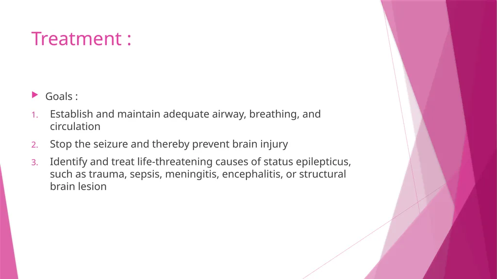
Treatment : Goals :
Establish and maintain adequate airway, breathing, and circulation
Stop the seizure and thereby prevent brain injury
Identify and treat life-threatening causes of status epilepticus, such as trauma, sepsis, meningitis, encephalitis, or structural brain lesion<br>
Establish and maintain adequate airway, breathing, and circulation
Stop the seizure and thereby prevent brain injury
Identify and treat life-threatening causes of status epilepticus, such as trauma, sepsis, meningitis, encephalitis, or structural brain lesion<br>
55
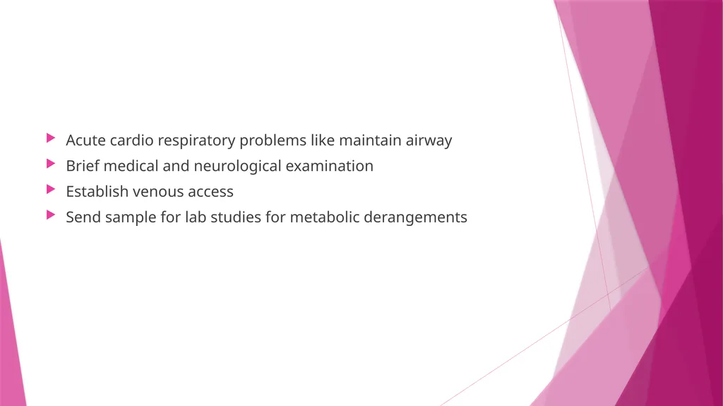
Acute cardio respiratory problems like maintain airway
Brief medical and neurological examination
Establish venous access
Send sample for lab studies for metabolic derangements<br>
Brief medical and neurological examination
Establish venous access
Send sample for lab studies for metabolic derangements<br>
56
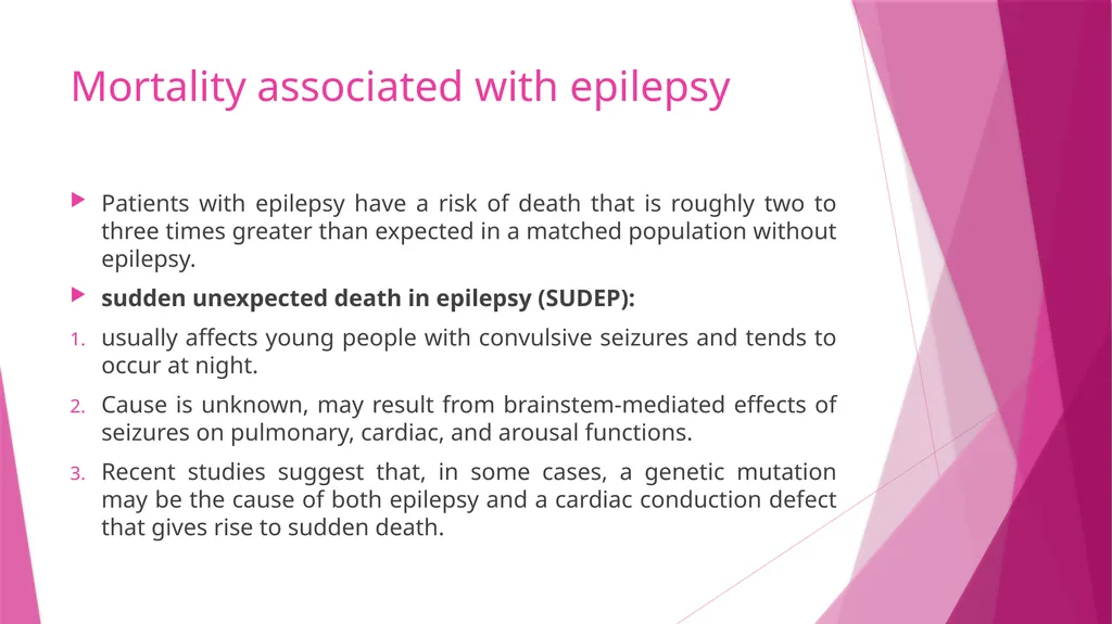
Mortality associated with epilepsy Patients with epilepsy have a risk of death that is roughly two to three times greater than expected in a matched population without epilepsy.
sudden unexpected death in epilepsy (SUDEP):
usually affects young people with convulsive seizures and tends to occur at night.
Cause is unknown, may result from brainstem-mediated effects of seizures on pulmonary, cardiac, and arousal functions.
Recent studies suggest that, in some cases, a genetic mutation may be the cause of both epilepsy and a cardiac conduction defect that gives rise to sudden death.<br>
sudden unexpected death in epilepsy (SUDEP):
usually affects young people with convulsive seizures and tends to occur at night.
Cause is unknown, may result from brainstem-mediated effects of seizures on pulmonary, cardiac, and arousal functions.
Recent studies suggest that, in some cases, a genetic mutation may be the cause of both epilepsy and a cardiac conduction defect that gives rise to sudden death.<br>
57
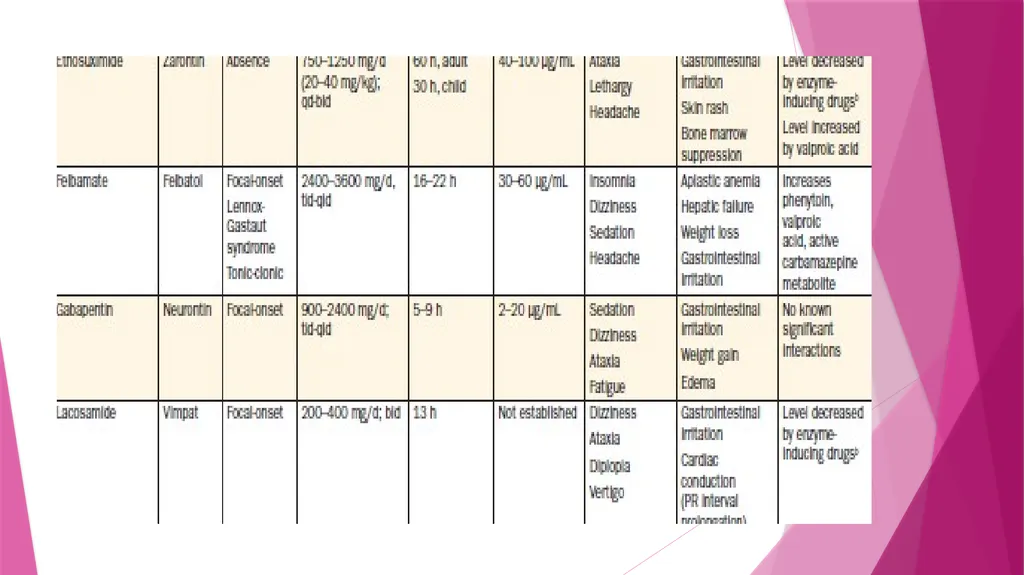
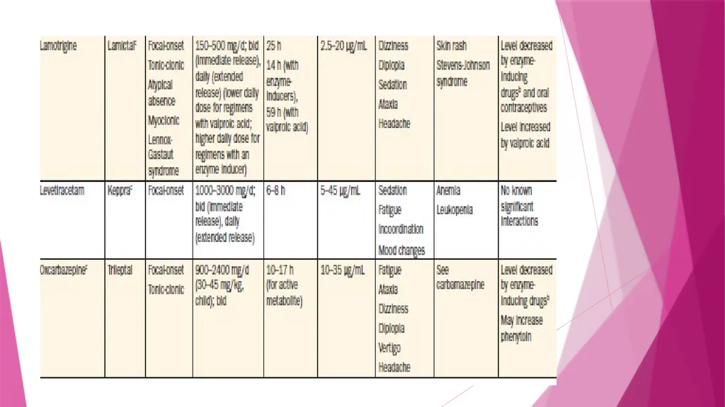
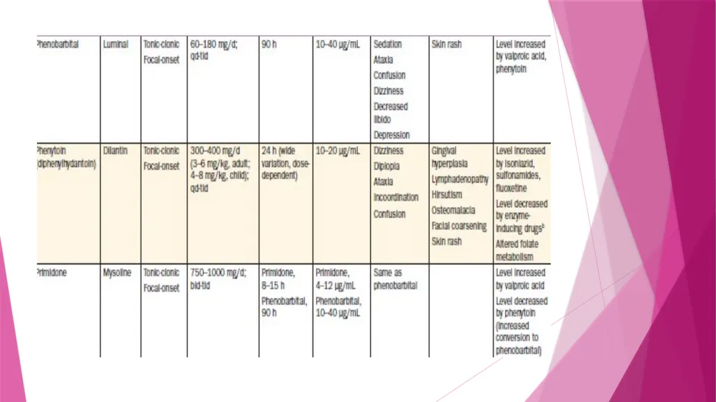
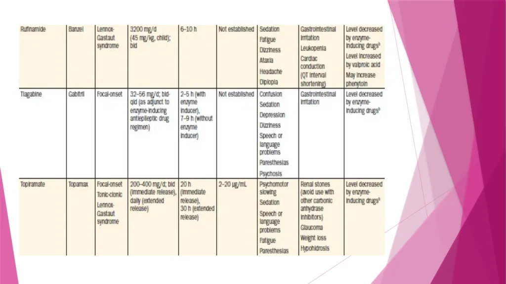
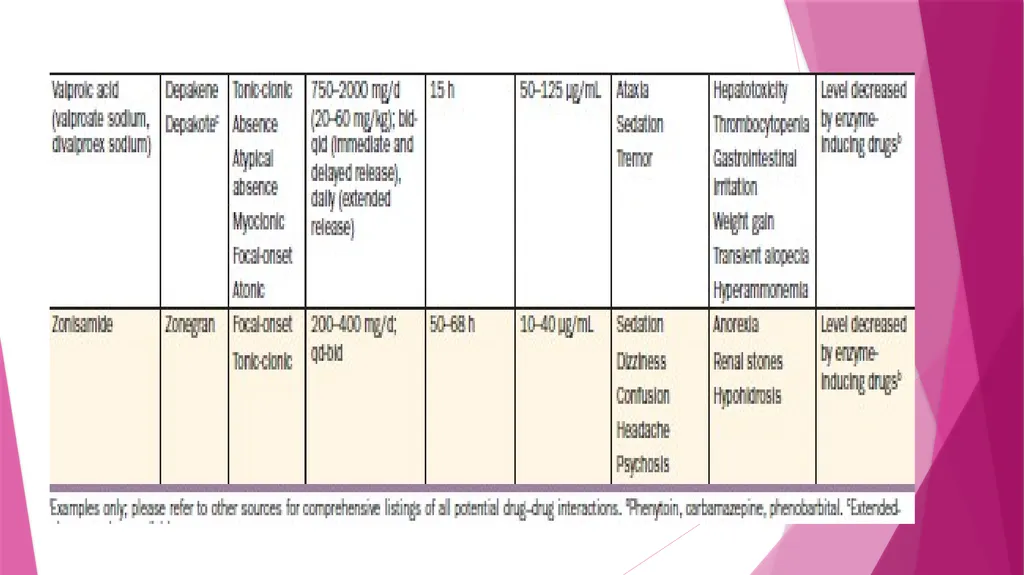
Common antiepileptic drugs<br>
58
THANK YOU<br>