
Spore-forming G positive bacilli - Aerobic
LD
Published · 15 slides · 0 views

1 / 1
Description
Spore-forming G positive bacilli - Aerobic (Bacillus) These bacilli are ubiquitous because they form spores they can survive in the environment for many years. They are strictly aerobic or facultative anaerobic, most are catalase positive
Related Topics
Share
Embed code
Download this presentation From Below
"Spore-forming G positive bacilli - Aerobic" is the property of its rightful owner. Permission is granted to download and print the materials on this website for personal, non-commercial use only, and to display it on your personal computer provided you do not modify the materials and that you retain all copyright notices contained in the materials. By downloading content from our website, you accept the terms of this agreement.
Presentation Transcript
01
Spore-forming G positive bacilli - Aerobic (Bacillus)
These bacilli are ubiquitous & because they form spores they can survive in the environment for many years. They are strictly aerobic or facultative anaerobic, most are catalase positive & motile. Several species causes important disease in human. Only few species are animal pathogens.
Bacillus
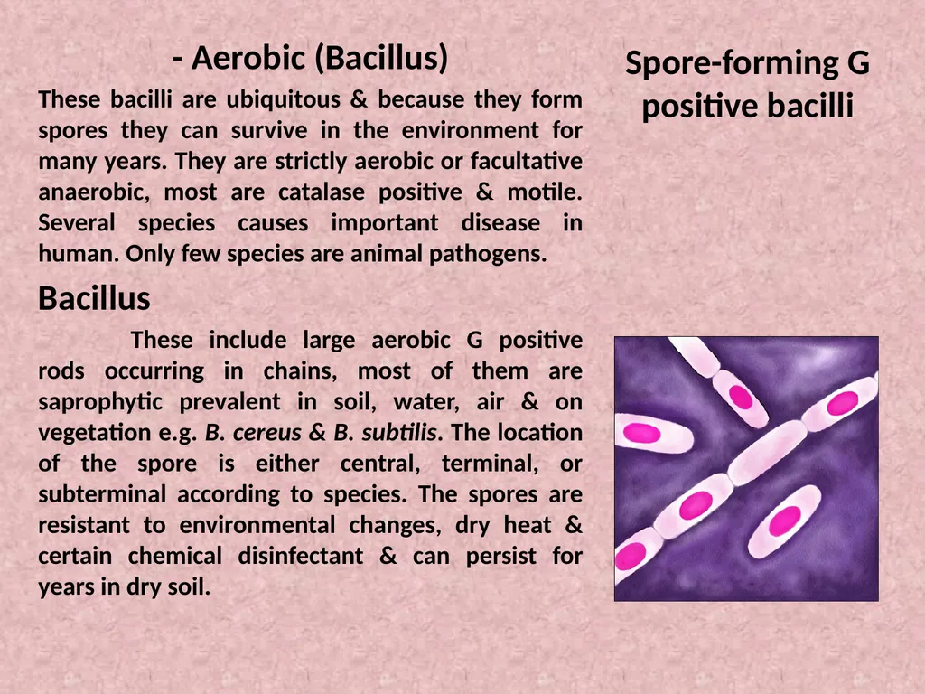
These include large aerobic G positive rods occurring in chains, most of them are saprophytic prevalent in soil, water, air & on vegetation e.g. B. cereus & B. subtilis. The location of the spore is either central, terminal, or subterminal according to species. The spores are resistant to environmental changes, dry heat & certain chemical disinfectant & can persist for years in dry soil.<br>
These bacilli are ubiquitous & because they form spores they can survive in the environment for many years. They are strictly aerobic or facultative anaerobic, most are catalase positive & motile. Several species causes important disease in human. Only few species are animal pathogens.
Bacillus
These include large aerobic G positive rods occurring in chains, most of them are saprophytic prevalent in soil, water, air & on vegetation e.g. B. cereus & B. subtilis. The location of the spore is either central, terminal, or subterminal according to species. The spores are resistant to environmental changes, dry heat & certain chemical disinfectant & can persist for years in dry soil.<br>
02
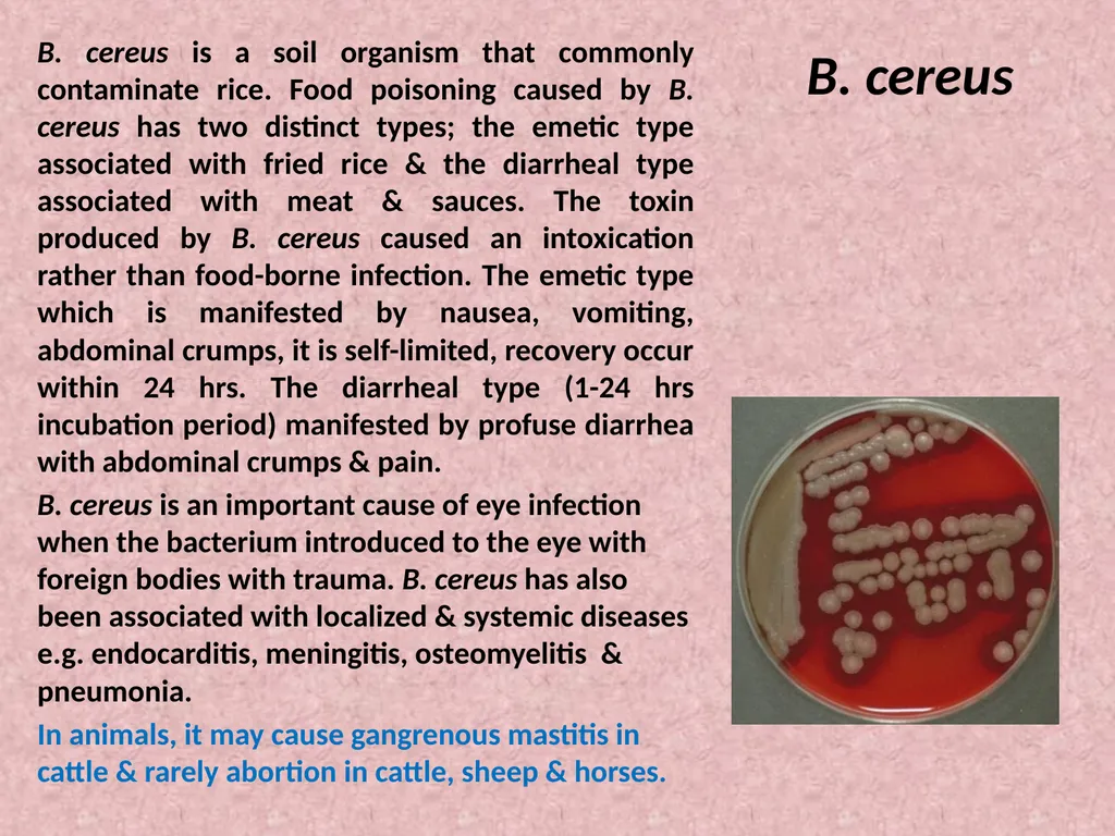
B. cereus B. cereus is a soil organism that commonly contaminate rice. Food poisoning caused by B. cereus has two distinct types; the emetic type associated with fried rice & the diarrheal type associated with meat & sauces. The toxin produced by B. cereus caused an intoxication rather than food-borne infection. The emetic type which is manifested by nausea, vomiting, abdominal crumps, it is self-limited, recovery occur within 24 hrs. The diarrheal type (1-24 hrs incubation period) manifested by profuse diarrhea with abdominal crumps & pain.
B. cereus is an important cause of eye infection when the bacterium introduced to the eye with foreign bodies with trauma. B. cereus has also been associated with localized & systemic diseases e.g. endocarditis, meningitis, osteomyelitis & pneumonia.
In animals, it may cause gangrenous mastitis in cattle & rarely abortion in cattle, sheep & horses.<br>
B. cereus is an important cause of eye infection when the bacterium introduced to the eye with foreign bodies with trauma. B. cereus has also been associated with localized & systemic diseases e.g. endocarditis, meningitis, osteomyelitis & pneumonia.
In animals, it may cause gangrenous mastitis in cattle & rarely abortion in cattle, sheep & horses.<br>
03
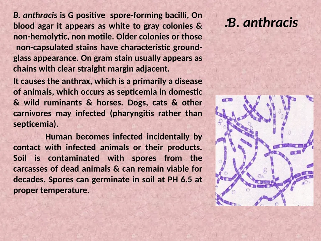
B. anthracis: B. anthracis is G positive spore-forming bacilli, On blood agar it appears as white to gray colonies & non-hemolytic, non motile. Older colonies or those non-capsulated stains have characteristic ground-glass appearance. On gram stain usually appears as chains with clear straight margin adjacent.
It causes the anthrax, which is a primarily a disease of animals, which occurs as septicemia in domestic & wild ruminants & horses. Dogs, cats & other carnivores may infected (pharyngitis rather than septicemia).
Human becomes infected incidentally by contact with infected animals or their products. Soil is contaminated with spores from the carcasses of dead animals & can remain viable for decades. Spores can germinate in soil at PH 6.5 at proper temperature.<br>
It causes the anthrax, which is a primarily a disease of animals, which occurs as septicemia in domestic & wild ruminants & horses. Dogs, cats & other carnivores may infected (pharyngitis rather than septicemia).
Human becomes infected incidentally by contact with infected animals or their products. Soil is contaminated with spores from the carcasses of dead animals & can remain viable for decades. Spores can germinate in soil at PH 6.5 at proper temperature.<br>
04
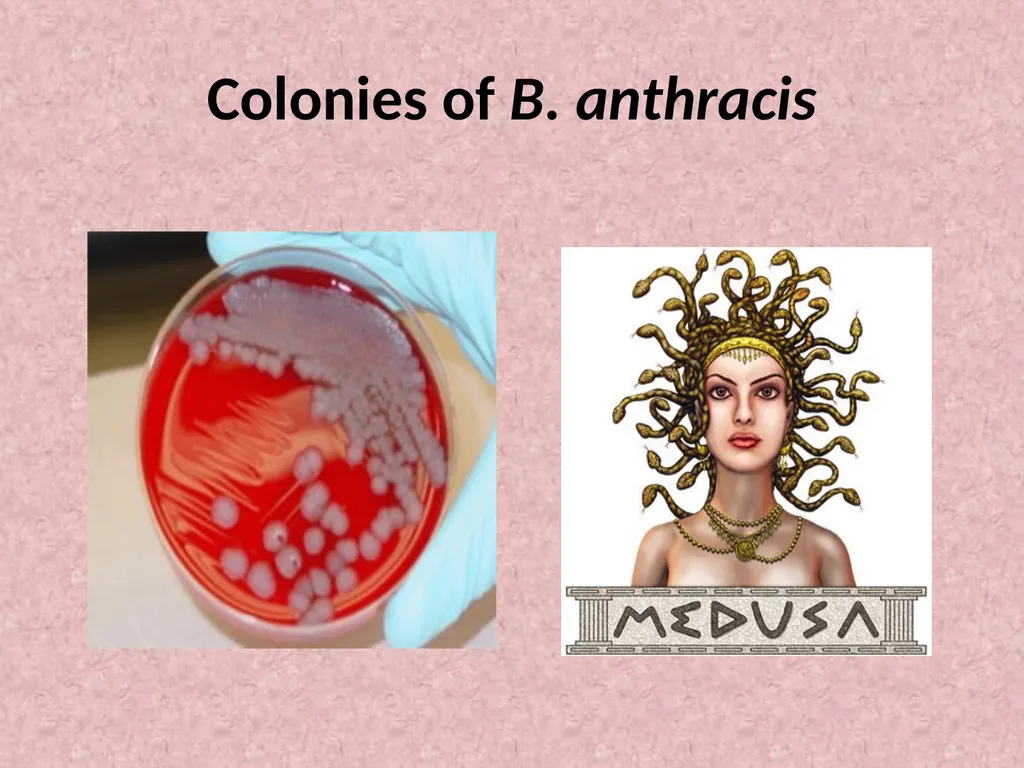
Colonies of B. anthracis<br>
05
Pathogenesis
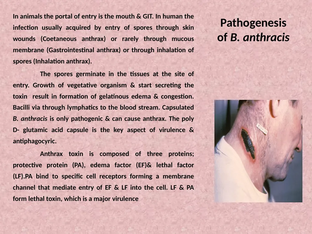
of B. anthracis In animals the portal of entry is the mouth & GIT. In human the infection usually acquired by entry of spores through skin wounds (Coetaneous anthrax) or rarely through mucous membrane (Gastrointestinal anthrax) or through inhalation of spores (Inhalation anthrax).
The spores germinate in the tissues at the site of entry. Growth of vegetative organism & start secreting the toxin result in formation of gelatinous edema & congestion. Bacilli via through lymphatics to the blood stream. Capsulated B. anthracis is only pathogenic & can cause anthrax. The poly D- glutamic acid capsule is the key aspect of virulence & antiphagocyric.
Anthrax toxin is composed of three proteins; protective protein (PA), edema factor (EF)& lethal factor (LF).PA bind to specific cell receptors forming a membrane channel that mediate entry of EF & LF into the cell. LF & PA form lethal toxin, which is a major virulence<br>
The spores germinate in the tissues at the site of entry. Growth of vegetative organism & start secreting the toxin result in formation of gelatinous edema & congestion. Bacilli via through lymphatics to the blood stream. Capsulated B. anthracis is only pathogenic & can cause anthrax. The poly D- glutamic acid capsule is the key aspect of virulence & antiphagocyric.
Anthrax toxin is composed of three proteins; protective protein (PA), edema factor (EF)& lethal factor (LF).PA bind to specific cell receptors forming a membrane channel that mediate entry of EF & LF into the cell. LF & PA form lethal toxin, which is a major virulence<br>
06
Pathogenesis
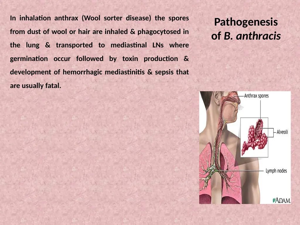
of B. anthracis In inhalation anthrax (Wool sorter disease) the spores from dust of wool or hair are inhaled & phagocytosed in the lung & transported to mediastinal LNs where germination occur followed by toxin production & development of hemorrhagic mediastinitis & sepsis that are usually fatal.<br>
07
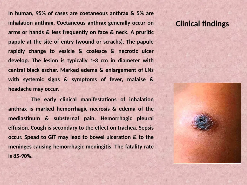
Clinical findings In human, 95% of cases are coetaneous anthrax & 5% are inhalation anthrax. Coetaneous anthrax generally occur on arms or hands & less frequently on face & neck. A pruritic papule at the site of entry (wound or scrachs). The papule rapidly change to vesicle & coalesce & necrotic ulcer develop. The lesion is typically 1-3 cm in diameter with central black eschar. Marked edema & enlargement of LNs with systemic signs & symptoms of fever, malaise & headache may occur.
The early clinical manifestations of inhalation anthrax is marked hemorrhagic necrosis & edema of the mediastinum & substernal pain. Hemorrhagic pleural effusion. Cough is secondary to the effect on trachea. Sepsis occur. Spead to GIT may lead to bowel ulceration & to the meninges causing hemorrhagic meningitis. The fatality rate is 85-90%.<br>
The early clinical manifestations of inhalation anthrax is marked hemorrhagic necrosis & edema of the mediastinum & substernal pain. Hemorrhagic pleural effusion. Cough is secondary to the effect on trachea. Sepsis occur. Spead to GIT may lead to bowel ulceration & to the meninges causing hemorrhagic meningitis. The fatality rate is 85-90%.<br>
08
Animal anthrax The incubation period of anthrax is variable, which is usually 3-7 days, but may be short 24 hrs or longer 2 weeks.
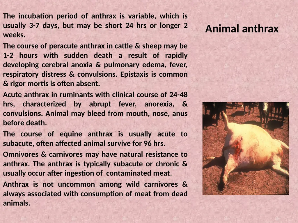
The course of peracute anthrax in cattle & sheep may be 1-2 hours with sudden death a result of rapidly developing cerebral anoxia & pulmonary edema, fever, respiratory distress & convulsions. Epistaxis is common & rigor mortis is often absent.
Acute anthrax in ruminants with clinical course of 24-48 hrs, characterized by abrupt fever, anorexia, & convulsions. Animal may bleed from mouth, nose, anus before death.
The course of equine anthrax is usually acute to subacute, often affected animal survive for 96 hrs.
Omnivores & carnivores may have natural resistance to anthrax. The anthrax is typically subacute or chronic & usually occur after ingestion of contaminated meat.
Anthrax is not uncommon among wild carnivores & always associated with consumption of meat from dead animals.<br>
The course of peracute anthrax in cattle & sheep may be 1-2 hours with sudden death a result of rapidly developing cerebral anoxia & pulmonary edema, fever, respiratory distress & convulsions. Epistaxis is common & rigor mortis is often absent.
Acute anthrax in ruminants with clinical course of 24-48 hrs, characterized by abrupt fever, anorexia, & convulsions. Animal may bleed from mouth, nose, anus before death.
The course of equine anthrax is usually acute to subacute, often affected animal survive for 96 hrs.
Omnivores & carnivores may have natural resistance to anthrax. The anthrax is typically subacute or chronic & usually occur after ingestion of contaminated meat.
Anthrax is not uncommon among wild carnivores & always associated with consumption of meat from dead animals.<br>
09
Laboratory diagnosis - specimens : fluid or pus from local lesion. Blood, sputum.
- Gram- stained smear usually revealed chains of large G positive rods. Dried smear may be stained by fluorescent stain.
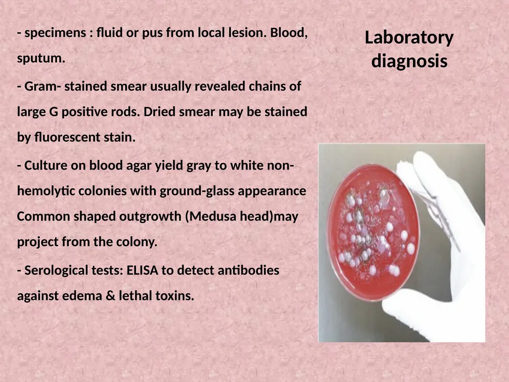
- Culture on blood agar yield gray to white non-hemolytic colonies with ground-glass appearance Common shaped outgrowth (Medusa head)may project from the colony.
- Serological tests: ELISA to detect antibodies against edema & lethal toxins.<br>
- Gram- stained smear usually revealed chains of large G positive rods. Dried smear may be stained by fluorescent stain.
- Culture on blood agar yield gray to white non-hemolytic colonies with ground-glass appearance Common shaped outgrowth (Medusa head)may project from the colony.
- Serological tests: ELISA to detect antibodies against edema & lethal toxins.<br>
10
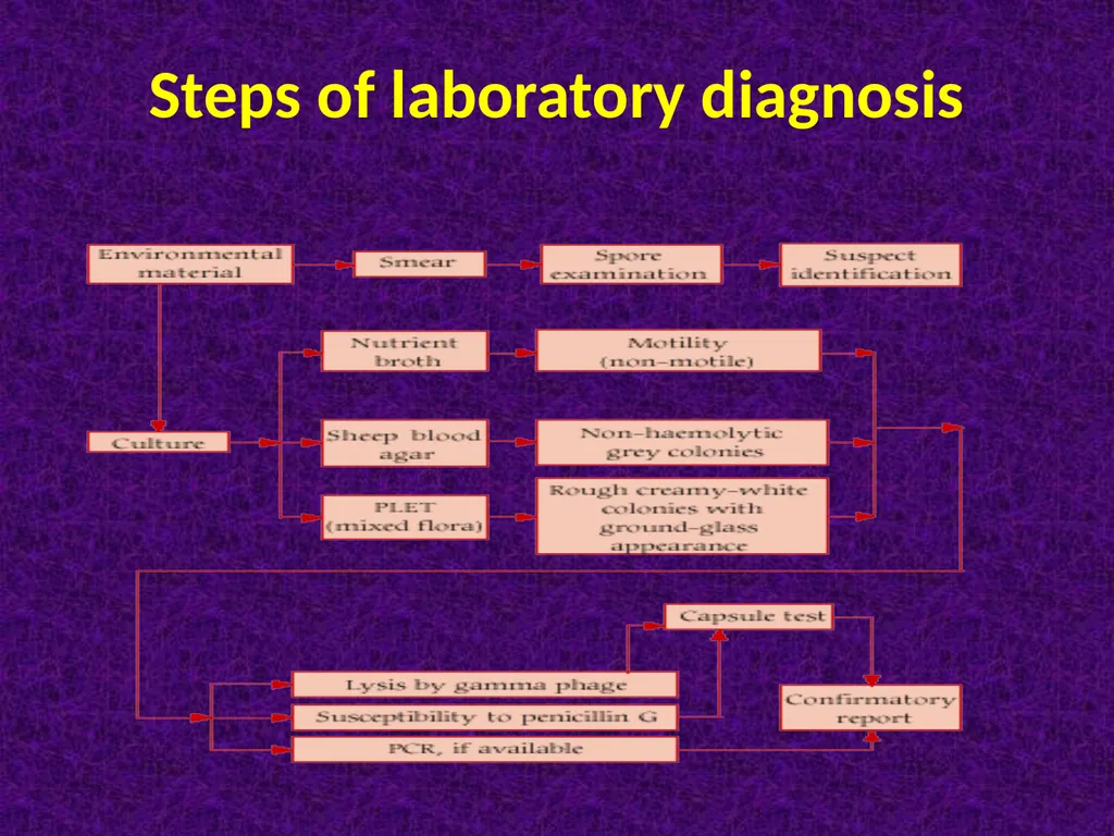
Steps of laboratory diagnosis<br>
11
Clostridia These are large anaerobic G positive rods. Their natural habitat is the soil ( exogenous) & intestine of animals & man (endogenous). Most pathogenic clostridia produce one or more toxins, which linked to their pathogenesis.
animal pathogenic clostridia can be categorized as neurotoxic (Cl. Botulinum, Cl. Tetani), enteric (Cl. Sordellii, Cl. Perfringens, Cl. Difficile), and histotoxic (Cl. Perfringens, Cl. Septicum, Cl. Chauvoei & Cl. novyi).
Morphology & identification:
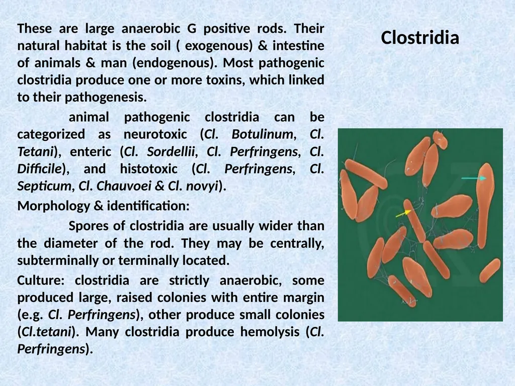
Spores of clostridia are usually wider than the diameter of the rod. They may be centrally, subterminally or terminally located.
Culture: clostridia are strictly anaerobic, some produced large, raised colonies with entire margin (e.g. Cl. Perfringens), other produce small colonies (Cl.tetani). Many clostridia produce hemolysis (Cl. Perfringens).<br>
animal pathogenic clostridia can be categorized as neurotoxic (Cl. Botulinum, Cl. Tetani), enteric (Cl. Sordellii, Cl. Perfringens, Cl. Difficile), and histotoxic (Cl. Perfringens, Cl. Septicum, Cl. Chauvoei & Cl. novyi).
Morphology & identification:
Spores of clostridia are usually wider than the diameter of the rod. They may be centrally, subterminally or terminally located.
Culture: clostridia are strictly anaerobic, some produced large, raised colonies with entire margin (e.g. Cl. Perfringens), other produce small colonies (Cl.tetani). Many clostridia produce hemolysis (Cl. Perfringens).<br>
12
Neurotoxic clostridia
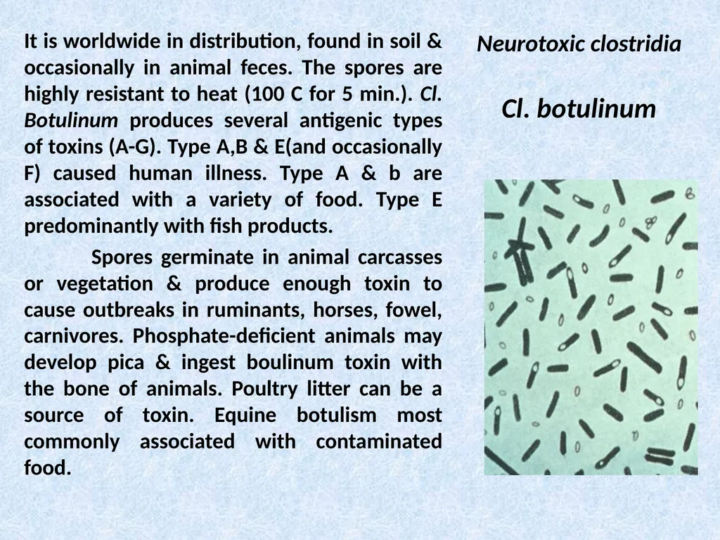
Cl. botulinum It is worldwide in distribution, found in soil & occasionally in animal feces. The spores are highly resistant to heat (100 C for 5 min.). Cl. Botulinum produces several antigenic types of toxins (A-G). Type A,B & E(and occasionally F) caused human illness. Type A & b are associated with a variety of food. Type E predominantly with fish products.
Spores germinate in animal carcasses or vegetation & produce enough toxin to cause outbreaks in ruminants, horses, fowel, carnivores. Phosphate-deficient animals may develop pica & ingest boulinum toxin with the bone of animals. Poultry litter can be a source of toxin. Equine botulism most commonly associated with contaminated food.<br>
Spores germinate in animal carcasses or vegetation & produce enough toxin to cause outbreaks in ruminants, horses, fowel, carnivores. Phosphate-deficient animals may develop pica & ingest boulinum toxin with the bone of animals. Poultry litter can be a source of toxin. Equine botulism most commonly associated with contaminated food.<br>
13
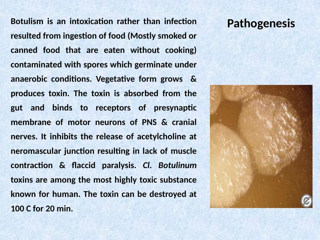
Pathogenesis Botulism is an intoxication rather than infection resulted from ingestion of food (Mostly smoked or canned food that are eaten without cooking) contaminated with spores which germinate under anaerobic conditions. Vegetative form grows & produces toxin. The toxin is absorbed from the gut and binds to receptors of presynaptic membrane of motor neurons of PNS & cranial nerves. It inhibits the release of acetylcholine at neromascular junction resulting in lack of muscle contraction & flaccid paralysis. Cl. Botulinum toxins are among the most highly toxic substance known for human. The toxin can be destroyed at 100 C for 20 min.<br>
14
Clinical findings Symptoms begin 18-24 hrs. after ingestion of toxic food with visual disturbances, inability to swallow & speech difficulty. Death occurs due to respiratory paralysis or cardiac arrest. The mortality rate is high. Recovered patients do not develop serum antitoxin.
Infant botulism is more common than classical paralysis botulism associated with ingestion of toxin-contaminated food. Infants in the first months of life develop poor feeding, weakness & signs of paralysis (floppy baby). Infant botulism may be one of the causes of sudden infant death syndrome.
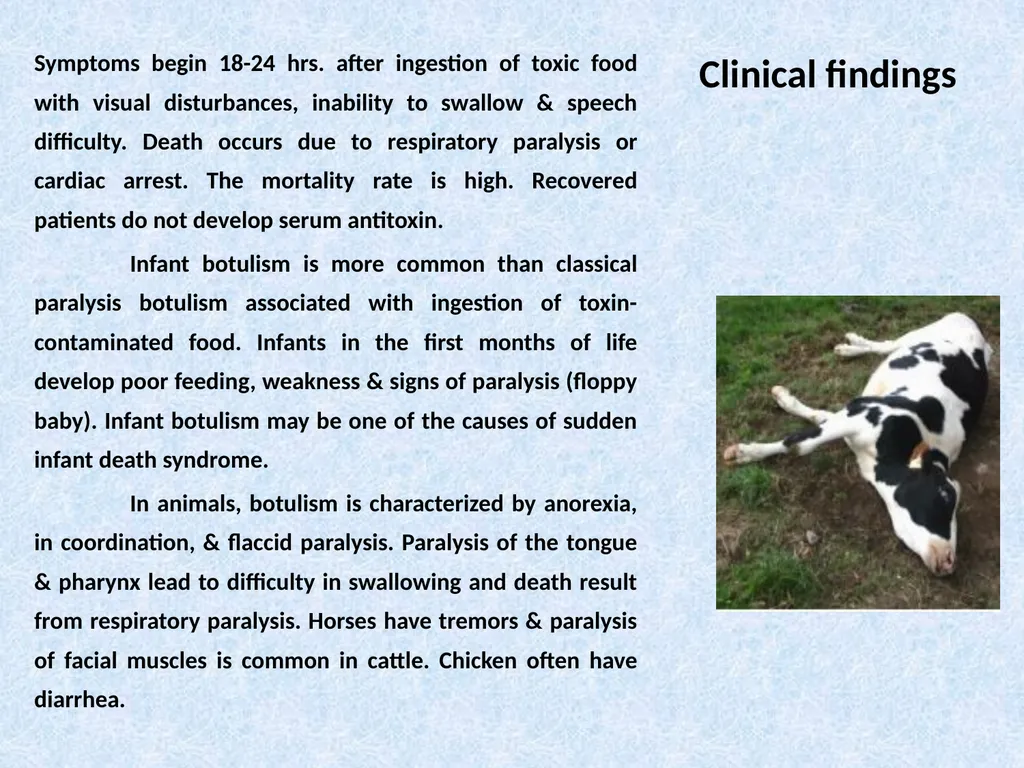
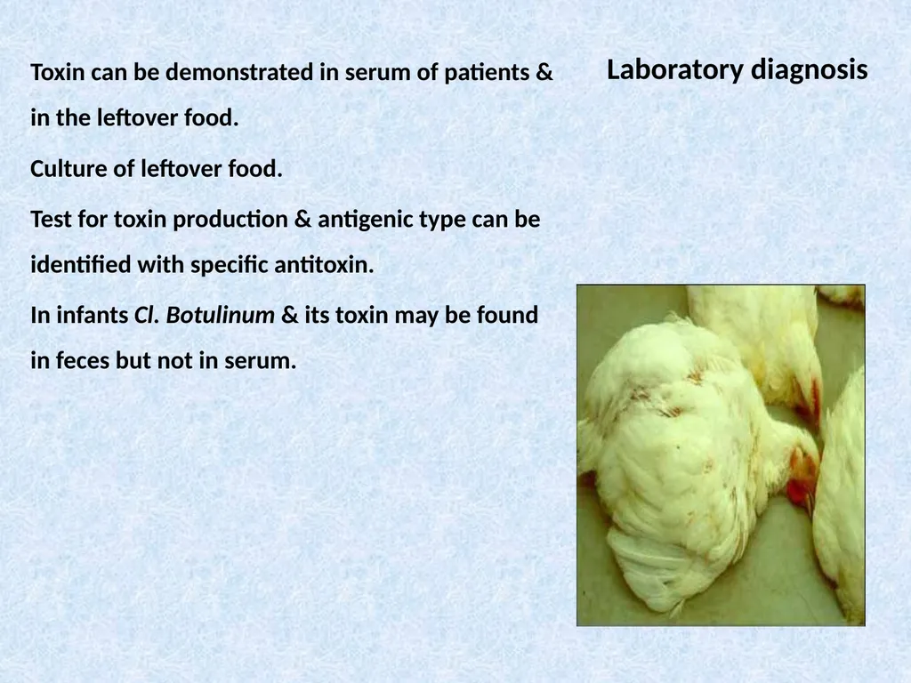
In animals, botulism is characterized by anorexia, in coordination, & flaccid paralysis. Paralysis of the tongue & pharynx lead to difficulty in swallowing and death result from respiratory paralysis. Horses have tremors & paralysis of facial muscles is common in cattle. Chicken often have diarrhea.<br>
Infant botulism is more common than classical paralysis botulism associated with ingestion of toxin-contaminated food. Infants in the first months of life develop poor feeding, weakness & signs of paralysis (floppy baby). Infant botulism may be one of the causes of sudden infant death syndrome.
In animals, botulism is characterized by anorexia, in coordination, & flaccid paralysis. Paralysis of the tongue & pharynx lead to difficulty in swallowing and death result from respiratory paralysis. Horses have tremors & paralysis of facial muscles is common in cattle. Chicken often have diarrhea.<br>
15
Laboratory diagnosis Toxin can be demonstrated in serum of patients & in the leftover food.
Culture of leftover food.
Test for toxin production & antigenic type can be identified with specific antitoxin.
In infants Cl. Botulinum & its toxin may be found in feces but not in serum.<br>
Culture of leftover food.
Test for toxin production & antigenic type can be identified with specific antitoxin.
In infants Cl. Botulinum & its toxin may be found in feces but not in serum.<br>