
The Meniscus: Injuries, Management and








Description: The Meniscus: Injuries, Management and Interventions in Rehabilitation Zac Snow PT, DPT Director of Rehabilitation Advanced Orthopaedic Specialists powered by Incite Rehab Fayetteville Rogers, Arkansas 2018 Razorback Sports Medicine
Related Topics
Download Presentation
"The Meniscus: Injuries, Management and" is the property of its rightful owner. Permission is granted to download and print the materials on this website for personal, non-commercial use only, and to display it on your personal computer provided you do not modify the materials and that you retain all copyright notices contained in the materials. By downloading content from our website, you accept the terms of this agreement.
Presentation Transcript
slide1. The Meniscus: Injuries, Management and Interventions in Rehabilitation Zac Snow PT, DPT
Director of Rehabilitation
Advanced Orthopaedic Specialists powered by Incite Rehab
Fayetteville & Rogers, Arkansas 2018 Razorback Sports Medicine Symposium
Saturday, February 24th<br>
slide2. Objectives Anatomy
Identify anatomical structures of the menisci and related structures
Evaluation
Recognize the mechanism of injury
Understand the common history and diagnostics
Operative Management/Post-Operative Management
Comprehend post-operative implications
Apply implications to rehabilitation timeline and goals
Apply knowledge of exercise to restore functional movement of the patient
Non-Operative Management
Use prior knowledge of anatomy and mechanism of injury for outcomes
Apply knowledge of exercise to restore functional movement of the patient<br>
slide3. Anatomy Medial and Lateral Menisci<br>
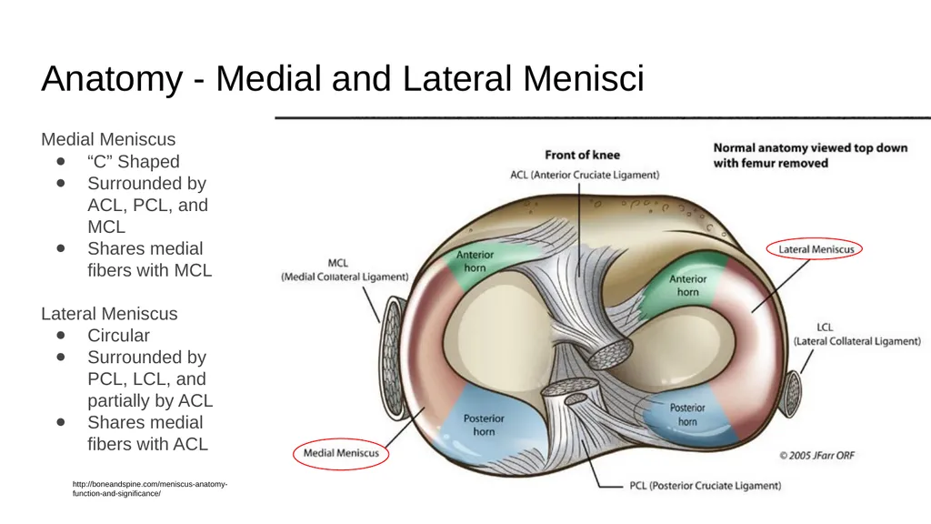
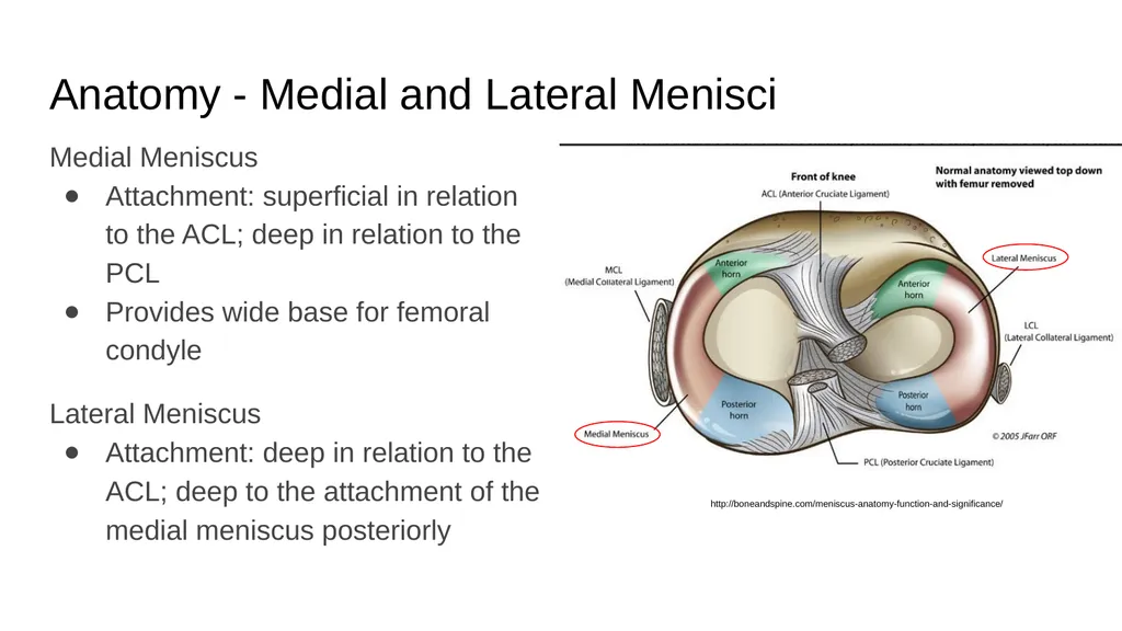
slide4. Anatomy - Medial and Lateral Menisci Medial Meniscus
“C” Shaped
Surrounded by ACL, PCL, and MCL
Shares medial fibers with MCL
Lateral Meniscus
Circular
Surrounded by PCL, LCL, and partially by ACL
Shares medial fibers with ACL http://boneandspine.com/meniscus-anatomy-function-and-significance/<br>
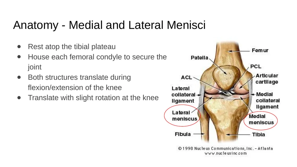
slide5. Anatomy - Medial and Lateral Menisci Rest atop the tibial plateau
House each femoral condyle to secure the joint
Both structures translate during flexion/extension of the knee
Translate with slight rotation at the knee<br>
slide6. Anatomy - Medial and Lateral Menisci Medial Meniscus
Attachment: superficial in relation to the ACL; deep in relation to the PCL
Provides wide base for femoral condyle
Lateral Meniscus
Attachment: deep in relation to the ACL; deep to the attachment of the medial meniscus posteriorly http://boneandspine.com/meniscus-anatomy-function-and-significance/<br>
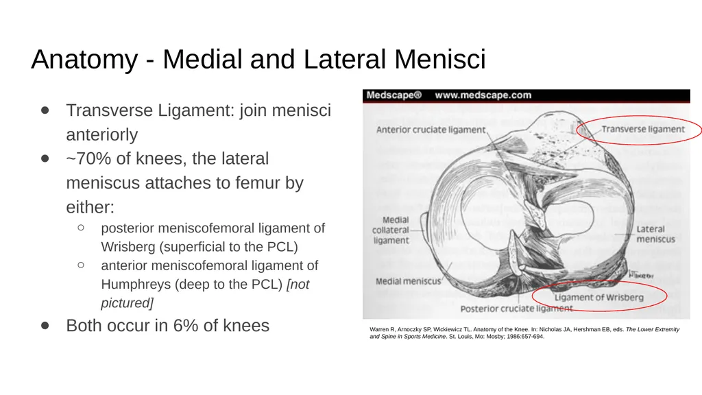
slide7. Anatomy - Medial and Lateral Menisci Transverse Ligament: join menisci anteriorly
~70% of knees, the lateral meniscus attaches to femur by either:
posterior meniscofemoral ligament of Wrisberg (superficial to the PCL)
anterior meniscofemoral ligament of Humphreys (deep to the PCL) [not pictured]
Both occur in 6% of knees Warren R, Arnoczky SP, Wickiewicz TL. Anatomy of the Knee. In: Nicholas JA, Hershman EB, eds. The Lower Extremity and Spine in Sports Medicine. St. Louis, Mo: Mosby; 1986:657-694.<br>
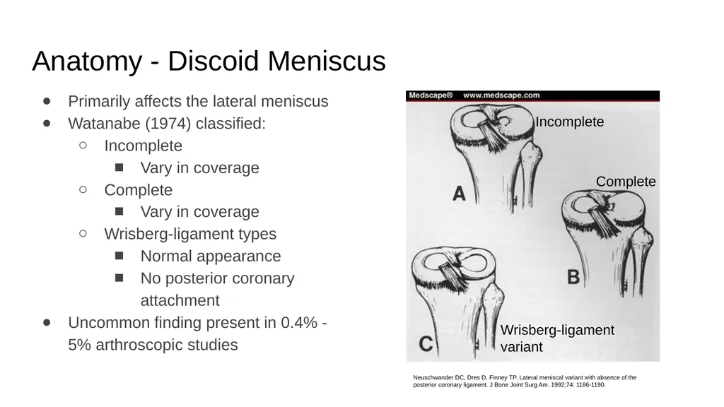
slide8. Anatomy - Discoid Meniscus Primarily affects the lateral meniscus
Watanabe (1974) classified:
Incomplete
Vary in coverage
Complete
Vary in coverage
Wrisberg-ligament types
Normal appearance
No posterior coronary attachment
Uncommon finding present in 0.4% - 5% arthroscopic studies Neuschwander DC, Dres D, Finney TP. Lateral meniscal variant with absence of the posterior coronary ligament. J Bone Joint Surg Am. 1992;74: 1186-1190. Incomplete Complete Wrisberg-ligament variant<br>
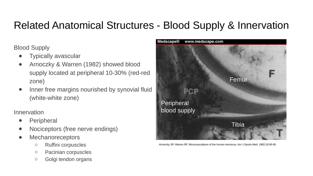
slide9. Related Anatomical Structures - Blood Supply & Innervation Blood Supply
Typically avascular
Arnoczky & Warren (1982) showed blood supply located at peripheral 10-30% (red-red zone)
Inner free margins nourished by synovial fluid (white-white zone)
Innervation
Peripheral
Nociceptors (free nerve endings)
Mechanoreceptors
Ruffini corpuscles
Pacinian corpuscles
Golgi tendon organs Arnoczky SP, Warren RF. Microvasculature of the human meniscus. Am J Sports Med. 1982;10:90-95. Femur Tibia Peripheral blood supply<br>
slide10. Related Anatomy Collateral Ligaments
Cruciate Ligaments
Synovium
“Hoop” Stress Principle<br>
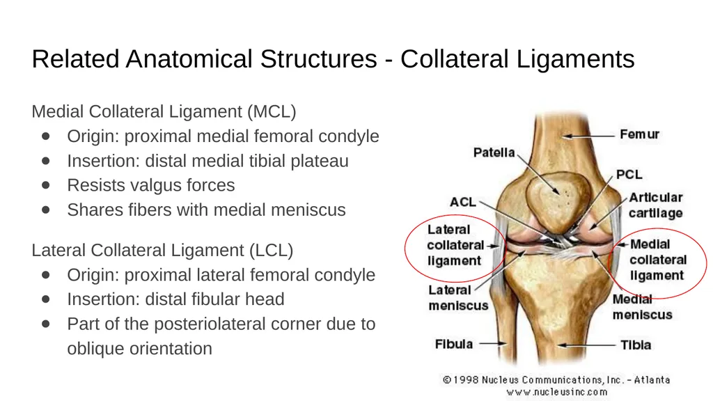
slide11. Related Anatomical Structures - Collateral Ligaments Medial Collateral Ligament (MCL)
Origin: proximal medial femoral condyle
Insertion: distal medial tibial plateau
Resists valgus forces
Shares fibers with medial meniscus
Lateral Collateral Ligament (LCL)
Origin: proximal lateral femoral condyle
Insertion: distal fibular head
Part of the posteriolateral corner due to oblique orientation<br>
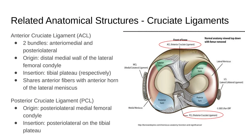
slide12. Related Anatomical Structures - Cruciate Ligaments Anterior Cruciate Ligament (ACL)
2 bundles: anteriomedial and posteriolateral
Origin: distal medial wall of the lateral femoral condyle
Insertion: tibial plateau (respectively)
Shares anterior fibers with anterior horn of the lateral meniscus
Posterior Cruciate Ligament (PCL)
Origin: posteriolateral medial femoral condyle
Insertion: posteriolateral on the tibial plateau http://boneandspine.com/meniscus-anatomy-function-and-significance/<br>
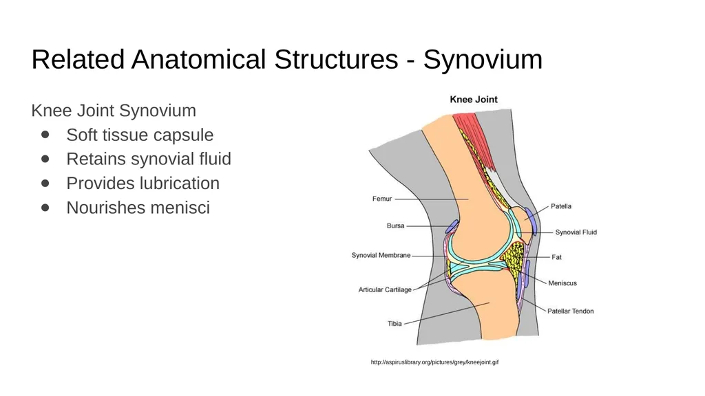
slide13. Related Anatomical Structures - Synovium Knee Joint Synovium
Soft tissue capsule
Retains synovial fluid
Provides lubrication
Nourishes menisci http://aspiruslibrary.org/pictures/grey/kneejoint.gif<br>
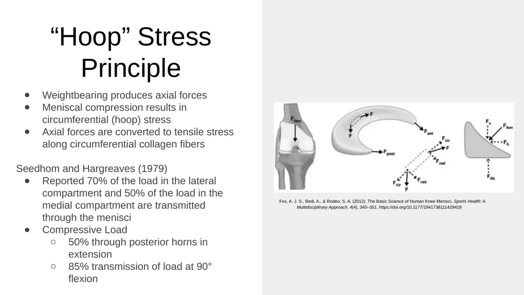
slide14. “Hoop” Stress Principle Weightbearing produces axial forces
Meniscal compression results in circumferential (hoop) stress
Axial forces are converted to tensile stress along circumferential collagen fibers
Seedhom and Hargreaves (1979)
Reported 70% of the load in the lateral compartment and 50% of the load in the medial compartment are transmitted through the menisci
Compressive Load
50% through posterior horns in extension
85% transmission of load at 90° flexion Fox, A. J. S., Bedi, A., & Rodeo, S. A. (2012). The Basic Science of Human Knee Menisci. Sports Health: A Multidisciplinary Approach, 4(4), 340–351. https://doi.org/10.1177/1941738111429419<br>
slide15. Evaluation Mechanism of Injury
Tear Patterns
History
Diagnostics<br>
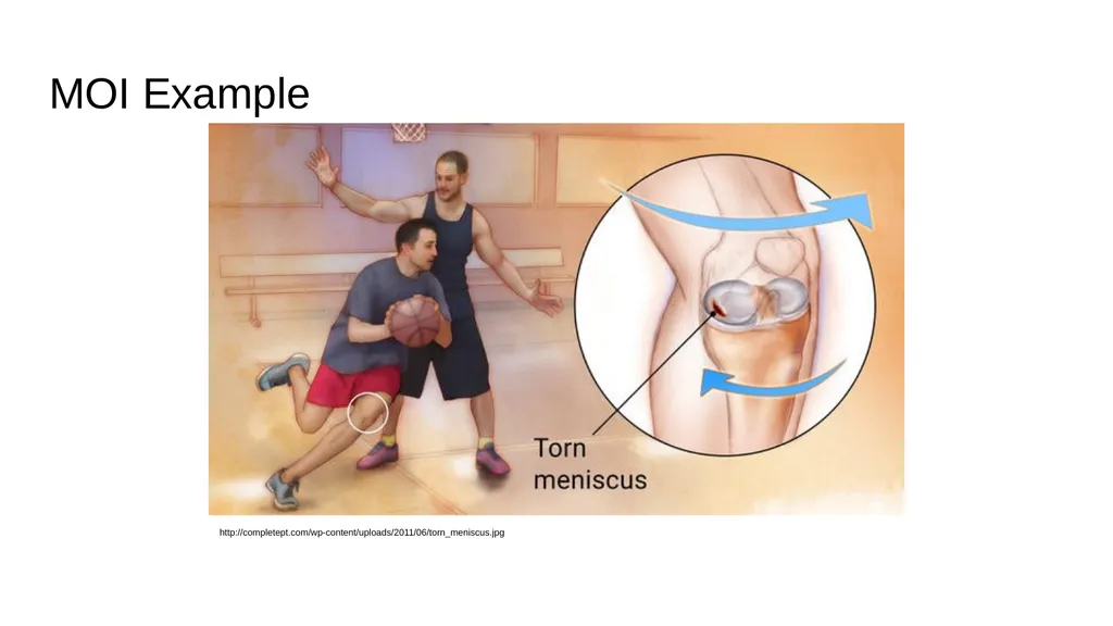
slide16. Mechanism of Injury (MOI) Internal or external rotation of the knee upon a flexed knee during a weightbearing task with or without ligamentous injury
Can be an excessive force on a healthy meniscus or a normal force on a degenerative meniscus<br>
slide17. MOI Example http://completept.com/wp-content/uploads/2011/06/torn_meniscus.jpg<br>
slide18. Types of Tears
&
Differentiating Tear Patterns<br>
slide19. Acute/Traumatic Tears Commonly the result of physical activity
Men present with overall higher incidence; often with bucket-handle tears
Women present with more peripheral detachment
Have a specific pattern (horizontal, vertical, radial, oblique or complex)
Often treated with surgery (meniscal repair, meniscectomy, meniscal allograft) followed by physical therapy Commonly observed in older individuals (55+)
Requires minimal stress or trauma
Can be managed with a combination of anti-inflammatories and physical therapy Chronic/Degenerative Tears<br>
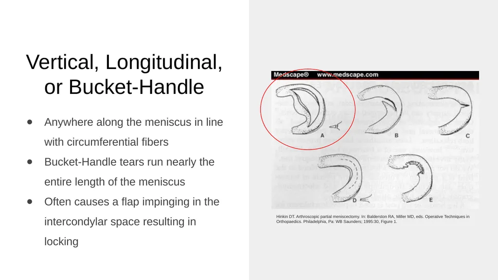
slide20. Vertical, Longitudinal, or Bucket-Handle Anywhere along the meniscus in line with circumferential fibers
Bucket-Handle tears run nearly the entire length of the meniscus
Often causes a flap impinging in the intercondylar space resulting in locking Hinkin DT. Arthroscopic partial meniscectomy. In: Balderston RA, Miller MD, eds. Operative Techniques in Orthopaedics. Philadelphia, Pa: WB Saunders; 1995:30, Figure 1.<br>
slide21. Flap, Oblique, or Parrot Beak Most common
Occurs at the posterior and middle thirds of the meniscus Hinkin DT. Arthroscopic partial meniscectomy. In: Balderston RA, Miller MD, eds. Operative Techniques in Orthopaedics. Philadelphia, Pa: WB Saunders; 1995:30, Figure 1.<br>
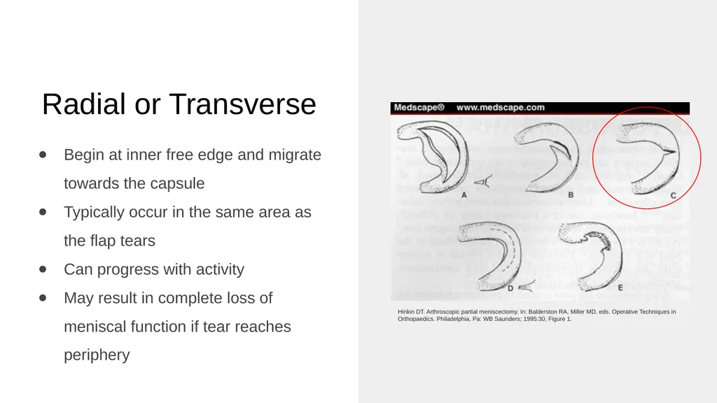
slide22. Radial or Transverse Begin at inner free edge and migrate towards the capsule
Typically occur in the same area as the flap tears
Can progress with activity
May result in complete loss of meniscal function if tear reaches periphery Hinkin DT. Arthroscopic partial meniscectomy. In: Balderston RA, Miller MD, eds. Operative Techniques in Orthopaedics. Philadelphia, Pa: WB Saunders; 1995:30, Figure 1.<br>
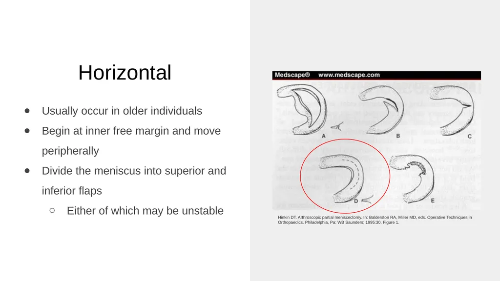
slide23. Horizontal Usually occur in older individuals
Begin at inner free margin and move peripherally
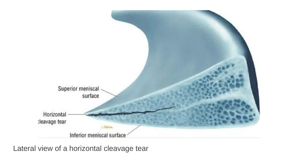
Divide the meniscus into superior and inferior flaps
Either of which may be unstable Hinkin DT. Arthroscopic partial meniscectomy. In: Balderston RA, Miller MD, eds. Operative Techniques in Orthopaedics. Philadelphia, Pa: WB Saunders; 1995:30, Figure 1.<br>
slide24. Lateral view of a horizontal cleavage tear<br>
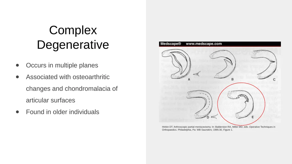
slide25. Complex Degenerative Occurs in multiple planes
Associated with osteoarthritic changes and chondromalacia of articular surfaces
Found in older individuals Hinkin DT. Arthroscopic partial meniscectomy. In: Balderston RA, Miller MD, eds. Operative Techniques in Orthopaedics. Philadelphia, Pa: WB Saunders; 1995:30, Figure 1.<br>
slide26. Exam & Diagnostics<br>
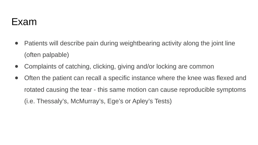
slide27. Exam Patients will describe pain during weightbearing activity along the joint line (often palpable)
Complaints of catching, clicking, giving and/or locking are common
Often the patient can recall a specific instance where the knee was flexed and rotated causing the tear - this same motion can cause reproducible symptoms (i.e. Thessaly’s, McMurray’s, Ege’s or Apley’s Tests)<br>
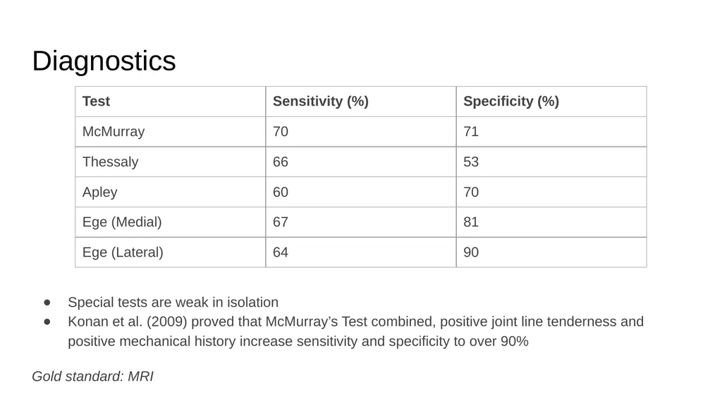
slide28. Diagnostics Special tests are weak in isolation
Konan et al. (2009) proved that McMurray’s Test combined, positive joint line tenderness and positive mechanical history increase sensitivity and specificity to over 90%
Gold standard: MRI<br>
slide29. Management Operative
Post-Operative
Non-Operative<br>
slide30. Management Observation
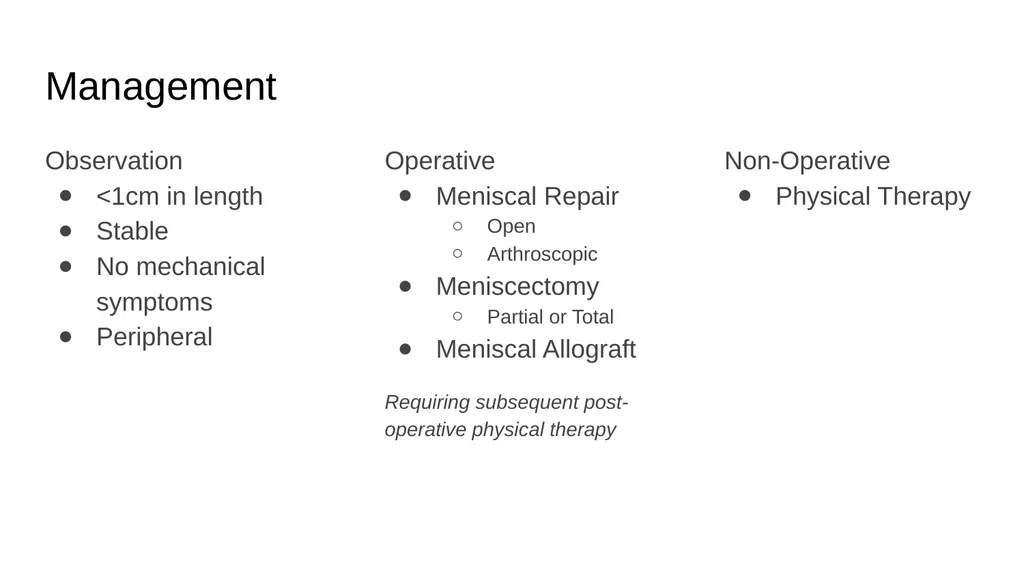
<1cm in length
Stable
No mechanical symptoms
Peripheral Operative
Meniscal Repair
Open
Arthroscopic
Meniscectomy
Partial or Total
Meniscal Allograft
Requiring subsequent post-operative physical therapy Non-Operative
Physical Therapy<br>
slide31. Meniscal Repair<br>
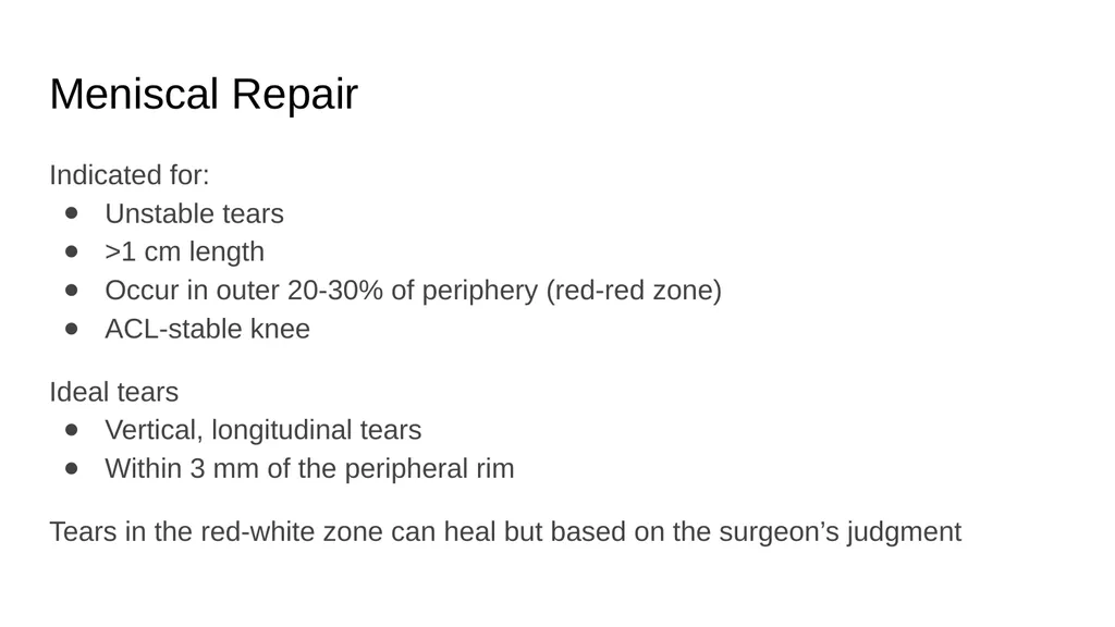
slide32. Meniscal Repair Indicated for:
Unstable tears
>1 cm length
Occur in outer 20-30% of periphery (red-red zone)
ACL-stable knee
Ideal tears
Vertical, longitudinal tears
Within 3 mm of the peripheral rim
Tears in the red-white zone can heal but based on the surgeon’s judgment<br>
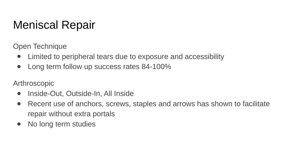
slide33. Meniscal Repair Open Technique
Limited to peripheral tears due to exposure and accessibility
Long term follow up success rates 84-100%
Arthroscopic
Inside-Out, Outside-In, All Inside
Recent use of anchors, screws, staples and arrows has shown to facilitate repair without extra portals
No long term studies<br>
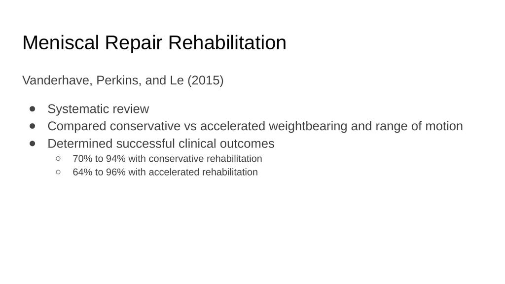
slide34. Meniscal Repair Rehabilitation Vanderhave, Perkins, and Le (2015)
Systematic review
Compared conservative vs accelerated weightbearing and range of motion
Determined successful clinical outcomes
70% to 94% with conservative rehabilitation
64% to 96% with accelerated rehabilitation<br>
slide35. Meniscal Repair Rehabilitation Immediate weightbearing and range of motion shows no significant difference in outcomes compared to delayed range of motion and weightbearing<br>
slide36. Meniscal Repair Rehabilitation Lind et al. (2013)
60 meniscal repairs
Age 18-50
2 groups
Restricted rehab (n=28)
Free rehab (n=32)
Similar knee arthritis outcome, Tegner and patient satisfaction scores at every follow-up<br>
slide37. Meniscal Repair Rehabilitation Lee and Diduch. (2005)
32 ACL-R with meniscal repair of vertical or longitudinal tears in the red-red or red-white zones
Allowed immediate full weightbearing and full range of motion
At a 2.3 year follow-up, 90% were deemed successful based on a lack of joint line effusion or tenderness, no mechanical symptoms and no meniscectomy
At a 6.6 year follow-up, 28 patients were available and yielded a 71% success rate<br>
slide38. Meniscal Repair Rehabilitation Mariani et al. (1996)
22 meniscal repairs began immediate full weightbearing and range of motion
MRI were conducted at 28 month follow-up
3 of the 22 showed clinical signs of retear<br>
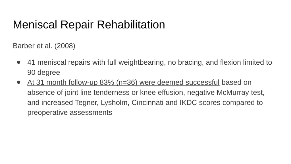
slide39. Meniscal Repair Rehabilitation Barber et al. (2008)
41 meniscal repairs with full weightbearing, no bracing, and flexion limited to 90 degree
At 31 month follow-up 83% (n=36) were deemed successful based on absence of joint line tenderness or knee effusion, negative McMurray test, and increased Tegner, Lysholm, Cincinnati and IKDC scores compared to preoperative assessments<br>
slide40. Meniscectomy<br>
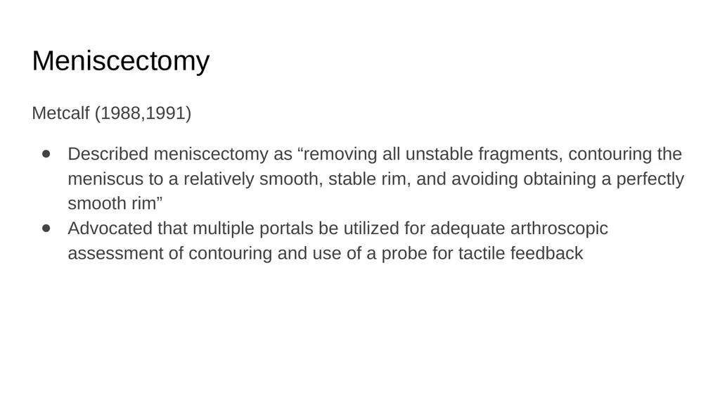
slide41. Meniscectomy Metcalf (1988,1991)
Described meniscectomy as “removing all unstable fragments, contouring the meniscus to a relatively smooth, stable rim, and avoiding obtaining a perfectly smooth rim”
Advocated that multiple portals be utilized for adequate arthroscopic assessment of contouring and use of a probe for tactile feedback<br>
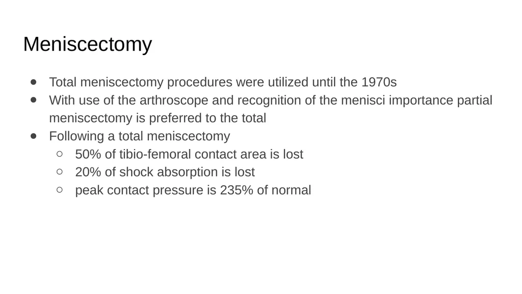
slide42. Meniscectomy Total meniscectomy procedures were utilized until the 1970s
With use of the arthroscope and recognition of the menisci importance partial meniscectomy is preferred to the total
Following a total meniscectomy
50% of tibio-femoral contact area is lost
20% of shock absorption is lost
peak contact pressure is 235% of normal<br>
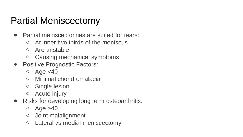
slide43. Partial Meniscectomy Partial meniscectomies are suited for tears:
At inner two thirds of the meniscus
Are unstable
Causing mechanical symptoms
Positive Prognostic Factors:
Age <40
Minimal chondromalacia
Single lesion
Acute injury
Risks for developing long term osteoarthritis:
Age >40
Joint malalignment
Lateral vs medial meniscectomy<br>
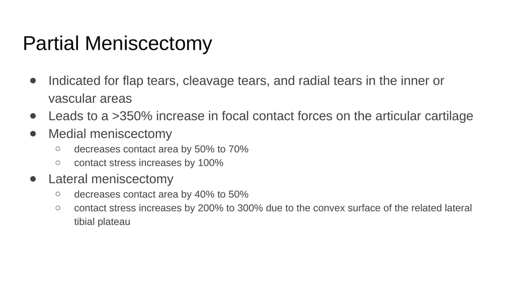
slide44. Partial Meniscectomy Indicated for flap tears, cleavage tears, and radial tears in the inner or vascular areas
Leads to a >350% increase in focal contact forces on the articular cartilage
Medial meniscectomy
decreases contact area by 50% to 70%
contact stress increases by 100%
Lateral meniscectomy
decreases contact area by 40% to 50%
contact stress increases by 200% to 300% due to the convex surface of the related lateral tibial plateau<br>
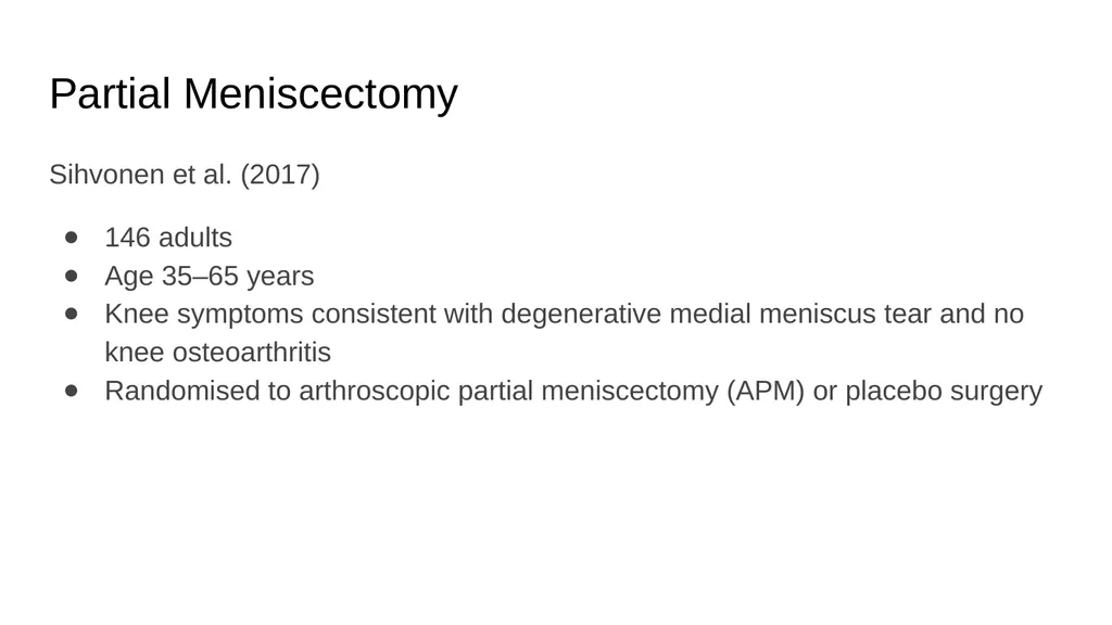
slide45. Partial Meniscectomy Sihvonen et al. (2017)
146 adults
Age 35–65 years
Knee symptoms consistent with degenerative medial meniscus tear and no knee osteoarthritis
Randomised to arthroscopic partial meniscectomy (APM) or placebo surgery<br>
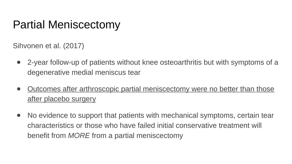
slide46. Partial Meniscectomy Sihvonen et al. (2017)
2-year follow-up of patients without knee osteoarthritis but with symptoms of a degenerative medial meniscus tear
Outcomes after arthroscopic partial meniscectomy were no better than those after placebo surgery
No evidence to support that patients with mechanical symptoms, certain tear characteristics or those who have failed initial conservative treatment will benefit from MORE from a partial meniscectomy<br>
slide47. Meniscal Allograft Transplantation<br>
slide48. Meniscal Allograft Harvested from a donor
Procured according to standards of the American Association of Tissue Banks
Long term studies display that allografts healed peripherally similar to meniscal repairs
Long term function of transplanted tissues has not been established<br>
slide49. Meniscal Allograft Technique:
Anterior to posterior tibial width is measured by lateral x-ray
Arthroscopic procedure
Performed with or without bone plugs
using bone plugs increases stability of graft and bone to bone healing
Once fixed, the meniscal repair technique of choice is performed<br>
slide50. Meniscal Allograft Indicated for:
Previous subtotal/total meniscectomy
Compartmental pain
Early osteoarthritis
Contraindicated for:
Advanced osteoarthritis
Excessive knee varus or valgus Procedure difficulties:
Graft processing
Donor cell preservation
Immunogenecity
Sterilization<br>
slide51. Meniscal Allograft Rehabilitation No consensus on weightbearing or range of motion following meniscal allograft transplant<br>
slide52. Meniscal Allograft Rehabilitation Paul C. Rijk’s (2004)
Systematic review
Found “full weight bearing immediately after operation showed uneventful healing of transplanted meniscal allografts in several experimental studies”
This was in animal models<br>
slide53. Meniscal Allograft Rehabilitation ElAttar et al. (2011)
Found most authors agreed upon immediate range of motion
Weightbearing varied depending on the trial
As tolerated immediately with crutches
Delayed 6 weeks
Delayed up to 14 weeks<br>
slide54. Meniscal Allograft Rehabilitation Advanced Orthopaedic Specialists
0-4 weeks
NWB
No ROM
5 weeks
50% WB
ROM as tolerated
6 weeks
WBAT Brigham and Women’s Hospital
0-2 weeks
PWB (<50%)
ROM 0-90 degrees, NWB only
2-6 weeks
WBAT, discontinue crutches at 4 weeks
2-8 weeks
ROM as tolerated, NWB only
8 weeks
Full ROM OrthoIndy
0-2 weeks
ROM 0-60 CPM only
2-4 weeks
ROM 0-90 CPM only
4 weeks
Full ROM
0-6 weeks
Weight bearing
Increase as tolerated after suture removal
No WB with flexion >60 degrees<br>
slide55. Post-Op Implications Arthrogenic Muscle Inhibition
Interventions<br>
slide56. Arthrogenic Muscle Inhibition (AMI)<br>
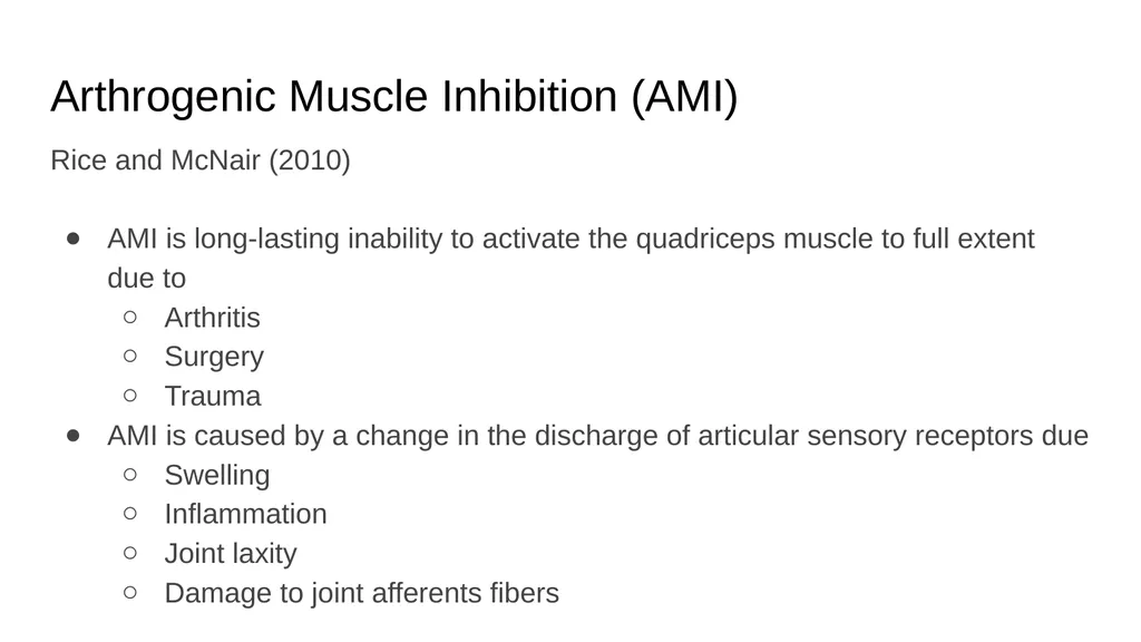
slide57. Arthrogenic Muscle Inhibition (AMI) Rice and McNair (2010)
AMI is long-lasting inability to activate the quadriceps muscle to full extent due to
Arthritis
Surgery
Trauma
AMI is caused by a change in the discharge of articular sensory receptors due
Swelling
Inflammation
Joint laxity
Damage to joint afferents fibers<br>
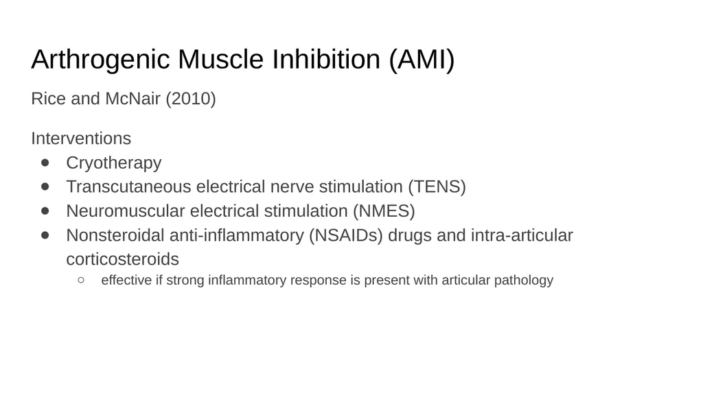
slide58. Arthrogenic Muscle Inhibition (AMI) Rice and McNair (2010)
Interventions
Cryotherapy
Transcutaneous electrical nerve stimulation (TENS)
Neuromuscular electrical stimulation (NMES)
Nonsteroidal anti-inflammatory (NSAIDs) drugs and intra-articular corticosteroids
effective if strong inflammatory response is present with articular pathology<br>
slide59. Post-Op Interventions<br>
slide60. Post-Op Interventions Meniscal Repair
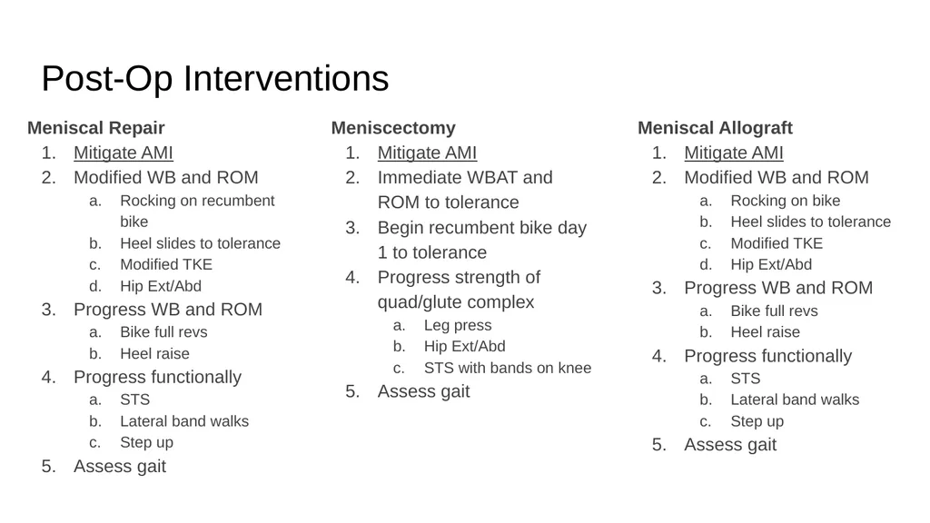
Mitigate AMI
Modified WB and ROM
Rocking on recumbent bike
Heel slides to tolerance
Modified TKE
Hip Ext/Abd
Progress WB and ROM
Bike full revs
Heel raise
Progress functionally
STS
Lateral band walks
Step up
Assess gait Meniscal Allograft
Mitigate AMI
Modified WB and ROM
Rocking on bike
Heel slides to tolerance
Modified TKE
Hip Ext/Abd
Progress WB and ROM
Bike full revs
Heel raise
Progress functionally
STS
Lateral band walks
Step up
Assess gait Meniscectomy
Mitigate AMI
Immediate WBAT and ROM to tolerance
Begin recumbent bike day 1 to tolerance
Progress strength of quad/glute complex
Leg press
Hip Ext/Abd
STS with bands on knee
Assess gait<br>
slide61. Post-Op Interventions AMI can last years depending on the patient and the intensity of the surgery.
Encourage patients to come back for a bout of PT annually until they are satisfied with their functional state following a meniscal repair or allograft.
Adjunct Techniques:
Manual Therapy
Dry Needling<br>
slide62. Non-Operative Management Physical Therapy<br>
slide63. Non-Operative Rehabilitation Physical Therapy can provide relief for meniscal tears.
If injury is due to trauma the PT still must address (or at least consider) that AMI is occurring.<br>
slide64. Non-Operative Rehabilitation Kise et al. (2016)
140 adults
Mean age 49.5 years
Degenerative medial meniscal tear verified by MRI
No radiological osteoarthritis
Performed supervised exercise therapy 2-3x/wk for 12 weeks or arthroscopic medial meniscectomy<br>
slide65. Non-Operative Rehabilitation Kise et al. (2016)
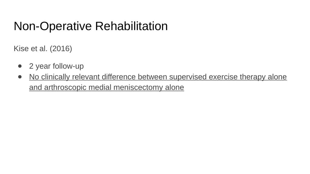
2 year follow-up
No clinically relevant difference between supervised exercise therapy alone and arthroscopic medial meniscectomy alone<br>
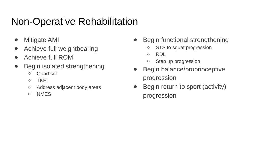
slide66. Non-Operative Rehabilitation Mitigate AMI
Achieve full weightbearing
Achieve full ROM
Begin isolated strengthening
Quad set
TKE
Address adjacent body areas
NMES Begin functional strengthening
STS to squat progression
RDL
Step up progression
Begin balance/proprioceptive progression
Begin return to sport (activity) progression<br>
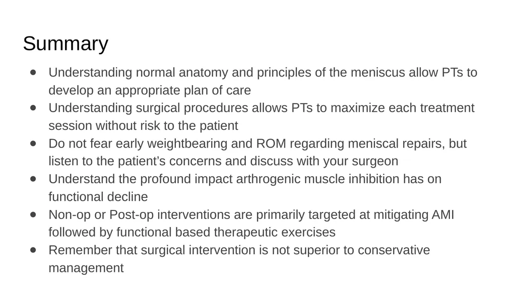
slide67. Summary Understanding normal anatomy and principles of the meniscus allow PTs to develop an appropriate plan of care
Understanding surgical procedures allows PTs to maximize each treatment session without risk to the patient
Do not fear early weightbearing and ROM regarding meniscal repairs, but listen to the patient’s concerns and discuss with your surgeon
Understand the profound impact arthrogenic muscle inhibition has on functional decline
Non-op or Post-op interventions are primarily targeted at mitigating AMI followed by functional based therapeutic exercises
Remember that surgical intervention is not superior to conservative management<br>
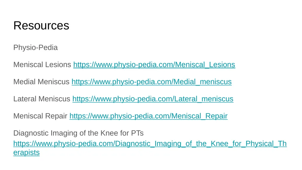
slide68. Resources Physio-Pedia
Meniscal Lesions https://www.physio-pedia.com/Meniscal_Lesions
Medial Meniscus https://www.physio-pedia.com/Medial_meniscus
Lateral Meniscus https://www.physio-pedia.com/Lateral_meniscus
Meniscal Repair https://www.physio-pedia.com/Meniscal_Repair
Diagnostic Imaging of the Knee for PTs https://www.physio-pedia.com/Diagnostic_Imaging_of_the_Knee_for_Physical_Therapists<br>
slide69. Resources Arthrex
Provides animated and cadaveric procedure videos
https://www.arthrex.com/knee/meniscal-tear-deficiency/?types=vid,ani&locales=en&taxonomy=meniscal_tear_deficiency&time=0&sort=relevance<br>
slide70. Sources Anatomy
T. Liu-Ambrose, MSc, PT, PhD (C). The anterior cruciate ligament and functional stability of the knee joint. BCMJ, Vol. 45, No. 10, December, 2003, page(s) 495-499 — Articles.
A review of the anatomical, biomechanical and kinematic findings of posterior cruciate ligament injury with respect to non-operative management
Chandrasekaran, Sivashankar et al. The Knee , Volume 19 , Issue 6 , 738 - 745
Moatshe, Gilbert et al. “Posterior Meniscal Root Injuries: A Comprehensive Review from Anatomy to Surgical Treatment.” Acta Orthopaedica 87.5 (2016): 452–458. PMC. Web. 21 Jan. 2018.
Thieme Atlas of Anatomy: General Anatomy and Musculoskeletal System. Thieme. 2006. pp. 393–395. ISBN 1-58890-419-9.
Platzer, Werner (2004). Color Atlas of Human Anatomy, Vol. 1: Locomotor System (5th ed.). Thieme. ISBN 3-13-533305-1.
Thieme Atlas of Anatomy: General Anatomy and Musculoskeletal System. Thieme. 2006. ISBN 1-58890-419-9.
Watanabe M. Arthroscopy of the knee joint. In: Helfet AJ, ed. Disorders of the Knee. Philadelphia, Pa.: Lippincott; 1974:45.
Arnoczky SP, Warren RF. Microvasculature of the human meniscus. Am J Sports Med. 1982;10:90-95.
Gray JC: Neural and vascular anatomy of the menisci of the human knee. J Orthop Sports Phys Ther 29:23–30, 1999.
Mine T, Kimura M, Sakka A, et al.: Innervation of nociceptors in the menisci of the knee joint: An immunohistochemical study. Arch Orthop Trauma Surg 120:201–204, 2000.
Fox, A. J. S., Bedi, A., & Rodeo, S. A. (2012). The Basic Science of Human Knee Menisci: Structure, Composition, and Function. Sports Health, 4(4), 340–351. http://doi.org/10.1177/1941738111429419<br>
slide71. Sources Exam
Hede, A., Jensen, D. B., Blyme, P., & Sonne-Holm, S. (1990). Epidemiology of meniscal lesions in the knee: 1,215 open operations in Copenhagen 1982-84.Acta orthopaedica Scandinavica, 61(5), 435 437.
Miller RH III (2003) Knee injuries. In: Canale ST (ed) Campbell’s operative orthopaedics. Mosby Elsevier, St. Louis
Milne JC, Marder RA (2001) Meniscus tears. In: Chapman MW (ed) Chapman’s orthopaedic surgery. Lippincott Williams & Wilkins, Philadelphia
Singh K, Helms CA, Jacobs MT, Higgins LD. MRI Appearance of Wrisberg Variant of Discoid Lateral Meniscus. AJR. Aug 2006 vol. 187 no. 2 384-387. (level: C)
Metcalf RW. Arthroscopic meniscal surgery. In: McGinty JB, ed. Operative Arthroscopy. New York, N.Y.: Raven Press; 1991:203-236.
Herschmiller T.A et al. The Trapped Medial Meniscus Tear: An Examination Maneuver Helps Predict Arthroscopic Findings; OJSM 2015
Akseki D, Özcan Ö, Boya H, Pınar H. New Weight-Bearing Meniscal Test and a Comparison With McMurray’s Test and Joint Line Tenderness. Arthroscopy: The Journal of Arthroscopic and Related Surgery 2004; Vol 20; 9:951-958
Hing W, White S, Reid D, Marshall R. Validity of the McMurray's Test and Modified Versions of the Test: A Systematic Literature Review. The Journal of Manual & Manipulative Therapy [2009, 17(1):22-35]
Daniel Bossen and Marcel Jurad. The Accuracy of Physical Examination Techniques in Diagnosing Meniscus Lesions. A Systematic Review
Konan S, Rayan F, Haddad FS. Do physical diagnostic tests accurately detect meniscal tears? Knee Surg Sports Traumatol Arthrosc. 2009;17:806–811<br>
slide72. Sources Exam
Goossens P, Keijsers E, Van Geenen RJ, Zijta A, Van den Broek M, Verhagen AP, Scholten-Peeters GG. Validity of the Thessaly test in evaluating meniscal tears compared with arthroscopy: a diagnostic accuracy study. journal of orthopaedic & sports physical therapy. 2015 Jan;45(1):18-24.
Snoeker BA, Lindeboom R, Zwinderman AH, Vincken PW, Jansen JA, Lucas C. Detecting Meniscal Tears in Primary Care: Reproducibility and Accuracy of 2 Weight-Bearing Tests and 1 Non–Weight-Bearing Test. The Journal of orthopaedic and sports physical therapy. 2015 Sep 1;45(9):693-702.
Blyth M, Anthony I, Francq B, Brooksbank K, Downie P, Powell A, Jones B, MacLean A, McConnachie A, Norrie J. Diagnostic accuracy of the Thessaly test, standardised clinical history and other clinical examination tests (Apley's, McMurray's and joint line tenderness) for meniscal tears in comparison with magnetic resonance imaging diagnosis. Health Technology Assessment. 2015.
Patrick J. McMahon (2006). Current diagnosis & treatment in sports medicine. McGraw-Hill Medical.
Scholten RJ, Deville WL, Opstelten W, Bijl D, van der Plas CG, Bouter LM. The accuracy of physical diagnostic tests for assessing meniscal lesions of the knee: a meta-analysis. J Fam Pract. 2001; 50:938-944.
Hegedus EJ, Cook C, Hasselblad V, Goode A, McCrory DC. Physical examination tests for assessing a torn meniscus in the knee: a systematic review with meta-analysis. Journal of Orthopaedic and Sports Physical Therapy, 2007; 37(9), 541-50.
Meserve BB, Cleland JA, Boucher TR. (2008) A meta-analysis examining clinical test utilities for assessing meniscal injury. Clinical Rehabilitation, 22(2), 143-61.
Hegedus EJ, Cook C, Hasselblad V, Goode A, McCrory DC. (2007)Physical examination tests for assessing a torn meniscus in the knee: a systematic review with meta-analysis. Journal of Orthopaedic and Sports Physical Therapy, 37(9), 541-50<br>
slide73. Sources Management
Metcalf RW. Arthroscopic meniscal surgery. In: McGinty JB, ed. Operative Arthroscopy. New York, N.Y.: Raven Press; 1991:203-236.
Garrett JC, Stevenson RN. Meniscal transplantation of the human knee. A preliminary report. Arthroscopy. 1991;7:57-62.
Metcalf RW. The torn medial meniscus. In: Parisien JS, ed. Arthroscopic Surgery. New York, NY: McGraw Hill; 1988:96-98.
Lind M, Nielsen T, Faune P, Lund B, Christiansen SE. Free rehabilitation is safe after isolated meniscus repair. Am J Sports Med. 2013;41:2753-2758
Mariani PP, Santori N, Adriani E, Mastantuono M. Accelerated rehabilitation after arthroscopic meniscal repair: a clinical and magnetic resonance imaging evaluation. Arthroscopy. 1996:12:680-686
Lee GP, Diduch DR. Deteriorating outcomes after meniscal repair using the meniscus arrow in knees undergoing concurrent anterior cruciate ligament reconstruction: increased failure rate with long-term follow-up. Am J Sports Med. 2005;33:1138-1141
Barber FA, Schroeder FA, Oro FB, Beavis RC. Fast-Fix meniscal repair: mid-term results. Arthroscopy. 2008;24:1342-1348
Rijk, P. C. (2004). Meniscal allograft transplantation - Part I: Background, results, graft selection and preservation, and surgical considerations. Arthroscopy - Journal of Arthroscopic and Related Surgery, 20(7), 728–743. https://doi.org/10.1016/j.arthro.2004.06.015
Sihvonen, R., Paavola, M., Malmivaara, A., Itälä, A., Joukainen, A., Nurmi, H., … Järvinen, T. L. N. (2017). Arthroscopic partial meniscectomy versus placebo surgery for a degenerative meniscus tear: A 2-year follow-up of the randomised controlled trial. Annals of the Rheumatic Diseases, 188–195. https://doi.org/10.1136/annrheumdis-2017-211172
Kise, N. J., Risberg, M. A., Stensrud, S., Ranstam, J., Engebretsen, L., & Roos, E. M. (2016). Exercise therapy versus arthroscopic partial meniscectomy for degenerative meniscal tear in middle aged patients: Randomised controlled trial with two year follow-up. BMJ (Online), 354. https://doi.org/10.1136/bmj.i3740
Rice, D. A., & McNair, P. J. (2010). Quadriceps Arthrogenic Muscle Inhibition: Neural Mechanisms and Treatment Perspectives. Seminars in Arthritis and Rheumatism, 40(3), 250–266. https://doi.org/10.1016/j.semarthrit.2009.10.001<br>
slide74. Questions? Fire away or email me at zsnow39@gmail.com<br>
Director of Rehabilitation
Advanced Orthopaedic Specialists powered by Incite Rehab
Fayetteville & Rogers, Arkansas 2018 Razorback Sports Medicine Symposium
Saturday, February 24th<br>
slide2. Objectives Anatomy
Identify anatomical structures of the menisci and related structures
Evaluation
Recognize the mechanism of injury
Understand the common history and diagnostics
Operative Management/Post-Operative Management
Comprehend post-operative implications
Apply implications to rehabilitation timeline and goals
Apply knowledge of exercise to restore functional movement of the patient
Non-Operative Management
Use prior knowledge of anatomy and mechanism of injury for outcomes
Apply knowledge of exercise to restore functional movement of the patient<br>
slide3. Anatomy Medial and Lateral Menisci<br>
slide4. Anatomy - Medial and Lateral Menisci Medial Meniscus
“C” Shaped
Surrounded by ACL, PCL, and MCL
Shares medial fibers with MCL
Lateral Meniscus
Circular
Surrounded by PCL, LCL, and partially by ACL
Shares medial fibers with ACL http://boneandspine.com/meniscus-anatomy-function-and-significance/<br>
slide5. Anatomy - Medial and Lateral Menisci Rest atop the tibial plateau
House each femoral condyle to secure the joint
Both structures translate during flexion/extension of the knee
Translate with slight rotation at the knee<br>
slide6. Anatomy - Medial and Lateral Menisci Medial Meniscus
Attachment: superficial in relation to the ACL; deep in relation to the PCL
Provides wide base for femoral condyle
Lateral Meniscus
Attachment: deep in relation to the ACL; deep to the attachment of the medial meniscus posteriorly http://boneandspine.com/meniscus-anatomy-function-and-significance/<br>
slide7. Anatomy - Medial and Lateral Menisci Transverse Ligament: join menisci anteriorly
~70% of knees, the lateral meniscus attaches to femur by either:
posterior meniscofemoral ligament of Wrisberg (superficial to the PCL)
anterior meniscofemoral ligament of Humphreys (deep to the PCL) [not pictured]
Both occur in 6% of knees Warren R, Arnoczky SP, Wickiewicz TL. Anatomy of the Knee. In: Nicholas JA, Hershman EB, eds. The Lower Extremity and Spine in Sports Medicine. St. Louis, Mo: Mosby; 1986:657-694.<br>
slide8. Anatomy - Discoid Meniscus Primarily affects the lateral meniscus
Watanabe (1974) classified:
Incomplete
Vary in coverage
Complete
Vary in coverage
Wrisberg-ligament types
Normal appearance
No posterior coronary attachment
Uncommon finding present in 0.4% - 5% arthroscopic studies Neuschwander DC, Dres D, Finney TP. Lateral meniscal variant with absence of the posterior coronary ligament. J Bone Joint Surg Am. 1992;74: 1186-1190. Incomplete Complete Wrisberg-ligament variant<br>
slide9. Related Anatomical Structures - Blood Supply & Innervation Blood Supply
Typically avascular
Arnoczky & Warren (1982) showed blood supply located at peripheral 10-30% (red-red zone)
Inner free margins nourished by synovial fluid (white-white zone)
Innervation
Peripheral
Nociceptors (free nerve endings)
Mechanoreceptors
Ruffini corpuscles
Pacinian corpuscles
Golgi tendon organs Arnoczky SP, Warren RF. Microvasculature of the human meniscus. Am J Sports Med. 1982;10:90-95. Femur Tibia Peripheral blood supply<br>
slide10. Related Anatomy Collateral Ligaments
Cruciate Ligaments
Synovium
“Hoop” Stress Principle<br>
slide11. Related Anatomical Structures - Collateral Ligaments Medial Collateral Ligament (MCL)
Origin: proximal medial femoral condyle
Insertion: distal medial tibial plateau
Resists valgus forces
Shares fibers with medial meniscus
Lateral Collateral Ligament (LCL)
Origin: proximal lateral femoral condyle
Insertion: distal fibular head
Part of the posteriolateral corner due to oblique orientation<br>
slide12. Related Anatomical Structures - Cruciate Ligaments Anterior Cruciate Ligament (ACL)
2 bundles: anteriomedial and posteriolateral
Origin: distal medial wall of the lateral femoral condyle
Insertion: tibial plateau (respectively)
Shares anterior fibers with anterior horn of the lateral meniscus
Posterior Cruciate Ligament (PCL)
Origin: posteriolateral medial femoral condyle
Insertion: posteriolateral on the tibial plateau http://boneandspine.com/meniscus-anatomy-function-and-significance/<br>
slide13. Related Anatomical Structures - Synovium Knee Joint Synovium
Soft tissue capsule
Retains synovial fluid
Provides lubrication
Nourishes menisci http://aspiruslibrary.org/pictures/grey/kneejoint.gif<br>
slide14. “Hoop” Stress Principle Weightbearing produces axial forces
Meniscal compression results in circumferential (hoop) stress
Axial forces are converted to tensile stress along circumferential collagen fibers
Seedhom and Hargreaves (1979)
Reported 70% of the load in the lateral compartment and 50% of the load in the medial compartment are transmitted through the menisci
Compressive Load
50% through posterior horns in extension
85% transmission of load at 90° flexion Fox, A. J. S., Bedi, A., & Rodeo, S. A. (2012). The Basic Science of Human Knee Menisci. Sports Health: A Multidisciplinary Approach, 4(4), 340–351. https://doi.org/10.1177/1941738111429419<br>
slide15. Evaluation Mechanism of Injury
Tear Patterns
History
Diagnostics<br>
slide16. Mechanism of Injury (MOI) Internal or external rotation of the knee upon a flexed knee during a weightbearing task with or without ligamentous injury
Can be an excessive force on a healthy meniscus or a normal force on a degenerative meniscus<br>
slide17. MOI Example http://completept.com/wp-content/uploads/2011/06/torn_meniscus.jpg<br>
slide18. Types of Tears
&
Differentiating Tear Patterns<br>
slide19. Acute/Traumatic Tears Commonly the result of physical activity
Men present with overall higher incidence; often with bucket-handle tears
Women present with more peripheral detachment
Have a specific pattern (horizontal, vertical, radial, oblique or complex)
Often treated with surgery (meniscal repair, meniscectomy, meniscal allograft) followed by physical therapy Commonly observed in older individuals (55+)
Requires minimal stress or trauma
Can be managed with a combination of anti-inflammatories and physical therapy Chronic/Degenerative Tears<br>
slide20. Vertical, Longitudinal, or Bucket-Handle Anywhere along the meniscus in line with circumferential fibers
Bucket-Handle tears run nearly the entire length of the meniscus
Often causes a flap impinging in the intercondylar space resulting in locking Hinkin DT. Arthroscopic partial meniscectomy. In: Balderston RA, Miller MD, eds. Operative Techniques in Orthopaedics. Philadelphia, Pa: WB Saunders; 1995:30, Figure 1.<br>
slide21. Flap, Oblique, or Parrot Beak Most common
Occurs at the posterior and middle thirds of the meniscus Hinkin DT. Arthroscopic partial meniscectomy. In: Balderston RA, Miller MD, eds. Operative Techniques in Orthopaedics. Philadelphia, Pa: WB Saunders; 1995:30, Figure 1.<br>
slide22. Radial or Transverse Begin at inner free edge and migrate towards the capsule
Typically occur in the same area as the flap tears
Can progress with activity
May result in complete loss of meniscal function if tear reaches periphery Hinkin DT. Arthroscopic partial meniscectomy. In: Balderston RA, Miller MD, eds. Operative Techniques in Orthopaedics. Philadelphia, Pa: WB Saunders; 1995:30, Figure 1.<br>
slide23. Horizontal Usually occur in older individuals
Begin at inner free margin and move peripherally
Divide the meniscus into superior and inferior flaps
Either of which may be unstable Hinkin DT. Arthroscopic partial meniscectomy. In: Balderston RA, Miller MD, eds. Operative Techniques in Orthopaedics. Philadelphia, Pa: WB Saunders; 1995:30, Figure 1.<br>
slide24. Lateral view of a horizontal cleavage tear<br>
slide25. Complex Degenerative Occurs in multiple planes
Associated with osteoarthritic changes and chondromalacia of articular surfaces
Found in older individuals Hinkin DT. Arthroscopic partial meniscectomy. In: Balderston RA, Miller MD, eds. Operative Techniques in Orthopaedics. Philadelphia, Pa: WB Saunders; 1995:30, Figure 1.<br>
slide26. Exam & Diagnostics<br>
slide27. Exam Patients will describe pain during weightbearing activity along the joint line (often palpable)
Complaints of catching, clicking, giving and/or locking are common
Often the patient can recall a specific instance where the knee was flexed and rotated causing the tear - this same motion can cause reproducible symptoms (i.e. Thessaly’s, McMurray’s, Ege’s or Apley’s Tests)<br>
slide28. Diagnostics Special tests are weak in isolation
Konan et al. (2009) proved that McMurray’s Test combined, positive joint line tenderness and positive mechanical history increase sensitivity and specificity to over 90%
Gold standard: MRI<br>
slide29. Management Operative
Post-Operative
Non-Operative<br>
slide30. Management Observation
<1cm in length
Stable
No mechanical symptoms
Peripheral Operative
Meniscal Repair
Open
Arthroscopic
Meniscectomy
Partial or Total
Meniscal Allograft
Requiring subsequent post-operative physical therapy Non-Operative
Physical Therapy<br>
slide31. Meniscal Repair<br>
slide32. Meniscal Repair Indicated for:
Unstable tears
>1 cm length
Occur in outer 20-30% of periphery (red-red zone)
ACL-stable knee
Ideal tears
Vertical, longitudinal tears
Within 3 mm of the peripheral rim
Tears in the red-white zone can heal but based on the surgeon’s judgment<br>
slide33. Meniscal Repair Open Technique
Limited to peripheral tears due to exposure and accessibility
Long term follow up success rates 84-100%
Arthroscopic
Inside-Out, Outside-In, All Inside
Recent use of anchors, screws, staples and arrows has shown to facilitate repair without extra portals
No long term studies<br>
slide34. Meniscal Repair Rehabilitation Vanderhave, Perkins, and Le (2015)
Systematic review
Compared conservative vs accelerated weightbearing and range of motion
Determined successful clinical outcomes
70% to 94% with conservative rehabilitation
64% to 96% with accelerated rehabilitation<br>
slide35. Meniscal Repair Rehabilitation Immediate weightbearing and range of motion shows no significant difference in outcomes compared to delayed range of motion and weightbearing<br>
slide36. Meniscal Repair Rehabilitation Lind et al. (2013)
60 meniscal repairs
Age 18-50
2 groups
Restricted rehab (n=28)
Free rehab (n=32)
Similar knee arthritis outcome, Tegner and patient satisfaction scores at every follow-up<br>
slide37. Meniscal Repair Rehabilitation Lee and Diduch. (2005)
32 ACL-R with meniscal repair of vertical or longitudinal tears in the red-red or red-white zones
Allowed immediate full weightbearing and full range of motion
At a 2.3 year follow-up, 90% were deemed successful based on a lack of joint line effusion or tenderness, no mechanical symptoms and no meniscectomy
At a 6.6 year follow-up, 28 patients were available and yielded a 71% success rate<br>
slide38. Meniscal Repair Rehabilitation Mariani et al. (1996)
22 meniscal repairs began immediate full weightbearing and range of motion
MRI were conducted at 28 month follow-up
3 of the 22 showed clinical signs of retear<br>
slide39. Meniscal Repair Rehabilitation Barber et al. (2008)
41 meniscal repairs with full weightbearing, no bracing, and flexion limited to 90 degree
At 31 month follow-up 83% (n=36) were deemed successful based on absence of joint line tenderness or knee effusion, negative McMurray test, and increased Tegner, Lysholm, Cincinnati and IKDC scores compared to preoperative assessments<br>
slide40. Meniscectomy<br>
slide41. Meniscectomy Metcalf (1988,1991)
Described meniscectomy as “removing all unstable fragments, contouring the meniscus to a relatively smooth, stable rim, and avoiding obtaining a perfectly smooth rim”
Advocated that multiple portals be utilized for adequate arthroscopic assessment of contouring and use of a probe for tactile feedback<br>
slide42. Meniscectomy Total meniscectomy procedures were utilized until the 1970s
With use of the arthroscope and recognition of the menisci importance partial meniscectomy is preferred to the total
Following a total meniscectomy
50% of tibio-femoral contact area is lost
20% of shock absorption is lost
peak contact pressure is 235% of normal<br>
slide43. Partial Meniscectomy Partial meniscectomies are suited for tears:
At inner two thirds of the meniscus
Are unstable
Causing mechanical symptoms
Positive Prognostic Factors:
Age <40
Minimal chondromalacia
Single lesion
Acute injury
Risks for developing long term osteoarthritis:
Age >40
Joint malalignment
Lateral vs medial meniscectomy<br>
slide44. Partial Meniscectomy Indicated for flap tears, cleavage tears, and radial tears in the inner or vascular areas
Leads to a >350% increase in focal contact forces on the articular cartilage
Medial meniscectomy
decreases contact area by 50% to 70%
contact stress increases by 100%
Lateral meniscectomy
decreases contact area by 40% to 50%
contact stress increases by 200% to 300% due to the convex surface of the related lateral tibial plateau<br>
slide45. Partial Meniscectomy Sihvonen et al. (2017)
146 adults
Age 35–65 years
Knee symptoms consistent with degenerative medial meniscus tear and no knee osteoarthritis
Randomised to arthroscopic partial meniscectomy (APM) or placebo surgery<br>
slide46. Partial Meniscectomy Sihvonen et al. (2017)
2-year follow-up of patients without knee osteoarthritis but with symptoms of a degenerative medial meniscus tear
Outcomes after arthroscopic partial meniscectomy were no better than those after placebo surgery
No evidence to support that patients with mechanical symptoms, certain tear characteristics or those who have failed initial conservative treatment will benefit from MORE from a partial meniscectomy<br>
slide47. Meniscal Allograft Transplantation<br>
slide48. Meniscal Allograft Harvested from a donor
Procured according to standards of the American Association of Tissue Banks
Long term studies display that allografts healed peripherally similar to meniscal repairs
Long term function of transplanted tissues has not been established<br>
slide49. Meniscal Allograft Technique:
Anterior to posterior tibial width is measured by lateral x-ray
Arthroscopic procedure
Performed with or without bone plugs
using bone plugs increases stability of graft and bone to bone healing
Once fixed, the meniscal repair technique of choice is performed<br>
slide50. Meniscal Allograft Indicated for:
Previous subtotal/total meniscectomy
Compartmental pain
Early osteoarthritis
Contraindicated for:
Advanced osteoarthritis
Excessive knee varus or valgus Procedure difficulties:
Graft processing
Donor cell preservation
Immunogenecity
Sterilization<br>
slide51. Meniscal Allograft Rehabilitation No consensus on weightbearing or range of motion following meniscal allograft transplant<br>
slide52. Meniscal Allograft Rehabilitation Paul C. Rijk’s (2004)
Systematic review
Found “full weight bearing immediately after operation showed uneventful healing of transplanted meniscal allografts in several experimental studies”
This was in animal models<br>
slide53. Meniscal Allograft Rehabilitation ElAttar et al. (2011)
Found most authors agreed upon immediate range of motion
Weightbearing varied depending on the trial
As tolerated immediately with crutches
Delayed 6 weeks
Delayed up to 14 weeks<br>
slide54. Meniscal Allograft Rehabilitation Advanced Orthopaedic Specialists
0-4 weeks
NWB
No ROM
5 weeks
50% WB
ROM as tolerated
6 weeks
WBAT Brigham and Women’s Hospital
0-2 weeks
PWB (<50%)
ROM 0-90 degrees, NWB only
2-6 weeks
WBAT, discontinue crutches at 4 weeks
2-8 weeks
ROM as tolerated, NWB only
8 weeks
Full ROM OrthoIndy
0-2 weeks
ROM 0-60 CPM only
2-4 weeks
ROM 0-90 CPM only
4 weeks
Full ROM
0-6 weeks
Weight bearing
Increase as tolerated after suture removal
No WB with flexion >60 degrees<br>
slide55. Post-Op Implications Arthrogenic Muscle Inhibition
Interventions<br>
slide56. Arthrogenic Muscle Inhibition (AMI)<br>
slide57. Arthrogenic Muscle Inhibition (AMI) Rice and McNair (2010)
AMI is long-lasting inability to activate the quadriceps muscle to full extent due to
Arthritis
Surgery
Trauma
AMI is caused by a change in the discharge of articular sensory receptors due
Swelling
Inflammation
Joint laxity
Damage to joint afferents fibers<br>
slide58. Arthrogenic Muscle Inhibition (AMI) Rice and McNair (2010)
Interventions
Cryotherapy
Transcutaneous electrical nerve stimulation (TENS)
Neuromuscular electrical stimulation (NMES)
Nonsteroidal anti-inflammatory (NSAIDs) drugs and intra-articular corticosteroids
effective if strong inflammatory response is present with articular pathology<br>
slide59. Post-Op Interventions<br>
slide60. Post-Op Interventions Meniscal Repair
Mitigate AMI
Modified WB and ROM
Rocking on recumbent bike
Heel slides to tolerance
Modified TKE
Hip Ext/Abd
Progress WB and ROM
Bike full revs
Heel raise
Progress functionally
STS
Lateral band walks
Step up
Assess gait Meniscal Allograft
Mitigate AMI
Modified WB and ROM
Rocking on bike
Heel slides to tolerance
Modified TKE
Hip Ext/Abd
Progress WB and ROM
Bike full revs
Heel raise
Progress functionally
STS
Lateral band walks
Step up
Assess gait Meniscectomy
Mitigate AMI
Immediate WBAT and ROM to tolerance
Begin recumbent bike day 1 to tolerance
Progress strength of quad/glute complex
Leg press
Hip Ext/Abd
STS with bands on knee
Assess gait<br>
slide61. Post-Op Interventions AMI can last years depending on the patient and the intensity of the surgery.
Encourage patients to come back for a bout of PT annually until they are satisfied with their functional state following a meniscal repair or allograft.
Adjunct Techniques:
Manual Therapy
Dry Needling<br>
slide62. Non-Operative Management Physical Therapy<br>
slide63. Non-Operative Rehabilitation Physical Therapy can provide relief for meniscal tears.
If injury is due to trauma the PT still must address (or at least consider) that AMI is occurring.<br>
slide64. Non-Operative Rehabilitation Kise et al. (2016)
140 adults
Mean age 49.5 years
Degenerative medial meniscal tear verified by MRI
No radiological osteoarthritis
Performed supervised exercise therapy 2-3x/wk for 12 weeks or arthroscopic medial meniscectomy<br>
slide65. Non-Operative Rehabilitation Kise et al. (2016)
2 year follow-up
No clinically relevant difference between supervised exercise therapy alone and arthroscopic medial meniscectomy alone<br>
slide66. Non-Operative Rehabilitation Mitigate AMI
Achieve full weightbearing
Achieve full ROM
Begin isolated strengthening
Quad set
TKE
Address adjacent body areas
NMES Begin functional strengthening
STS to squat progression
RDL
Step up progression
Begin balance/proprioceptive progression
Begin return to sport (activity) progression<br>
slide67. Summary Understanding normal anatomy and principles of the meniscus allow PTs to develop an appropriate plan of care
Understanding surgical procedures allows PTs to maximize each treatment session without risk to the patient
Do not fear early weightbearing and ROM regarding meniscal repairs, but listen to the patient’s concerns and discuss with your surgeon
Understand the profound impact arthrogenic muscle inhibition has on functional decline
Non-op or Post-op interventions are primarily targeted at mitigating AMI followed by functional based therapeutic exercises
Remember that surgical intervention is not superior to conservative management<br>
slide68. Resources Physio-Pedia
Meniscal Lesions https://www.physio-pedia.com/Meniscal_Lesions
Medial Meniscus https://www.physio-pedia.com/Medial_meniscus
Lateral Meniscus https://www.physio-pedia.com/Lateral_meniscus
Meniscal Repair https://www.physio-pedia.com/Meniscal_Repair
Diagnostic Imaging of the Knee for PTs https://www.physio-pedia.com/Diagnostic_Imaging_of_the_Knee_for_Physical_Therapists<br>
slide69. Resources Arthrex
Provides animated and cadaveric procedure videos
https://www.arthrex.com/knee/meniscal-tear-deficiency/?types=vid,ani&locales=en&taxonomy=meniscal_tear_deficiency&time=0&sort=relevance<br>
slide70. Sources Anatomy
T. Liu-Ambrose, MSc, PT, PhD (C). The anterior cruciate ligament and functional stability of the knee joint. BCMJ, Vol. 45, No. 10, December, 2003, page(s) 495-499 — Articles.
A review of the anatomical, biomechanical and kinematic findings of posterior cruciate ligament injury with respect to non-operative management
Chandrasekaran, Sivashankar et al. The Knee , Volume 19 , Issue 6 , 738 - 745
Moatshe, Gilbert et al. “Posterior Meniscal Root Injuries: A Comprehensive Review from Anatomy to Surgical Treatment.” Acta Orthopaedica 87.5 (2016): 452–458. PMC. Web. 21 Jan. 2018.
Thieme Atlas of Anatomy: General Anatomy and Musculoskeletal System. Thieme. 2006. pp. 393–395. ISBN 1-58890-419-9.
Platzer, Werner (2004). Color Atlas of Human Anatomy, Vol. 1: Locomotor System (5th ed.). Thieme. ISBN 3-13-533305-1.
Thieme Atlas of Anatomy: General Anatomy and Musculoskeletal System. Thieme. 2006. ISBN 1-58890-419-9.
Watanabe M. Arthroscopy of the knee joint. In: Helfet AJ, ed. Disorders of the Knee. Philadelphia, Pa.: Lippincott; 1974:45.
Arnoczky SP, Warren RF. Microvasculature of the human meniscus. Am J Sports Med. 1982;10:90-95.
Gray JC: Neural and vascular anatomy of the menisci of the human knee. J Orthop Sports Phys Ther 29:23–30, 1999.
Mine T, Kimura M, Sakka A, et al.: Innervation of nociceptors in the menisci of the knee joint: An immunohistochemical study. Arch Orthop Trauma Surg 120:201–204, 2000.
Fox, A. J. S., Bedi, A., & Rodeo, S. A. (2012). The Basic Science of Human Knee Menisci: Structure, Composition, and Function. Sports Health, 4(4), 340–351. http://doi.org/10.1177/1941738111429419<br>
slide71. Sources Exam
Hede, A., Jensen, D. B., Blyme, P., & Sonne-Holm, S. (1990). Epidemiology of meniscal lesions in the knee: 1,215 open operations in Copenhagen 1982-84.Acta orthopaedica Scandinavica, 61(5), 435 437.
Miller RH III (2003) Knee injuries. In: Canale ST (ed) Campbell’s operative orthopaedics. Mosby Elsevier, St. Louis
Milne JC, Marder RA (2001) Meniscus tears. In: Chapman MW (ed) Chapman’s orthopaedic surgery. Lippincott Williams & Wilkins, Philadelphia
Singh K, Helms CA, Jacobs MT, Higgins LD. MRI Appearance of Wrisberg Variant of Discoid Lateral Meniscus. AJR. Aug 2006 vol. 187 no. 2 384-387. (level: C)
Metcalf RW. Arthroscopic meniscal surgery. In: McGinty JB, ed. Operative Arthroscopy. New York, N.Y.: Raven Press; 1991:203-236.
Herschmiller T.A et al. The Trapped Medial Meniscus Tear: An Examination Maneuver Helps Predict Arthroscopic Findings; OJSM 2015
Akseki D, Özcan Ö, Boya H, Pınar H. New Weight-Bearing Meniscal Test and a Comparison With McMurray’s Test and Joint Line Tenderness. Arthroscopy: The Journal of Arthroscopic and Related Surgery 2004; Vol 20; 9:951-958
Hing W, White S, Reid D, Marshall R. Validity of the McMurray's Test and Modified Versions of the Test: A Systematic Literature Review. The Journal of Manual & Manipulative Therapy [2009, 17(1):22-35]
Daniel Bossen and Marcel Jurad. The Accuracy of Physical Examination Techniques in Diagnosing Meniscus Lesions. A Systematic Review
Konan S, Rayan F, Haddad FS. Do physical diagnostic tests accurately detect meniscal tears? Knee Surg Sports Traumatol Arthrosc. 2009;17:806–811<br>
slide72. Sources Exam
Goossens P, Keijsers E, Van Geenen RJ, Zijta A, Van den Broek M, Verhagen AP, Scholten-Peeters GG. Validity of the Thessaly test in evaluating meniscal tears compared with arthroscopy: a diagnostic accuracy study. journal of orthopaedic & sports physical therapy. 2015 Jan;45(1):18-24.
Snoeker BA, Lindeboom R, Zwinderman AH, Vincken PW, Jansen JA, Lucas C. Detecting Meniscal Tears in Primary Care: Reproducibility and Accuracy of 2 Weight-Bearing Tests and 1 Non–Weight-Bearing Test. The Journal of orthopaedic and sports physical therapy. 2015 Sep 1;45(9):693-702.
Blyth M, Anthony I, Francq B, Brooksbank K, Downie P, Powell A, Jones B, MacLean A, McConnachie A, Norrie J. Diagnostic accuracy of the Thessaly test, standardised clinical history and other clinical examination tests (Apley's, McMurray's and joint line tenderness) for meniscal tears in comparison with magnetic resonance imaging diagnosis. Health Technology Assessment. 2015.
Patrick J. McMahon (2006). Current diagnosis & treatment in sports medicine. McGraw-Hill Medical.
Scholten RJ, Deville WL, Opstelten W, Bijl D, van der Plas CG, Bouter LM. The accuracy of physical diagnostic tests for assessing meniscal lesions of the knee: a meta-analysis. J Fam Pract. 2001; 50:938-944.
Hegedus EJ, Cook C, Hasselblad V, Goode A, McCrory DC. Physical examination tests for assessing a torn meniscus in the knee: a systematic review with meta-analysis. Journal of Orthopaedic and Sports Physical Therapy, 2007; 37(9), 541-50.
Meserve BB, Cleland JA, Boucher TR. (2008) A meta-analysis examining clinical test utilities for assessing meniscal injury. Clinical Rehabilitation, 22(2), 143-61.
Hegedus EJ, Cook C, Hasselblad V, Goode A, McCrory DC. (2007)Physical examination tests for assessing a torn meniscus in the knee: a systematic review with meta-analysis. Journal of Orthopaedic and Sports Physical Therapy, 37(9), 541-50<br>
slide73. Sources Management
Metcalf RW. Arthroscopic meniscal surgery. In: McGinty JB, ed. Operative Arthroscopy. New York, N.Y.: Raven Press; 1991:203-236.
Garrett JC, Stevenson RN. Meniscal transplantation of the human knee. A preliminary report. Arthroscopy. 1991;7:57-62.
Metcalf RW. The torn medial meniscus. In: Parisien JS, ed. Arthroscopic Surgery. New York, NY: McGraw Hill; 1988:96-98.
Lind M, Nielsen T, Faune P, Lund B, Christiansen SE. Free rehabilitation is safe after isolated meniscus repair. Am J Sports Med. 2013;41:2753-2758
Mariani PP, Santori N, Adriani E, Mastantuono M. Accelerated rehabilitation after arthroscopic meniscal repair: a clinical and magnetic resonance imaging evaluation. Arthroscopy. 1996:12:680-686
Lee GP, Diduch DR. Deteriorating outcomes after meniscal repair using the meniscus arrow in knees undergoing concurrent anterior cruciate ligament reconstruction: increased failure rate with long-term follow-up. Am J Sports Med. 2005;33:1138-1141
Barber FA, Schroeder FA, Oro FB, Beavis RC. Fast-Fix meniscal repair: mid-term results. Arthroscopy. 2008;24:1342-1348
Rijk, P. C. (2004). Meniscal allograft transplantation - Part I: Background, results, graft selection and preservation, and surgical considerations. Arthroscopy - Journal of Arthroscopic and Related Surgery, 20(7), 728–743. https://doi.org/10.1016/j.arthro.2004.06.015
Sihvonen, R., Paavola, M., Malmivaara, A., Itälä, A., Joukainen, A., Nurmi, H., … Järvinen, T. L. N. (2017). Arthroscopic partial meniscectomy versus placebo surgery for a degenerative meniscus tear: A 2-year follow-up of the randomised controlled trial. Annals of the Rheumatic Diseases, 188–195. https://doi.org/10.1136/annrheumdis-2017-211172
Kise, N. J., Risberg, M. A., Stensrud, S., Ranstam, J., Engebretsen, L., & Roos, E. M. (2016). Exercise therapy versus arthroscopic partial meniscectomy for degenerative meniscal tear in middle aged patients: Randomised controlled trial with two year follow-up. BMJ (Online), 354. https://doi.org/10.1136/bmj.i3740
Rice, D. A., & McNair, P. J. (2010). Quadriceps Arthrogenic Muscle Inhibition: Neural Mechanisms and Treatment Perspectives. Seminars in Arthritis and Rheumatism, 40(3), 250–266. https://doi.org/10.1016/j.semarthrit.2009.10.001<br>
slide74. Questions? Fire away or email me at zsnow39@gmail.com<br>